. 2025 May 9;74(7):193.
doi: 10.1007/s00262-025-04051-x.

Osteosarcoma cell-derived CCL2 facilitates lung metastasis via accumulation of tumor-associated macrophages

Affiliations

Osteosarcoma cell-derived CCL2 facilitates lung metastasis via accumulation of tumor-associated macrophages

Hiroya Kondo et al. Cancer Immunol Immunother. .

Abstract

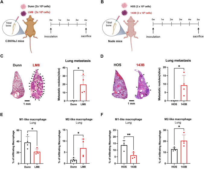
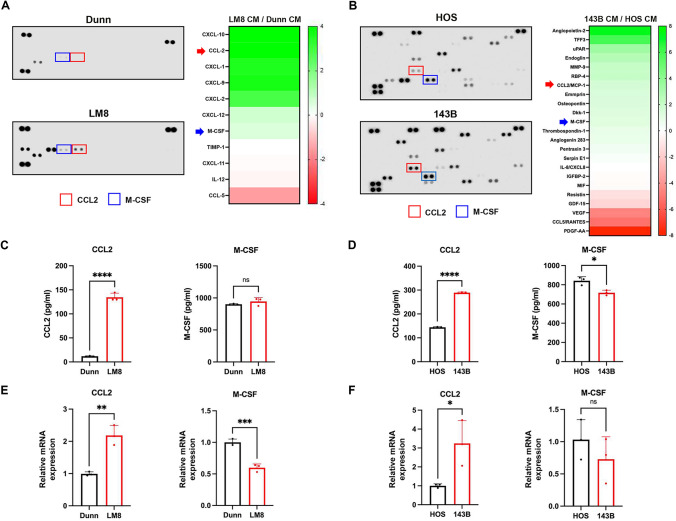
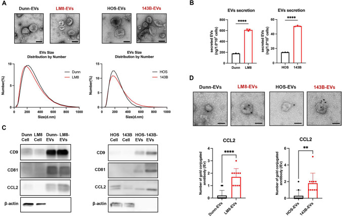
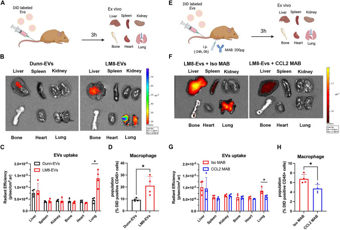
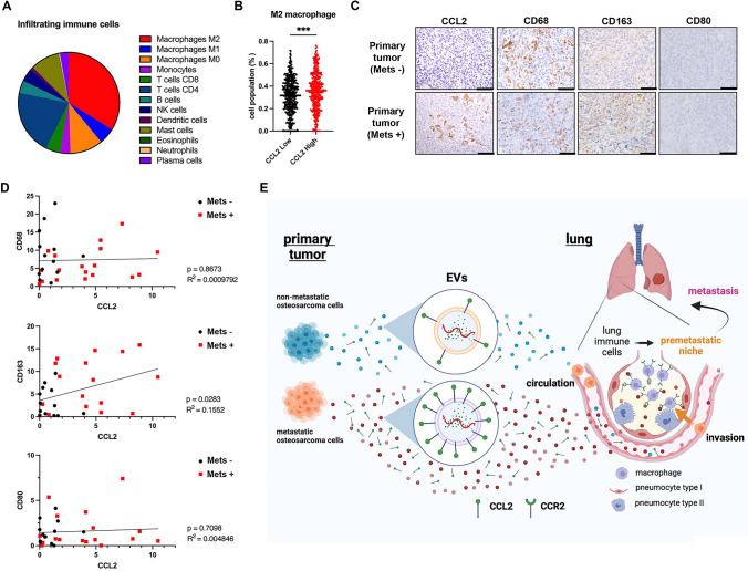
Osteosarcoma (OS) is the most common malignant tumor of bone in children and adolescents. Although lung metastasis is a major obstacle to improving the prognosis of OS patients, the underlying mechanism of lung metastasis of OS is poorly understood. Tumor-associated macrophages (TAMs) with M2-like characteristics are reportedly associated with lung metastasis and poor prognosis in OS patients. In this study, we investigated the metastasis-associated tumor microenvironment (TME) in orthotopic OS tumor models with non-metastatic and metastatic OS cells. Non-metastatic and metastatic tumor cells derived from mouse OS (Dunn and LM8) and human OS (HOS and 143B) were used to analyze the TME associated with lung metastasis in orthotopic OS tumor models. OS cell-derived secretion factors were identified by cytokine array and enzyme-linked immunosorbent assay (ELISA). Orthotopic tumor models with metastatic LM8 and 143B cells were analyzed to evaluate the therapeutic potential of a neutralizing antibody in the development of primary and metastatic tumors. Metastatic OS cells developed metastatic tumors with infiltration of M2-like TAMs in the lungs. Cytokine array and ELISA demonstrated that metastatic mouse and human OS cells commonly secreted CCL2, which was partially encapsulated in extracellular vesicles. In vivo experiments demonstrated that while primary tumor growth was unaffected, administration of CCL2-neutralizing antibody led to a significant suppression of lung metastasis and infiltration of M2-like TAMs in the lung tissue. Our results suggest that CCL2 plays a crucial role in promoting the lung metastasis of OS cells via accumulation of M2-like TAMs.

Keywords: CCL2; Extracellular vesicle; Lung metastasis; Osteosarcoma; Tumor-associated macrophage.


Conflict of interest statement

Declarations. Conflict of interest: The authors declare no competing interests.

Figures

Fig. 1
Fig. 1
Differential induction of lung metastasis and M2-like macrophage accumulation in orthotopic tumor model mice with non-metastatic and metastatic OS cells. A, B Mouse (A) and human (B) OS cells with different metastatic potentials (2 × 106 cells) were inoculated into the tibia of C3H/HeJ mice and athymic nude mice, respectively. Lung metastases and the immune microenvironment in mice that received orthotopic inoculation with mouse and human OS cells were assessed on days 28 and 35 after tumor inoculation, respectively. C, D Left panel: representative photographs of lung tissues stained with hematoxylin/eosin at low magnification. Right panel: number of metastatic nodules in lung tissues is shown as the mean ± SD (n = 4 in each group; *, P < 0.05). E, F Comparison of the polarization of macrophages in lung tissues. The percentages of M1-like and M2-like macrophages are shown as mean ± SD (n = 4 in each group; *, P < 0.05; **, P < 0.01). Statistical significance was determined using Student’s t-test. Figures were generated using BioRender
Fig. 2
Fig. 2
Identification of CCL2 commonly secreted by mouse and human OS cells with metastatic ability. A, B Conditioned medium (CM) of mouse (A) and human (B) OS cells was subjected to cytokine/chemokine array analysis. C, D The amounts of CCL2 and M-CSF in the CM were analyzed by ELISA. The mRNA expression of CCL2 and M-CSF in OS cells was analyzed by RT-PCR. Data are expressed as mean ± SD (n = 3 in each group; *, P < 0.05; **, P < 0.01; ***, P < 0.001; ****, P < 0.0001; ns, not significant). Statistical significance was determined using Student’s t-test
Fig. 3
Fig. 3
Evaluation of CCL2 expression on EVs secreted by murine and human OS cells with different metastatic potentials. A Upper panel: representative images of EVs isolated from murine and human OS cells analyzed by transmission electron microscopy. Lower panel: particle size distribution of EVs measured by dynamic light scattering. B Secretion of EVs from murine and human OS cells (1 × 107 cells) was quantified based on the amount of protein (n = 3 in each group; ****, P < 0.0001). Statistical significance was determined using Student’s t-test. C Cell lysates (Cell) and EV lysates (EVs) obtained from mouse and human OS cells were subjected to Western blotting of EV surface markers (CD9, CD81) and CCL2. D Upper panel: representative photographs of EVs labeled with anti-CCL2 antibody–conjugated gold particles. Lower panel: number of gold particles in EVs. Data are expressed as mean ± SD (n = 10 in each group; **, P < 0.01; ****, P < 0.0001). Scale bars, 100 nm
Fig. 4
Fig. 4
Biodistribution of DiD-labeled EVs secreted by murine OS cells with different metastatic abilities. A DiD-labeled EVs of mouse Dunn and LM8 cells (100 mg/mouse) were intravenously injected into the tail vein of mice. The biodistribution of fluorescently labeled EVs was assessed 3 h after injection using an In Vivo Image System. B Representative ex vivo images of fluorescence in various organs (liver, spleen, kidney, bone, heart, lung). C Quantification of DiD fluorescence in various organs. D Percentage of macrophages among DiD + CD45 + cells in lung tissues. E CCL2-neutralizing antibody (CCL2 MAB) and isotype control IgG (Iso MAB) (200 ng/mouse) were administered 24 h before and just before injection of EVs. Biodistribution of EVs was assessed 3 h after EV injection. F Representative ex vivo images of fluorescence in various organs (liver, spleen, kidney, bone, heart, lung). G Quantification of DiD fluorescence in various organs. H Percentage of macrophages among DiD + CD45 + cells in the lung tissues. Data are expressed as mean ± SD (n = 4 in each group; *, P < 0.05). Statistical significance was determined using Student’s t-test. Figures were generated using BioRender
Fig. 5
Fig. 5
CCL2-neutralizing antibody prevents lung metastasis via suppression of macrophage polarization in the lung. A, B Mouse LM8 cells (A) and human 143B cells (B) with metastatic potential (2 × 106 cells) were inoculated into the tibia of C3H/Hej mice and athymic nude mice, respectively. CCL2-neutralizing antibody (CCL2 MAB) and control isotype IgG (Iso MAB) (200 μg/mouse) were administered intraperitoneally twice per week, starting 3 days before inoculation. Lung metastases and the immune microenvironment in mice that received orthotopic inoculation with LM8 and 143B cells were assessed on days 21 and 28 after tumor inoculation, respectively. C, D Left panel: representative photographs of lung tissues stained with hematoxylin/eosin at low magnification. Right panel: number of metastatic nodules in the lung tissues is shown as mean ± SD (n = 5 in each group; *, P < 0.05; **, P < 0.01; ns, not significant). E, F Comparison of the polarization of macrophages in lung tissues. The percentages of M1-like and M2-like macrophages are shown as mean ± SD (n = 5 in each group; *, P < 0.05; ns, not significant). Statistical significance was determined using one-way ANOVA with Tukey’s post-test. Figures were generated using BioRender
Fig. 6
Fig. 6
Clinical relevance of the relationship between M2-like macrophages, CCL2, and lung metastasis in sarcomas. A Pie chart demonstrating that subsets of 13 immune cell types infiltrated in 858 sarcoma samples. B Dot plots showing the correlation between CCL2 expression and M2-like macrophages in sarcoma tissues. Statistical significance was determined using Student’s t-test. C Immunohistochemistry analysis of CCL2, CD68 (macrophages), CD163 (M2-like macrophages), and CD80 (M1-like macrophages) in primary OS tumors with or without lung metastasis. D Correlation between CCL2 expression and expression of CD68 (macrophages), CD163 (M2-like macrophages), and CD80 (M1-like macrophages). Statistical significance was determined using Pearson’s correlation coefficients. E Outline describing lung metastasis of OS cells via the accumulation of CCL2 and CCL2 + EVs secreted by metastatic OS cells. Figures were generated using BioRender

Similar articles

Cited by

References

    1. Rojas GA, Hubbard AK, Diessner BJ, Ribeiro KB, Spector LG (2021) International trends in incidence of osteosarcoma (1988–2012). Int J Cancer 149:1044–1053. 10.1002/ijc.33673 - PMC - PubMed
    1. Meltzer PS, Helman LJ (2021) New horizons in the treatment of osteosarcoma. N Engl J Med 385:2066–2076. 10.1056/NEJMra2103423 - PubMed
    1. Collins M, Wilhelm M, Conyers R et al (2013) Benefits and adverse events in younger versus older patients receiving neoadjuvant chemotherapy for osteosarcoma: findings from a meta-analysis. J Clin Oncol 31:2303–2312. 10.1200/JCO.2012.43.8598 - PubMed
    1. Kager L, Zoubek A, Potschger U et al (2003) Primary metastatic osteosarcoma: presentation and outcome of patients treated on neoadjuvant Cooperative Osteosarcoma Study Group protocols. J Clin Oncol 21:2011–2018. 10.1200/JCO.2003.08.132 - PubMed
    1. Mantovani A, Marchesi F, Malesci A, Laghi L, Allavena P (2017) Tumour-associated macrophages as treatment targets in oncology. Nat Rev Clin Oncol 14:399–416. 10.1038/nrclinonc.2016.217 - PMC - PubMed

MeSH terms

LinkOut - more resources