. 2025 May 9;74(7):192.
doi: 10.1007/s00262-025-04067-3.

HIF-PH inhibitors induce pseudohypoxia in T cells and suppress the growth of microsatellite stable colorectal cancer by enhancing antitumor immune responses

Affiliations

HIF-PH inhibitors induce pseudohypoxia in T cells and suppress the growth of microsatellite stable colorectal cancer by enhancing antitumor immune responses

Yuehua Chen et al. Cancer Immunol Immunother. .

Abstract

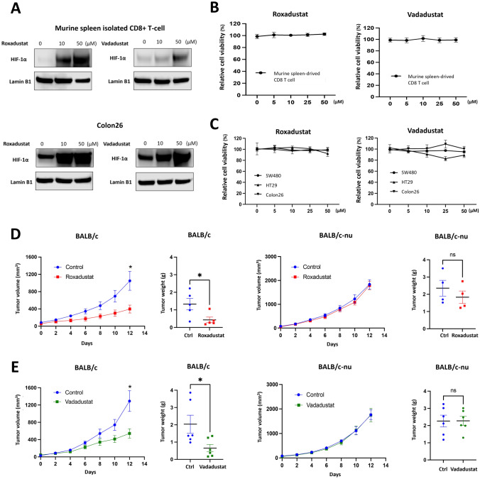
Background: Recent studies have revealed that CD8+ T cells can be activated via genetic upregulation of HIF-1α, thereby augmenting antitumor effector functions. HIF-1α upregulation can be attained by inhibiting HIF-prolyl hydroxylase (HIF-PH) under normoxic conditions, termed pseudohypoxia. This study investigated whether pseudohypoxia induced by HIF-PH inhibitors suppresses Microsatellite stable (MSS) colorectal cancer (CRC) by affecting tumor immune response.

Methods: The HIF-PH inhibitors Roxadustat and Vadadustat were utilized in this study. In vitro, we assessed the effects of HIF-PH inhibitors on human and murine colon cancer cell lines (SW480, HT29, Colon26) and murine T cells. In vivo experiments were performed with mice bearing Colon26 tumors to evaluate the effect of these inhibitors on tumor immune responses. Tumor and spleen samples were analyzed using immunohistochemistry, RT-qPCR, and flow cytometry to elucidate potential mechanisms.

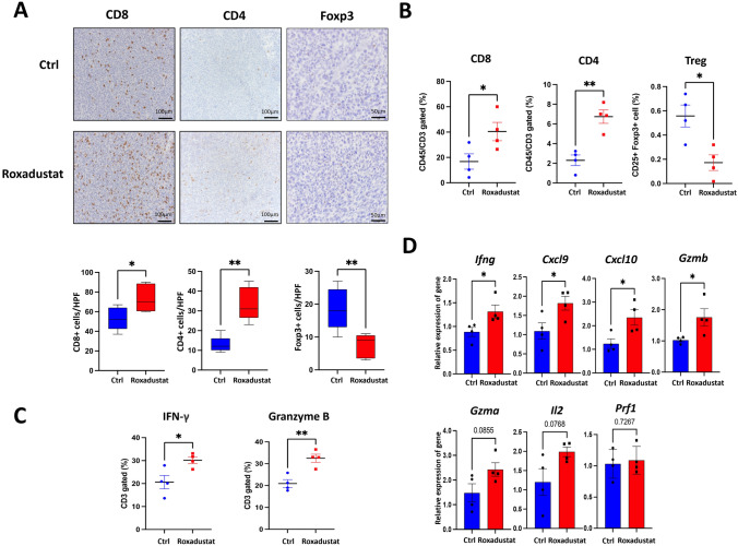
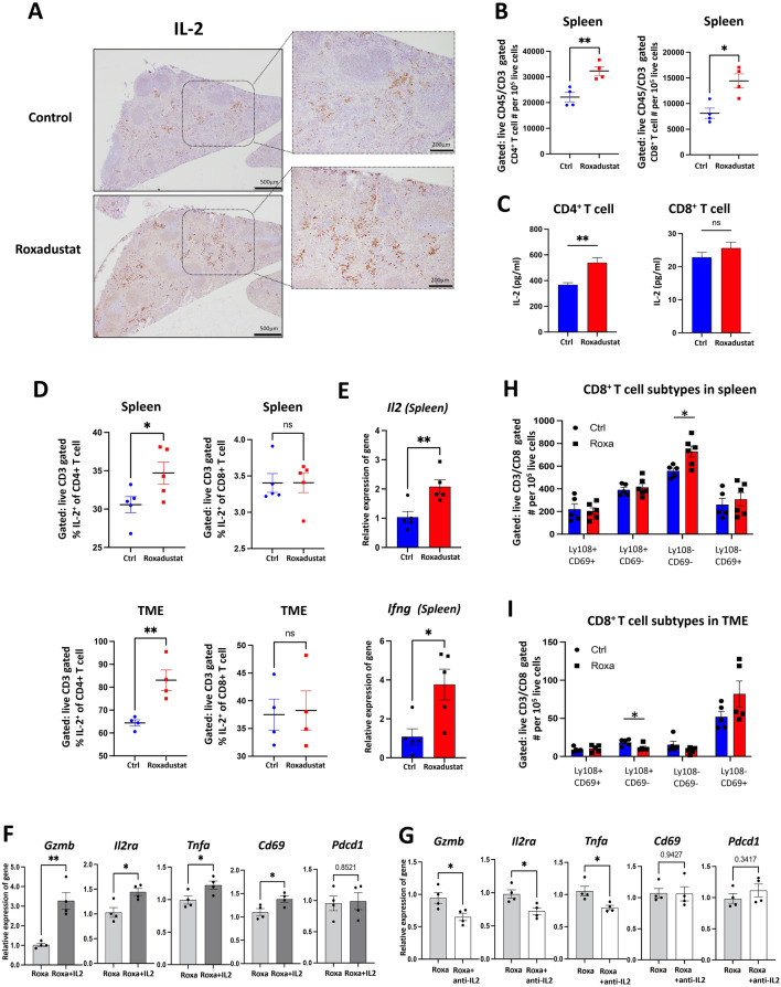
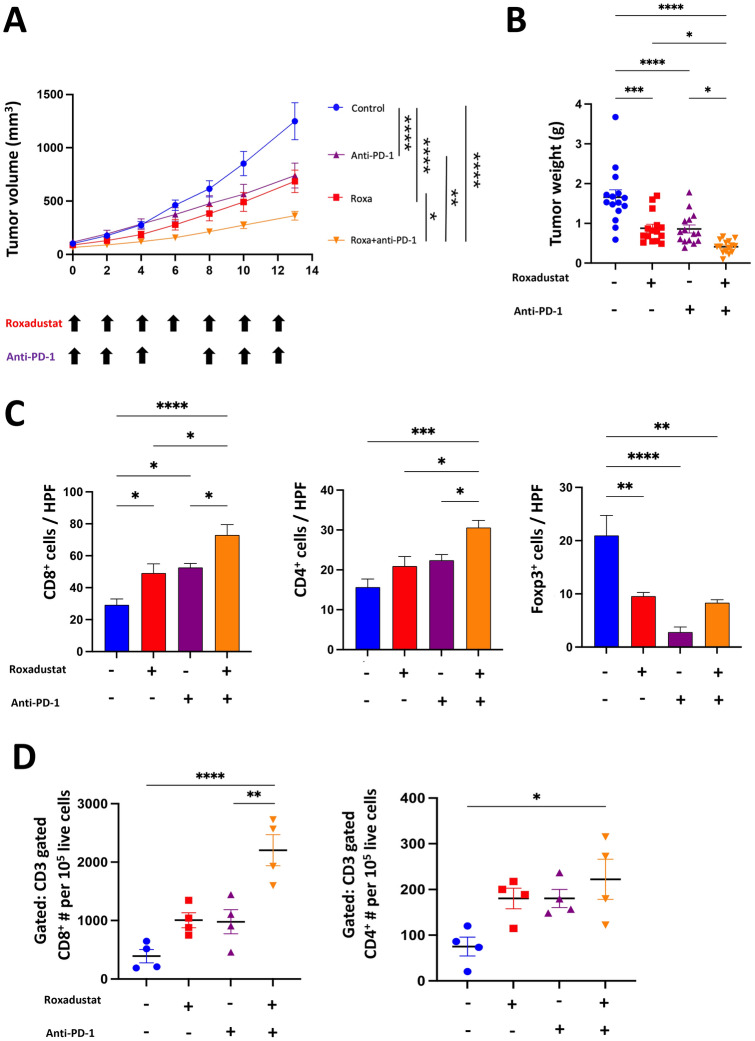
Results: HIF-PH inhibitors demonstrated antitumor effects in vivo but not in vitro. These inhibitors enhanced the tumor immune response by increasing the infiltration of CD8+ and CD4+ tumor-infiltrating lymphocytes (TILs). HIF-PH inhibitors induced IL-2 production in splenic and intratumoral CD4+ T cells, promoting T cell proliferation, differentiation, and immune responses. Roxadustat synergistically enhanced the efficacy of anti-PD-1 antibody for MSS cancer by increasing the recruitment of TILs and augmenting effector-like CD8+ T cells.

Conclusion: Pseudohypoxia induced by HIF-PH inhibitors activates antitumor immune responses, at least in part, through the induction of IL-2 secretion from CD4+ T cells in the spleen and tumor microenvironment, thereby enhancing immune efficacy against MSS CRC.

Keywords: Colorectal cancer; Hypoxia-inducible factor; Immune checkpoint inhibitors; Microsatellite stable.


Conflict of interest statement

Declarations. Conflict of interest: The authors declare no competing interests. Ethical approval: All experiments involving animals were conducted according to the ethical policies and procedures approved by The Animal Care and Use Committee of Okayama University (Approved No. OKU-2021900, OKU- 2022723, OKU- 2022879, OKU- 2023535). Consent for publication: Not applicable.

Figures

Fig. 1
Fig. 1
HIF-PH inhibitors activate immune system to suppress the tumor. A Murine spleen-derived CD8+ T cells and Colon26 were treated with different HIF-PH inhibitors for 48 h. HIF-1α was measured using western blotting. B, C Murine spleen-derived CD8+ T cells and MSS colon cancer cell lines (SW480, HT29, Colon26) were treated with different concentrations of HIF-PH inhibitors for 48 h. Cell viability was assessed by XTT assay. D, E 7 days post Colon26 injection, BALB/c and BALB/c-nude mice were randomized into two groups: (i) Control; (ii) Roxadustat/Vadadustat; PBS and Roxadustat/Vadadustat were administered via oral route (Roxadustat: 50 mg/kg. every other day. Vadadustat: 150 mg/kg. every day.). Tumor volumes and weights of BALB/c and BALB/c-nude mice were measured at the indicated day under HIF-PH inhibitors treatment (Roxadustat’s n = 5, Vadadustat’s n = 6). The results are presented as the means ± S.E.M. of a representative experiment performed in triplicate. Student’s t test. *P < 0.05, **P < 0.01, ***P < 0.001
Fig. 2
Fig. 2
HIF-PH inhibitors enhance T cell efficacy, promote CD4+ and CD8+ T cell infiltration, and reduce regulatory T cell populations. A Representative images and positive cell number of CD8+, CD4+ and Foxp3+ cells stained from Colon26 tumor tissues of control and Roxadustat treatment group mice. The number of positive cells was manually counted from five selected high-power fields (HPFs) per tumor. (n = 5; scale bar,100 and 50 μm; quantitative data, blow). B, C Frequencies of the indicated cells (n = 4) for TME collected from tumor-bearing mice. D The mRNA levels of indicated markers were measured by RT-qPCR. Total mRNA was extracted from Colon26 tumor tissues (n = 4). The results are presented as the means ± S.E.M. of a representative experiment performed in triplicate. Student’s t test. *P < 0.05, **P < 0.01, ***P < 0.001
Fig. 3
Fig. 3
HIF-PH inhibitors elevate IL-2 levels in CD4+ T cell to boost T cell proliferation and CD8+ T cell activation. A Representative images of IL-2 protein in tumor-bearing mice spleen of control and Roxadustat group mice (scale bar, 500 μm; magnification scale bar, 200 μm). B Numbers of the indicated cells (n = 4) for spleen collected from tumor-bearing mice. C Concentrations of IL-2 from indicated murine spleen-derived cells stimulated with 5 μg/ml ConA and treated with 25 μM Roxadustat for 48 h. D IL-2 frequencies of the indicated cells (n = 4) for spleen and TME collected from tumor-bearing mice. E The mRNA levels of indicated markers were measured by RT-qPCR. Total mRNA was extracted from spleen of tumor-bearing mice (n = 5). F Isolated CD8⁺ T cells were stimulated with 5 μg/ml ConA and treated with 5 μM Roxadustat in the presence or absence of 50 ng/ml recombinant IL-2 for 12 h. mRNA expression levels were analyzed by RT-qPCR. G Isolated CD8⁺ T cells were cultured for 12 h in conditioned media (supernatants) collected from CD4⁺ T cells pre-treated with 5 μg/ml ConA and 5 μM Roxadustat, in the presence or absence of 2.5 μg/ml anti-IL-2 antibody. mRNA expression levels were analyzed by RT-qPCR. H, I Numbers of different CD8+ T cell subtypes (n = 5) for spleen and TME collected from tumor-bearing mice. The results are presented as the means ± S.E.M. of a representative experiment performed in triplicate. Student’s t test. *P < 0.05, **P < 0.01, ***P < 0.001
Fig. 4
Fig. 4
Roxadustat and anti-PD-1 combination therapy enhances MSS tumor suppression and increase TILs. AD 7 days after colon 26 inoculation, BALB/c mice were randomized into four groups: (i) Control; (ii) Roxadustat; (iii) aPD1; (iv) Roxadustat + aPD-1. Roxadustat (50 mg/kg) was administered orally, and aPD-1(7 mg/kg) intraperitoneally. (A-B) Tumor growth and weight of Colon26 tumors in BALB/c mice treated with PBS, Roxadustat, aPD-1, or Roxadustat + aPD-1. (n = 15/ group pooled from three experiments). C IHC was used to analyze the staining of CD8+, CD4+ and Foxp3+ cells for TME collected from tumor-bearing mice (n = 5). The number of positive cells was counted manually. D Numbers of CD4+ and CD8+ T cells (n = 5) for TME collected from tumor-bearing mice were measured by flow cytometry. The results are presented as the means ± S.E.M. of a representative experiment performed in triplicate. ANOVA followed by Tukey’s multiple comparison test was applied. *P < 0.05, **P < 0.01, ***P < 0.001
Fig. 5
Fig. 5
Synergistic increase of effector-like CD8+ T-cell population by Roxadustat combined with anti-PD-1 therapy. 14 days post treatment initiation, the indicated tissues from tumor-bearing BALB/c mice were examined by flow cytometry (n = 4/group). A, B Numbers of the indicated subtypes for CD8+ T cells collected from TME of tumor-bearing mice. C PD-1 MFI of CD8+ T cells collected from TME of tumor-bearing mice. D Representative images of IL-2 protein in tumor-bearing mice spleen of control and Roxadustat group mice (scale bar, 500 μm; magnification scale bar, 200 μm). E Numbers of the indicated T cells collected from spleen of tumor-bearing mice. F Kaplan–Meier shows progression-free survival of patients with MSS colon cancer classified by stable MSI and the level of HIF-1 expression: high (red) and low (black), measured with Affymetrix arrays (IDs: 220946_s_at). Data were from GSE143985 in GEO database. The results are presented as the means ± S.E.M. of a representative experiment performed in triplicate. ANOVA followed by Tukey’s multiple comparison test was applied. *P < 0.05, **P < 0.01, ***P < 0.001

References

    1. Tirendi S, Marengo B, Domenicotti C, Bassi AM, Almonti V, Vernazza S (2023) Colorectal cancer and therapy response: a focus on the main mechanisms involved. Front Oncol 13:1208140 - PMC - PubMed
    1. Franke AJ, Skelton WP, Starr JS, Parekh H, Lee JJ, Overman MJ, Allegra C, George TJ (2019) Immunotherapy for colorectal cancer: a review of current and novel therapeutic approaches. J Natl Cancer Inst 111(11):1131–1141 - PMC - PubMed
    1. Yu I, Dakwar A, Takabe K (2023) Immunotherapy: recent advances and its future as a neoadjuvant, adjuvant, and primary treatment in colorectal cancer. Cells 12(2):258 - PMC - PubMed
    1. Okuda T, Azuma T, Ohtani M, Masaki R, Ito Y, Yamazaki Y, Ito S, Kuriyama M (2005) Hypoxia-inducible factor 1 alpha and vascular endothelial growth factor overexpression in ischemic colitis. World J Gastroenterol 11(10):1535–1539 - PMC - PubMed
    1. Mortezaee K, Majidpoor J, Kharazinejad E (2023) The impact of hypoxia on tumor-mediated bypassing anti-PD-(L)1 therapy. Biomed Pharmacother 162:114646 - PubMed

MeSH terms