. 2025 May 9;38(4):98.
doi: 10.1007/s13577-025-01230-x.

Establishment and characterization of Cri Du Chat neuronal stem cells: a novel promising resource to study the syndrome

Affiliations

Establishment and characterization of Cri Du Chat neuronal stem cells: a novel promising resource to study the syndrome

Giovanna Piovani et al. Hum Cell. .

Abstract

The Cri Du Chat (CdC) Syndrome is a rare chromosome disease condition resulting from variable size deletion occurring on the short arm of one of the chromosomes 5. This disorder, which affects one in 50,000 births, is responsible for developmental retardation, the mechanism of which has remained unexplained. TERT, SEMA5 A, CTNND2, TPPP, mapped in chromosome 5 short arm, are known to be expressed in the brain, and to play a role in the development of the nervous system, oligodentrocytes and in the regulation of glutamatergic and dopaminergic synaptic transmission. It is critical to understand how their haploinsufficiency might affect the development and presentation of the disease. In the absence of an animal model and of significant accessible, human tissue, human pluripotent stem cells (iPSC) directly reprogrammed from patient somatic cells open a new area of disease modeling as they can virtually be differentiated into any cell type. Our study reports, for the first time, the generation of neuronal stem cells (NSCs) from CdC-iPSCs line and in addition, subsequent differentiation into a heterogeneous population of neurons. Gene expression of the mentioned and single copy deleted genes was also evaluated by comparing their expression level in iPSC, NSCs and neuron lines. The present research represents the first and the most innovative approach, to create an in vitro CdC neuronal model to have a new translational framework to study the pathologic processes.

Keywords: Cri du Chat neurons; Cri du Chat syndrome; Deletion chromosome 5p; Disease modeling; IPSCs-NSC.


Conflict of interest statement

Declarations. Conflict of interest: The authors declare that they have no known competing financial interests. Ethical approval: This study was approved by the A.B.C scientific committee. Informed consent: Not pertinent.

Figures

Fig. 1
Fig. 1
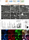
CdC-NSCs differentiation a Experimental workflow. b NSCs differentiation: iPSCs colonies were transferred to Matrigel-coated plates and culture in xeno- and serum-free Gibco® PSC Neural Induction Medium (ThermoFisher Scientific) for 7 days. At day 7 of neural induction, primitive NSCs were enzymatically dissociated and plated on Matrigel-coated dishes for expansion and cryopreservation. c Immunofluorescence assay performed on NSCs at p0 with antibodies against NSCs markers Nestin (red), and against Phalloidin (green), Magnification 20X. d q-PCR assay for NESTIN, SOX1, SOX2, OCT4 and PAX6 expression in NSCs p0 obtained. Data were normalized on ACTIN and calculated in relation to parental iPSCs line (black bars). NSCs show an increased gene expression of neural markers NESTIN, SOX1, SOX2, and PAX6. A reduced gene expression of pluripotency marker OCT4 indicates the absence of residual iPSCs not induced. The standard deviation was calculated from gene expression of three distinct HD-derived iPSCs clones (n = 3)
Fig. 2
Fig. 2
Karyotype and FISH a Karyotype, the arrow indicates chromosome 5 with the deletion of the short arm. b Metaphase stained with DAPI to counterstain the chromosomes. In the square bordered in white, an interphase is inserted; Merge in the metaphase plate and in the interphase within the white-bordered square, both control signals for the 5qter region appear in red, while only one signal for the candidate 5p (CdCCR) region is visible in green, highlighting the deletion of the short arm of chromosome 5. The red arrow indicates the deleted chromosome 5, which lacks the green signals. Magnification 100X
Fig. 3
Fig. 3
CdC-derived neurons a Experimental workflow. b Morphologic modifications occurred during differentiation into neurons. Scale bar of 200 μm. c qPCR analysis for DCX, MAP2, TUBB3, TH, GAD, CHAT, and GFAP gene expression in a CdC- and HD-derived neurons at day 15 and day 30 of differentiation. Data were normalized on ACTIN and calculated in relation to parental NSCs line at passage 0. The standard deviation was calculated from gene expression of three distinct HD-derived iPSCs clones (n = 3). d Western blot analysis and band densitometry graph of TH expression in CdC- and HD-derived neurons at day 30 of differentiation. TH signal was normalized to GAPDH. e Immunofluorescence images of healthy donor (HD) and CdCS derived neurons stained with antibodies against the neuronal marker MAP2 in red and the dopaminergic marker TH in green; nuclei are stained with DAPI in blue, Magnification 10X
Fig. 4
Fig. 4
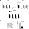
Expression deleted genes a Quantitative Real Time PCR (qPCR) analysis for TERT, CTNND2, SEMA5 A and TPPP gene expression in iPSCs. b in NSCs. c Neurons. Data were normalized on GAPDH and plotted in relation to values of HD as calibrator (black bars). The bars indicate the standard deviation calculated from three distinct experimental replicates (n = 3). Statistical significance was determined using an unpaired Student’s t test for each gene. The asterisks indicate significant differences between CdC-derived cells and healthy donor- derived cells (*p < 0.05, **p < 0.001). b Western blot analysis and band densitometry graph of DAT expression in CdC- and HD-derived neurons at day 30 of differentiation. DAT signal was normalized to GAPDH

References

    1. Lejeune J, Lafourcade J, Berger R, Vialatte J, Boeswillwald M, Seringe P. Turpin, R [3 cases of partial deletion of the short arm of a 5 chromosome.]. C R Hebd Seances Acad Sci. 1963;257:3098–102. - PubMed
    1. Rodriguez-Caballero A, Torres-Lagares D, Rodriguez-Perez A, Serrera-Figallo MA, Hernandez-Guisado JM, Machuca-Portillo G. Cri du chat syndrome: a critical review. Med Oral Patol Oral Cir Bucal. 2010;15:473–8. - PubMed
    1. Cerruti Mainardi P. Cri du chat syndrome. Orphanet J Rare Dis. 2006;1:33. - PMC - PubMed
    1. Nevado J, Bel-Fenellós C, Sandoval-Talamantes AK, Hernández A, Biencinto-López C, Martínez-Fernández ML, Barrúz P, Santos-Simarro F, Mori-Álvarez MÁ, Mansilla E, García-Santiago FA, Valcorba I, Sáenz-Rico B, Martínez-Frías ML, Lapunzina P. Phenotyping and genetic characterization of a cohort of 70 individuals with 5p minus syndrome. Front Genet. 2021;12:645595. - PMC - PubMed
    1. Cornish KM, Pigram J. Developmental and behavioural characteristics of cri du chat syndrome. Arch Dis Child. 1996;75:448–50. - PMC - PubMed

LinkOut - more resources