. 2025 May 9;21(5):e1013158.
doi: 10.1371/journal.ppat.1013158. eCollection 2025 May.

Evolution of primate T-cell leukemia virus type 1 accessory genes and functional divergence of its antisense proteins

Affiliations

Evolution of primate T-cell leukemia virus type 1 accessory genes and functional divergence of its antisense proteins

Osama Hussein et al. PLoS Pathog. .

Abstract

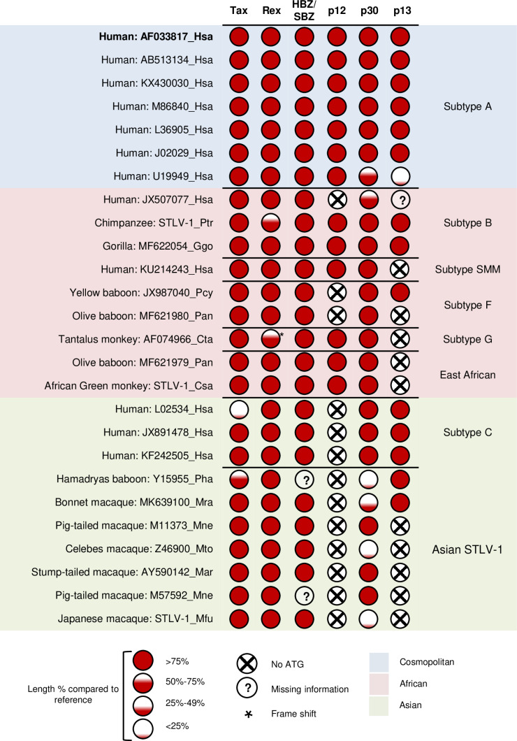
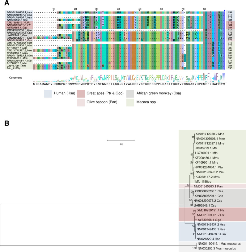
Human T-cell leukemia virus type 1 (HTLV-1) is derived from simian T-cell leukemia virus type 1 (STLV-1), and together they form a broader category known as primate T-cell leukemia virus type 1 (PTLV-1). PTLV-1 encodes multiple proteins from overlapping open reading frames (ORFs) in the pX region. This study aims to characterize the conservation of these proteins in different PTLV-1 subtypes and their role in pathogenesis. For the first time, we report the full-length proviral sequence of an STLV-1 strain isolated from chimpanzee and African green monkey. Phylogenetic analysis reveals high conservation of the accessory proteins p12, p30, and p13 in the HTLV-1a subtype. Conversely, some African PTLV-1 subtypes exhibit loss of ORFs for p12 or p13. For Asian subtypes, simian strains often lack p12, p13, or p30 proteins, whereas human strains retain the ORFs of p30 and p13 but not p12. To assess the infectivity of a simian strain of PTLV-1 lacking ORFs for p12, p13, and p30, we constructed a molecular clone from a naturally infected Japanese macaque (Mfu: Macaca fuscata) and compared it with HTLV-1a. Using a reporter assay and ELISA, we found similar infectivity to Jurkat T cells; however, STLV-1 Mfu exhibited impaired infectivity in the monocytic cell line THP-1. Additionally, despite the conservation of the HTLV-1/STLV-1 bZIP factor (HBZ/SBZ) ORFs, HBZ/SBZ proteins derived from HTLV-1a and African PTLV-1 subtypes induce significantly higher activation of the TGF-β/Smad signaling pathway than those from Asian subtypes. Collectively, our findings suggest that the acquisition of the accessory proteins by PTLV-1 subtypes potentially confers an advantageous adaptation of PTLV-1 during infection in apes, including humans. Moreover, among PTLV-1 strains, HBZ/SBZ had varying degrees of activity on the TGF-β/Smad pathway; this fact underscores the complex interplay between viral proteins and host signaling pathways, possibly influencing the viral pathogenicity in different species.


Conflict of interest statement

The authors have declared that no competing interests exist

Figures

Fig 1
Fig 1. PTLV-1 phylogenetic tree and subtype assignment of STLV-1 from chimpanzee, African green monkey, and Japanese macaque.
Maximum likelihood tree depicting the sequences (522 bp) corresponding to the Env coding region of PTLV-1. The tree comprises sequences from the publicly available database and newly sequenced STLV-1 from Pan troglodytes (STLV-1 Ptr), Chlorocebus sabaeus (STLV-1 Csa), and Macaca fuscata (STLV-1 Mfu). Each sequence is annotated with its NCBI accession number followed by a three-letter abbreviation denoting the genus and species (the first letter in the upper case refers to the genus, and the second and third letters in the lower case refer to the species name). The accession number for the new sequences is replaced by “STLV-1”. Fast-bootstrapping percentage values (1000 times) are indicated for each node. A scale bar for branch length is shown in the upper left of the figure. Information on the proviral sequences used for this analysis is given in S1 Table.
Fig 2
Fig 2. Coding potential of accessory and regulatory proteins in PTLV-1.
Analysis of the coding potential of pX region ORFs for the accessory and regulatory proteins in various HTLV-1/STLV-1 strains for which a full/nearly full pX region sequence is available. HTLV-1 subtype A (AF033817 Hsa) was used as the reference. The analysis accounted for the presence/absence of the initiation codon and the predicted length of the protein. Generally, the amino acid identity was considered relatively high (>75%) in comparison to reference proteins in HTLV-1a whenever an ORF was predicted to be conserved (S1, S2, and S3 Figs and S3 Table). Exons were defined by known splicing sites in HTLV-1 subtype A. The conservation of splicing acceptor/donor sites in a particular strain was not considered when assembling spliced mRNA and predicting ORFs. However, the nucleotides aligned to the reference’s splicing sites for each strain are shown in S3 Table. Information on the proviral sequences used for this analysis is given in S2 Table.
Fig 3
Fig 3. Effect of p12 on MHC-I surface expression in Jurkat Cells.
(A) Schematic illustration of the p12 expression constructs. (B) Western blot analysis of HA-tagged p12 (from HTLV-1a or STLV-1 Ptr) showing expression and successful cleavage of the 2A peptide. (C) Flow cytometry analysis of MHC-I surface expression in GFP-positive viable Jurkat cells 5 days post-electroporation with p12-expressing plasmids or an empty vector. Left, representative histogram showing MHC-I surface expression level. Right, median fluorescence intensity (MFI) of MHC-I. Error bars represent SD for three independent triplicates. P-values were determined using unpaired t-test, * P ≤ 0.05; ** P ≤ 0.01.
Fig 4
Fig 4. STLV-1 Mfu infectivity compares to that of HTLV-1a in T cells but not in monocytes.
(A) Titration of virus production from an HTLV-1a or STLV-1 Mfu full-length molecular clone transfected into HEK293T cells. Cell supernatant was collected and p19 levels were measured using ELISA. (B) Western blot analysis depicting Tax expression in HEK293T cells transfected with HTLV-1a or STLV-1 Mfu full-length molecular clone along with the naturally STLV-1 Mfu-infected cell line (Si-2). (C) Multiple sequence alignment of the Tax coding sequence from HTLV-1a and STLV-1 Mfu. Key functional domains are highlighted. (D) Jurkat or THP-1 cells were co-cultured with virus-producing HEK293T cells for 48 hours, and the virus infectivity was measured by LTR reporter luciferase assay. (E) HTLV p19 antigen ELISA measuring viral production in Jurkat and THP-1 cells following co-culture with HEK293T cells transfected with HTLV-1a or STLV-1 Mfu full-length molecular clones. Three independent experiments were performed. Error bars represent SD for three replicas. P-values were calculated using unpaired t-test. * P ≤ 0.05; ** P ≤ 0.01; ns: non-significant.
Fig 5
Fig 5. Sequence conservation but functional divergence of HBZ/SBZ.
(A) Multiple sequence alignment of the translated nucleotide sequences of HBZ/SBZ from different primate species. Sequences were aligned using MUSCLE, and visualized using Jalview software. (B) Schematic illustration of the TGF-β luciferase activity assay (Created in https://BioRender.com). (C) Luciferase activity of 3TP-Lux under the control of a TGF-β responsive element in cells expressing HBZ/SBZ proteins derived from different PTLV-1 strains. Hsa HTLV-1a: human (subtype A), Ptr: chimpanzee, Pan: olive baboon, Csa: African green monkey, Hsa HTLV-1c: human (subtype C), Mne: pig-tailed macaque, and Mfu: Japanese macaque. The experiment was performed in triplicate; representative data is shown. Error bars represent SD for three replicas. P-values were determined using unpaired t-test. ** P ≤ 0.01; *** P ≤ 0.001; **** P ≤ 0.0001.
Fig 6
Fig 6. Multiple sequence alignment and phylogenetic analysis of APOBEC3G.
(A) Multiple sequence alignment of the translated nucleotide sequences for APOBEC3G from various primate species. The first 90aa are shown here; the full-length sequences are shown in S5 Fig. Sequences from the publicly available database and an in-house sequenced Japanese macaque APOBEC3G (Mfu_395aa) were aligned using MUSCLE and visualized using Jalview software. (B) The maximum likelihood tree of the APOBEC3G gene comprising the full-length sequences shown in S5 Fig along with Mus musculus APOBEC3 protein as an outgroup. Each sequence is annotated with its NCBI accession number followed by a three-letter abbreviation denoting the genus and species. For the new sequences, the accession number is replaced by the genus and species abbreviation. Fast-bootstrapping percentage values (1000 times) are indicated for each node. A scale bar for the branch length is shown in the upper left of the figure.
Fig 7
Fig 7. TGF-β
/Smad signaling pathway activation by primate APOBEC3G. Luciferase activity of 3TP-Lux under the control of a TGF-β responsive element in cells expressing different APOBEC3G isoforms derived from different primate species. Human APOBEC3G (Hsa A3G 384aa) was used as a control. (A) Luciferase activity in cells expressing human APOBEC3G (Hsa A3G 373aa and Hsa A3G 384aa), chimpanzee APOBEC3G (Ptr A3G 384aa), and olive baboon APOBEC3G (Pan A3G 376aa). (B) Luciferase activity in cells expressing Hsa A3G 384aa and African green monkey APOBEC3G (Csa A3G 376aa, 383aa, and 394aa). (C) Luciferase activity in cells expressing Hsa A3G 384aa, rhesus macaque APOBEC3G (Mmu A3G 395aa), crab-eating macaque APOBEC3G (Mfa A3G 394aa), and Japanese macaque APOBEC3G (Mfu A3G 395aa). (D) Luciferase activity in cells expressing Hsa A3G 384aa and pig-tailed macaque APOBEC3G (Mne A3G 383aa and 394aa). Each experiment was performed in triplicate; representative data are shown. Error bars indicate SD for three replicas. P-values were determined using unpaired t-test. *** P ≤ 0.001; **** P ≤ 0.0001.
Fig 8
Fig 8. Synergistic activation of the TGF-β
/Smad signaling pathway by SBZ/HBZ and primate APOBEC3G. Luciferase activity of 3TP-Lux under the control of a TGF-β responsive element in cells expressing HBZ/SBZ and increasing concentrations of APOBEC3G derived from four different primate species. In each case, HBZ/SBZ from PTLV-1 isolated from a particular primate species is paired with APOBEC3G from that same species. (A) Luciferase activity in cells expressing HTLV-1a HBZ and human APOBEC3G 384aa (B) Luciferase activity in cells expressing STLV-1 Ptr SBZ and chimpanzee APOBEC3G 384aa. (C) Luciferase activity in cells expressing STLV-1 Pan SBZ and olive baboon APOBEC3G 131aa. (D) Luciferase activity in cells expressing HTLV-1c HBZ and human APOBEC3G 384aa. Each experiment was performed in triplicate; representative data are shown. Error bars indicate the SD for three replicas. P-values were determined using unpaired t-test. *P ≤ 0.05; **P ≤ 0.01.

Similar articles

Cited by

References

    1. Matsuoka M, Jeang KT. Human T-cell leukaemia virus type 1 (HTLV-1) infectivity and cellular transformation. Nat Rev Cancer. 2007;7:270–80. - PubMed
    1. Ayouba A, Michem A, Peeters M, Vercammen F. Full-genome characterization of simian T-cell leukemia virus Type 1 Subtype b from a wild-born Captive Gorilla gorilla gorilla with T-cell lymphoma. Genome Announc. 2017;5(43):e01117-17. doi: 10.1128/genomeA.01117-17 - DOI - PMC - PubMed
    1. Turpin J, Alais S, Marçais A, Bruneau J, Melamed A, Gadot N, et al.. Whole body clonality analysis in an aggressive STLV-1 associated leukemia (ATLL) reveals an unexpected clonal complexity. Cancer Lett. 2017;389:78–85. doi: 10.1016/j.canlet.2016.12.022 - DOI - PubMed
    1. Akari H, Ono F, Sakakibara I, Takahashi H, Murayama Y, Hiyaoka A, et al.. Simian T cell leukemia virus type I-induced malignant adult T cell leukemia-like disease in a naturally infected African green monkey: implication of CD8+ T cell leukemia. AIDS Res Hum Retroviruses. 1998;14(4):367–71. doi: 10.1089/aid.1998.14.367 - DOI - PubMed
    1. Tsujimoto H, Noda Y, Ishikawa K, Nakamura H, Fukasawa M, Sakakibara I, et al.. Development of adult T-cell leukemia-like disease in African green monkey associated with clonal integration of simian T-cell leukemia virus type I. Cancer Res. 1987;47(1):269–74. - PubMed

Substances