. 2025 Jul;12(28):e2503697.
doi: 10.1002/advs.202503697. Epub 2025 May 8.

Strong and Fatigue-Resistant Hydrogels via Poor Solvent Evaporation Assisted Hot-Stretching for Tendon Repair

Affiliations

Strong and Fatigue-Resistant Hydrogels via Poor Solvent Evaporation Assisted Hot-Stretching for Tendon Repair

Huamin Li et al. Adv Sci (Weinh). 2025 Jul.

Abstract

It is highly desirable but still remains challenging to develop high-performance hydrogels with satisfactory mechanical properties for tissue engineering. Here, anisotropic yet transparent hydrogels (AHs) are prepared for tendon repair via a facile "poor solvent evaporation assisted hot-stretching" strategy. AHs have great mechanical properties with tensile strength, toughness, and fracture energy as high as 33.14 ± 2.05 MPa, 44.1 ± 3.5 MJ m-3, and 106.18 ± 7.2 kJ m-2, respectively. Especially, AHs show unique flaw-insensitive characteristics, and cracks can only deflect along the fiber alignment direction rather than propagate transverse to this direction, showing an interesting self-protection function. The high strength, toughness, and fatigue resistance originate from the hierarchal structure of AHs, i.e., the densified polymeric network comprising fiber bundles and nanofibrils with aligned macromolecular chains, crystalline domains, and intermolecular hydrogen bonds. AHs with superior biocompatibility and swelling resistance can be used to repair rat tendons, and implantation of AHs can promote collagen regeneration for the tendon repair. This study provides a new method to fabricate strong and anti-fatigue hydrogels as a new class of promising materials for soft tissues.

Keywords: anisotropic hydrogels; flaw‐insensitivity; hot‐stretching; poor solvent evaporation; tendon repair.


Conflict of interest statement

The authors declare no conflict of interest.

Figures

Figure 1
Figure 1
Bioinspired design and structure of anisotropic polyvinyl alcohol (PVA) hydrogels (AHs). a) Schematic for the preparation of AHs by poor solvent evaporation assisted hot‐stretching. b) Poor solvent evaporation promoted macromolecular chain aggregation and alignment. c) Structures of natural tendon. d) SEM images and photographs of AH‐200 and tendon.
Figure 2
Figure 2
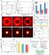
Mechanical properties of AHs. a) Stress‐strain curves, and summary of b) tensile strength and elongation at break, and c) elastic modulus and toughness of AHs prepared by different glycerol/ethanol ratios. d) Stress‐strain curves, and summary of e) tensile strength and elongation at break, and f) elastic modulus and toughness of AHs prepared with different pre‐stretching ratios (PSRs). g) Water content of hydrogels prepared by different PSRs. h) Fracture energy of AHs with different PSRs. i) Summary of different mechanical properties of isotropic PVA hydrogel (IH), AH‐100 and AH‐200. Comparison of j) tensile strength versus fracture strain, k) toughness versus tensile strength, and l) fracture energy versus tensile strength of AHs and other anisotropic hydrogels.
Figure 3
Figure 3
Structural characterization of IH, AH‐100, and AH‐200. a) XRD patterns. b) DSC thermographs, and c) corresponding calculated crystallinities. d) 2D WAXS and 2D SAXS patterns. e) Correlation of the azimuthally integrated intensity distribution of 2D WAXS patterns. f) Scattering intensity versus scattering vector q. g) Corrected scattering intensity Iq2 versus vector q. h) Calculated average distance between crystalline regions. i) Schematic illustration of intermolecular spacing, interlamellar spacing and intercrystal spacing of AHs.
Figure 4
Figure 4
Fatigue resistance of AHs. Crack propagation per cycle, dc/dN versus applied energy release rate G for a) IH and b) AH‐150 (notch parallel to aligned fiber direction), and c) summary of corresponding fatigue thresholds. d) Stress‐strain curve of AH‐150. e) Micrographs of the crack before and after 1000 and 10 000 cycles for AH‐150. Scale bar: 500 µm. f) Energy release rate (G) for AH‐150 in fibril alignment direction. Schematic for g) simplified tearing model of soft tissues, and h) anti‐crack propagation mechanism of AHs at different length scale.
Figure 5
Figure 5
a) Typical tensile stress‐strain curves of AH‐150 before and after swelling in water for 7 days, and b) retention coefficient of mechanical properties c) Stress versus strain curves over 1000 cyclic tensile tests of AH‐150 in a water bath. d) LIVE/DEAD staining images of NIH/3T3 cells upon contacting directly cell culture plate (control) and AH‐150 for 1, 3, and 7 d. e) Proliferation abilities. f) Fluorescence images of NlH‐3T3 fibroblasts cultured on different substrates and g) distribution of orientation angles for the Control group and AH‐150 (n ≥ 20), scale: 100 µm.
Figure 6
Figure 6
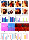
Tendon repair performance of AH‐150. a) Surgical procedures: I) peel off tendon, II) cut off mid‐portion of the tendon, III) suture the hydrogel to both sections of the severed tendon, and IV) suture the wound. Scale bar: 5 mm. b) Typical macroscopic appearance of repaired tendons. Scale bar: 5 mm. c,d) Thickness and width of the repaired tendon, n = 4. e) Macroscopic score of the two groups, n = 6. f) Histological score based on H&E and Masson staining, n = 6. g) H&E and Masson staining of tendon tissues at the 8th week. Blue arrows indicate densely aligned fibers and red arrows indicate muscle fibers. Scale bar: 100 µm. h) Tendon immunofluorescence staining images of COL1 (red), COL3 (green), and nucleus (blue) at 8 weeks and results statistics, white arrows indicate COL1, yellow arrows indicate densely COL3, orange arrow indicate nucleus. Scale bar: 100 µm, n = 6. i) Statistics of tendon immunofluorescence staining of COL1, COL3 and COL1/COL3 at 4 weeks. j) Statistics of tendon immunofluorescence staining of COL1, COL3 and COL1/COL3 at 8 weeks. (*) P < 0.05, (**) P < 0.01, and (***) P < 0.001.

Similar articles

References

    1. Wang Z., Xu X., Tan R., Zhang S., Zhang K., Hu J., Adv. Funct. Mater. 2024, 34, 2312667.
    1. Han S., Wu Q., Zhu J., Zhang J., Chen A., Su S., Liu J., Huang J., Yang X., Guan L., Mater. Horiz. 2023, 10, 1012. - PubMed
    1. Kong P., Dong J., Li W., Li Z., Gao R., Liu X., Wang J., Su Q., Wen B., Ouyang W., Wang S., Zhang F., Feng S., Zhuang D., Xie Y., Zhao G., Yi H., Feng Z., Wang W., Pan X., Adv. Sci. 2023, 10, 2301244. - PMC - PubMed
    1. Wang Z., Zhang Y., Yin Y., Liu J., Li P., Zhao Y., Bai D., Zhao H., Han X., Chen Q., Adv. Mater. 2022, 34, 2108300. - PubMed
    1. Wu Y., Zhang Y., Wu H., Wen J., Zhang S., Xing W., Zhang H., Xue H., Gao J., Mai Y., Adv. Mater. 2023, 35, 2210624. - PubMed

LinkOut - more resources