. 2025 Jul;12(27):e2502493.
doi: 10.1002/advs.202502493. Epub 2025 May 8.

DDX3 Regulates the Cap-Independent Translation of the Japanese Encephalitis Virus via Its Interactions with PABP1 and the Untranslated Regions of the Viral Genome

Affiliations

DDX3 Regulates the Cap-Independent Translation of the Japanese Encephalitis Virus via Its Interactions with PABP1 and the Untranslated Regions of the Viral Genome

Chenxi Li et al. Adv Sci (Weinh). 2025 Jul.

Abstract

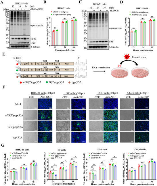
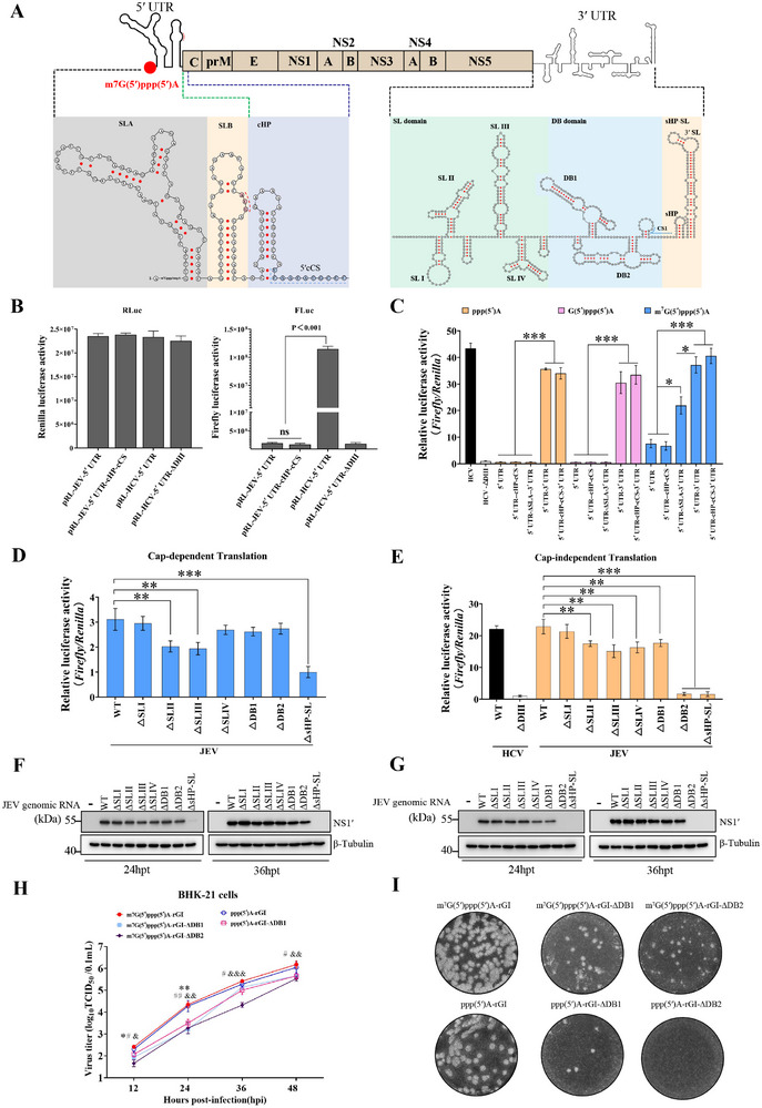
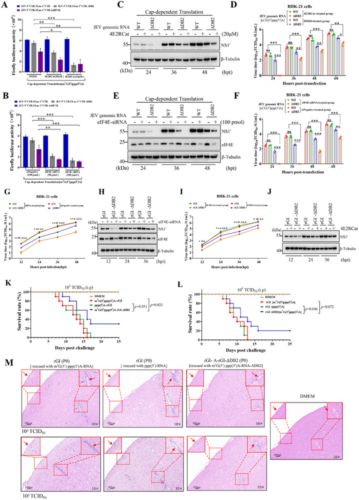
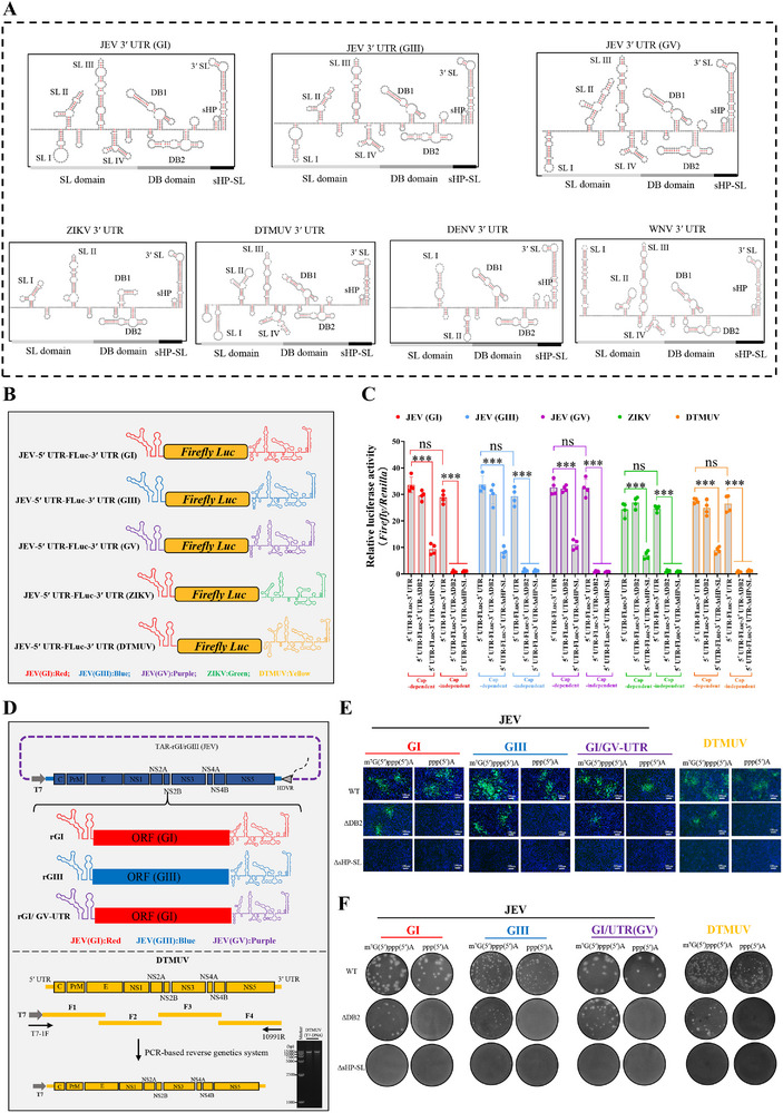
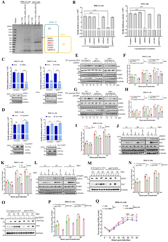
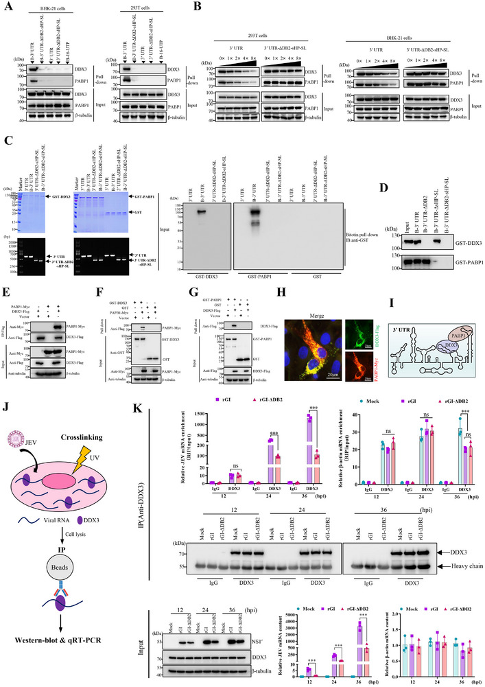
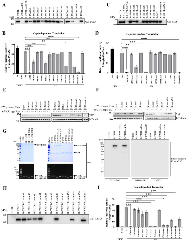
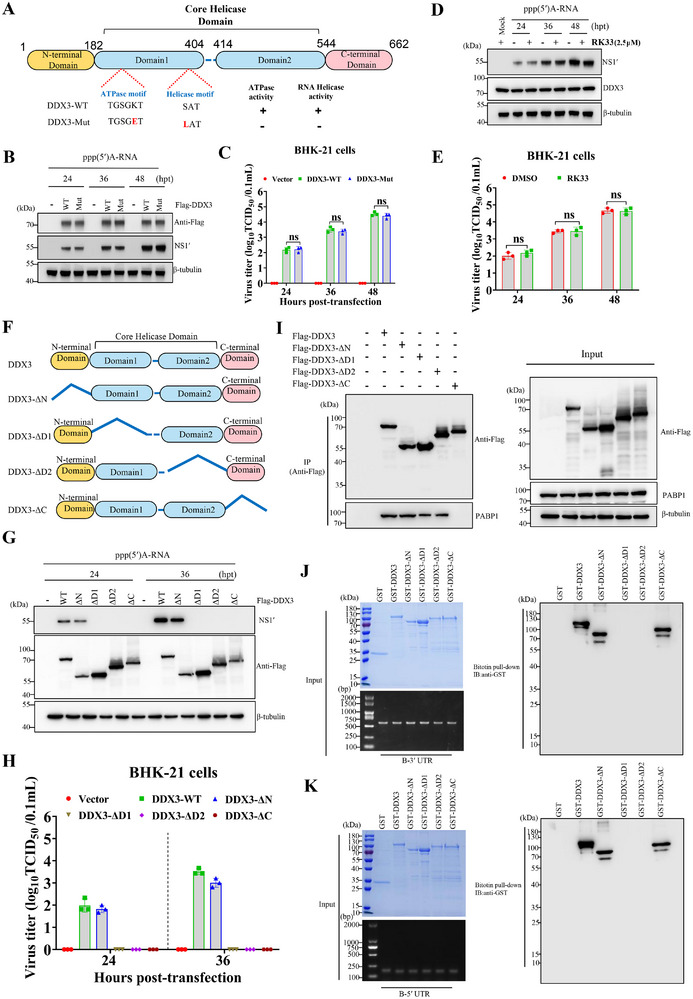
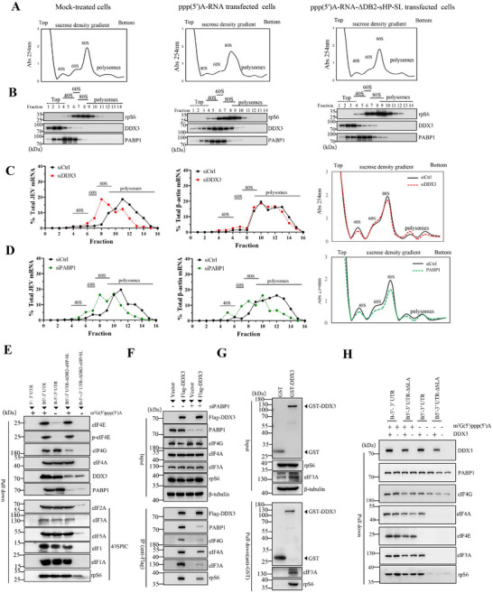
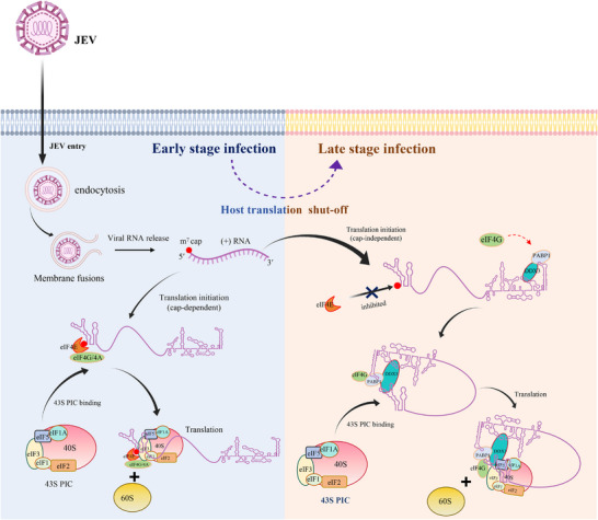
The translation of global cellular proteins is almost completely repressed in cells with flavivirus infection, while viral translation remains efficient. The mechanisms of flaviviruses evade host translational shutoff are largely unknown. Here, it is found that Japanese encephalitis virus (JEV) can adopt cap-independent (CI) translation to escape the host translational shutoff. Furthermore, the elements DB2 and sHP-SL within 3'UTR are involved in the regulation of CI translation, which is conserved in the genus Orthoflavivirus. By RNA affinity purification and mass spectrometry analysis, cellular DEAD-box protein 3 (DDX3) and poly(A)-binding protein 1 (PABP1) are identified as key factors in regulating CI translation of JEV via their interactions with DB2 and sHP-SL RNA structures. Mechanistically, it is revealed that DDX3 binds to both 5'UTR and 3'UTR of the JEV genome to establish a closed-loop architecture and recruit eIF4G/eIF4A to form the DDX3/PABP1/eIF4G/eIF4A tetrameric complex via its interaction with PABP1, thereby recruiting the ribosomal 43S preinitiation complex (PIC) to the 5'-end of the JEV genome to start translation. These findings demonstrate a noncanonical translation strategy employed by JEV and further reveal the regulatory roles of DDX3 and PABP1 in this mechanism. These results expand the knowledge of the translation initiation regulation in flaviviruses under the state of host translational shutoff, which provides a conserved antiviral target against orthoflavivirus.

Keywords: DDX3; Japanese encephalitis virus; cap‐independent translation.


Conflict of interest statement

The authors declare no conflict of interest.

Figures

Figure 1
Figure 1
Both CD and CI translation initiation strategies are involved in the expression of JEV proteins. A–D) BHK‐21 cells were respectively transfected with 100 pmol of a mixture of eIF4E‐specific siRNAs (Table S3, Supporting Information) or treated with 20 µm 4E2RCat for 12 h, and then infected with JEV at an MOI of 0.1. At different time points of post‐infection, the cells were labeled with puromycin for 30 min and harvested to analyze puromycin incorporation (A,C). The viral titers in culture supernatants of BHK‐21 cells treated with eIF4E‐specific siRNAs (B) or 4E2RCat (D) were measured by TCID50 assay (n = 3). E) Schematic diagram of the virus rescue using JEV RNA transcripts modified with three different 5′termini: m7G(5′)ppp(5′)A, G(5′)ppp(5′)A and ppp(5′)A. F) Cytopathic effects and immunofluorescence assay of cells transfected with full‐length viral RNA transcripts with different 5′ termini. G) The viral titers in culture supernatants of BHK‐21, ST, and DF‐1 cells transfected with 2 µg RNA transcripts at 24, 36, and 48 hpt, and the viral titers in culture supernatants of C6/36 cells transfected with 2 µg RNA transcripts at 48, 72, and 96 hpt, were measured by TCID50 assay on BHK‐21 cells (n = 3). *, p < 0.05; **, p < 0.01; ns, no statistical difference. Data are presented as mean ± standard deviation (SD) of three independent experiments and tested by Student's t‐test (B, D, and G).
Figure 2
Figure 2
Three cis‐acting elements in UTRs are crucial for the CI translation initiation of JEV mRNA. A) Secondary structure diagram of the 5′‐ and 3′‐ termini of JEV. B) BHK‐21 cells were respectively transfected with bicistronic constructs pRL‐JEV‐5′UTR, pRL‐JEV‐5′UTR‐cHP‐cCS, pRL‐HCV‐5′UTR, and pRL‐HCV‐5′UTR‐ΔDIII at a dose of 2 µg. At 24 h post‐transfection, the firefly luciferase and Renilla luciferase activities in BHK‐21 cells were determined by luciferase assay (n = 4). C–E) Monocistronic reporter RNA or its deletion mutants were generated via T7 promoter‐mediated in vitro transcription and then co‐transfected with a 5′capped‐RLuc mRNA into BHK‐21 cells. At 12 h post‐transfection, the firefly and Renilla luciferase activity in BHK‐21 cells was determined using a dual‐luciferase reporter assay. The relative luciferase activity was calculated by normalizing firefly luciferase activity to Renilla luciferase activity (n = 4; *, < 0.05, **, < 0.01, ***, < 0.001, ns, no significance; statistical significance determined by one‐way ANOVA). F,G) Western‐blot analysis of BHK‐21 cells transfected with the JEV genomic RNA with 5′termini m7G(5′)ppp(5′)A or ppp(5′)A of WT or the truncated. H) BHK cells were infected with viruses m7Gppp(5′)A‐rGI, ppp(5′)A‐rGI, m7G(5′)ppp(5′)A‐rGI‐ΔDB1, ppp(5′)A‐rGI‐ΔDB1 and m7G(5′)ppp(5′)A‐rGI‐ΔDB2 at an MOI of 0.05, respectively. At the indicated time points, culture supernatants were collected to determine virus titers by TCID50 assay (n = 3). Data are the means ± SD of three or four independent experiments, and statistical significance was tested by one‐way ANOVA analysis with Tukey's multiple comparison test. The significant differences between m7Gppp(5′)A‐rGI and m7G(5′)ppp(5′)A‐rGI‐ΔDB1 are labeled (*, < 0.05; **, < 0.01). The significant differences between m7Gppp(5′)A‐rGI and ppp(5′)A‐rGI‐ΔDB1 is marked ( # , < 0.05; ## ,< 0.01). The significant differences between m7Gppp(5′)A‐rGI and m7G(5′)ppp(5′)A‐rGI‐ΔDB2 is indicated (&, < 0.05; &&, < 0.01; &&&, < 0.001). I) Plaque morphology of the recombinant viruses in BHK‐21 cells.
Figure 3
Figure 3
The cis‐acting elements DB2 and sHP‐SL of 3′UTR determine the resistance of JEV to the suppression of CD translation initiation and the virulence of JEV in mice. A,B) The firefly luciferase activity assay of monocistronic RNA reporters with 5′terminal modified by either m7G(5′)ppp(5′)A or ppp(5′) in BHK‐21 cells treated with eIF4E‐specific siRNAs or 4E2RCat (n = 4). C–F) BHK‐21 cells were respectively treated with eIF4E‐specific siRNAs (100 pmol) or 4E2RCat (20 µm) and then transfected with JEV genomic RNA of WT or ΔDB2 mutant with a type I cap structure. At the indicated time points of post‐transfection, NS1′ protein was detected by immunoblotting (C,E), and viral titers in culture supernatants were measured by TCID50 assays (D and F) (n = 3). G–J) BHK‐21 cells were respectively treated with eIF4E‐specific siRNAs or 4E2RCat and then infected with rGI and rGI‐ΔDB2 at an MOI of 0.05. At the indicated time points of post‐infection, viral titers in culture supernatants were measured by TCID50 assays (G,I) (n = 3), and NS1′ protein was detected by immunoblotting (H,J). K,L) The survival rate of mice (n = 10) intraperitoneally mock‐infected or infected with the indicated JEV at doses of 103 and 105 TCID50. The significant differences were determined by the Kaplan–Meier analysis. M) Histopathological analysis of brain lesions of the dead mice. *, < 0.05; **, < 0.01; ***, < 0.001. Data are presented as mean ± SD and tested by one‐way ANOVA (A, B, D, F, G, and I).
Figure 4
Figure 4
The critical role of DB2 and sHP‐SL within 3′UTR in CI translation initiation is evolutionarily conserved in various flaviviruses. A) Secondary structure diagram of 3′UTR of various genotypes JEV, ZIKV, DENV, WNV and DTMUV. B) Diagrams of flaviviruses monocistronic reporter constructs controlled by a T7 promoter: JEV‐5′UTR‐FLuc‐3′UTR(GI), JEV‐5′UTR‐FLuc‐3′UTR (GIII), JEV‐5′UTR‐FLuc‐3′UTR (GV), ZIKV‐5′ UTR‐FLuc‐3′ UTR and DTMUV‐5′UTR‐FLuc‐3′UTR. C) Monocistronic RNA reporters with 5′terminal modified by either m7G(5′)ppp(5′)A or ppp(5′) were generated via T7 promoter‐mediated in vitro transcription, and then co‐transfected with a 5′‐capped‐RLuc mRNA into BHK‐21 cells. At 12 h post‐transfection, the cell samples were harvested for firefly and Renilla luciferase activity assay (n = 4). The relative luciferase activity was calculated by normalizing firefly luciferase activity to Renilla luciferase activity. ***, p < 0.001; **, p < 0.01; ns, no significance, tested by the one‐way ANOVA analysis with Tukey's multiple comparison test(C). Data are the means ± SD of the results of four independent experiments. D) Schematic illustration of constructed infectious clones of GI JEV, GIII JEV, and GI/GV‐UTR JEV, and a PCR‐based reverse genetics system of DTMUV. E) Immunofluorescence analysis of recombinant viruses infection in BHK‐21 cells. F) Plaque morphology of recombinant viruses in BHK‐21 cells.
Figure 5
Figure 5
DDX3 and PABP1 regulate CI translation initiation of JEV in vitro. A) Identification of cellular proteins associated with JEV 3′UTR and JEV 3′UTR‐ΔDB2‐sHP‐SL (left). Venn diagram of DB2 and sHP‐SL specific binding proteins in BHK‐21 and 293T cells detected by RNA pull‐down and mass spectrometry (right). B) The firefly luciferase activity assay using the noncapped monocistronic RNA reporter JEV‐5′UTR‐FLuc‐3′UTR in BHK‐21 and 293T cells with an indicated protein silenced using siRNAs (n = 3). C) BHK‐21 cells pre‐treated with 100 pmol of a mixture of DDX3‐specific siRNAs (left) or PABP1‐specific siRNAs (right) were respectively transfected with monocistronic RNA reporters JEV 5′ UTR‐FLuc‐3′ UTR with 5′terminal modified by either m7G(5′)ppp(5′)A or ppp(5′), and then harvested at 12 h post‐transfection for analysis of firefly luciferase activities (n = 4). The levels of endogenous DDX3 and PABP1 were detected by western blotting. D) The firefly luciferase activity assay of monocistronic RNA reporter JEV‐5′UTR‐FLuc‐3′UTR in BHK‐21 cells with Flag‐DDX3 (left) or Myc‐PABP1 (right) over‐expressed (n = 4). E–H) BHK‐21 cells with DDX3 or PABP1 silenced were transfected with the capped JEV genomic RNA of WT or ΔDB2 mutant, or noncapped WT genomic RNA. At the indicated time points post‐transfection, the expression of viral NS1′ protein, p‐eIF2α, DDX3, and PABP1 in BHK‐21 cells was analyzed by immunoblotting (E,G), and viral titers in culture supernatants were determined by TCID50 assays (F and H) (n = 3). I–L) BHK‐21 cells with DDX3 or PABP1 silenced were infected with rGI and rGI‐ΔDB2 at a dose of 0.05 MOI. Replication of rGI and rGI‐ΔDB2 was monitored by TCID50 assays (I and K) (= 3). The expression of viral NS1′ protein, p‐eIF2α, DDX3, and PABP1 in BHK‐21 cells was analyzed by immunoblotting (J,L). M–P) The translational activity of JEV genomic RNA in DDX3‐KO BHK‐21 cells. DDX3‐KO cells with or without DDX3‐Flag over‐expression were transfected with JEV genomic RNA m7G(5′)ppp(5′)A‐RNA, ppp(5′)A‐RNA, respectively. At different time points post‐transfection, immunoblotting analysis of viral NS1′ protein (M,O). Virus titers in culture supernatant were evaluated by TCID50 assays (N and P) (n = 3). Q) The replication ability of rGI and rGI‐ΔDB2 in WT and DDX3‐KO BHK‐21 cells (n = 3). The significant differences between WT and DDX3‐KO BHK‐21 cells are marked (*, < 0.05; **, < 0.01; **, < 0.001). *, < 0.05; **, < 0.01; ***, < 0.001; ns, no statistical differences. Data are presented as mean ± SD of three independent experiments (B, C, D, F, H, I, K, N, P, and Q).
Figure 6
Figure 6
The interactions among DDX3, PABP1, and JEV 3′UTR. A) The cell lysates of BHK‐21 and 293T cells were respectively incubated with biotinylated‐3′UTR (B‐3′UTR), biotinylated‐3′UTR‐ΔDB2‐sHP‐SL (B‐3′UTR‐ΔDB2‐sHP‐SL), nonbiotinylated‐3′UTR (3′UTR) or nonbiotinylated‐3′UTR‐ΔDB2‐sHP‐SL(3′UTR‐ΔDB2‐sHP‐SL). The bound complexes were analyzed by immunoblotting. B) The interactions between DDX3/PABP1 and JEV 3′UTR were confirmed by competition assays. C,D) RNA pulldown analysis of the interaction between nonbiotinylated or biotinylated 3′UTR and recombinant proteins GST‐DDX3, GST‐PABP1, or GST. The bound complexes were analyzed by immunoblotting, and recombinant proteins and RNA in the input were detected by Coomassie brilliant blue staining and RT‐PCR. E) Co‐immunoprecipitation analysis of the interaction between the ectopically expressed DDX3‐Flag and Myc‐PABP1 in HEK293T cells. F,G) GST pulldown analysis of the interaction between recombinant DDX3 and PABP1. H) Immunofluorescent analysis of the interaction between DDX3‐Flag and PABP1‐Myc in BHK‐21 cells. I) A model for the interactions among DDX3, PABP1, and JEV 3′UTR. J) Schematic diagram of the RIP assay. BHK‐21 cells are infected with rGI or rGI‐ΔDB2 at an MOI of 0.01. At 12, 24, and 36 h post‐infection, the cells are cross‐linked with short‐wave UV light to form RNA‐protein complexes and then lysed for RNA immunoprecipitation. K) Top panel: RIP‐qPCR analysis quantifying the immunoprecipitation efficiency of JEV and β‐actin mRNA using either anti‐IgG or anti‐DDX3 antibody‐conjugated beads. Results are presented as fold‐change values normalized to input samples, with the IgG control group set as 1 for comparative analysis. Bottom panel: Quantitative analysis showing: (Left) intracellular DDX3 protein expression levels by Western blot; (Middle) JEV mRNA and (Right) β‐actin mRNA levels by RT‐qPCR. *, < 0.05; **, < 0.01; ***, < 0.001; ns, no statistical differences. Data are presented as mean ± SD of three independent experiments.
Figure 7
Figure 7
The key RNA structures in UTRs bound by DDX3 and PABP1 are critical to the CD translation initiation of JEV. A,C) Mapping interaction regions in DB2 and sHP‐SL RNA structures for DDX3 and PABP1. Pulldown assays were performed with biotinylated‐JEV 3′UTR or 3′UTR mutants and the purified recombinant GST‐DDX3(A) or GST‐PABP1 (C). The bound complexes were analyzed by immunoblotting. B,D,I) The relative luciferase activity assay of JEV‐RLuc reporter mutants in BHK‐21 cells. BHK‐21 cells were co‐transfected with the indicated noncapped reporters and a 5′‐capped‐RLuc mRNA and then lysed at 12 h post‐transfection for analyzing the luciferase activities (n = 4). The relative luciferase activity was calculated by normalizing firefly luciferase activity to Renilla luciferase activity. E,F) Western‐blot analysis of BHK‐21 cells transfected with the capped and noncapped viral genomic RNAs carrying the corresponding DB2 and sHP‐SL mutations or deletions. G) RNA pulldown analysis of the interaction between nonbiotinylated or biotinylated 5′UTR and recombinant GST‐DDX3, GST‐PABP1, or GST. The bound complexes were analyzed by immunoblotting, and recombinant proteins and RNA in the input were detected by Coomassie brilliant blue staining and RT‐PCR. H) Mapping regions in 5′UTR bound by DDX3. **, p < 0.01; ***, p < 0.001. Data are presented as mean ± SD from four independent experiments (B, D, and I).
Figure 8
Figure 8
The regulation of JEV CI translation by DDX3 relies on its interaction with JEV UTRs and PABP1 but not its helicase activity. A) Schematic representation of the WT and mutant DDX3. The lysine (K) in the ATPase motif and the serine (S) in the helicase motif were respectively changed into glutamic acid (E) and leucine (L) in DDX3‐Mut. +, presence of ATPase or RNA helicase activities; ‐, absence of ATPase or RNA helicase activities. B,C) The translational activity of JEV genomic RNA in DDX3‐KO BHK‐21 cells. DDX3‐KO cells with DDX3‐WT‐Flag or DDX3‐Mut‐Flag overexpression were transfected with noncapped JEV genomic RNA. At different time points post‐transfection, immunoblotting analysis of viral NS1′ protein (B). Virus titers in culture supernatants were evaluated by TCID50 assays (C) (n = 3). D,E) BHK‐21 cells were treated with RK‐33 (2.5 µm) and then transfected with noncapped JEV genomic RNA of WT. At indicated time points post‐transfection, NS1′ protein was detected by immunoblotting (D), and viral titers in culture supernatants were determined by TCID50 assays (E) (n = 3). F) Schematic representation of the truncated mutants of DDX3. G,H) The translational activity of JEV genomic RNA in DDX3‐KO BHK‐21 cells with the overexpression of DDX3 or DDX3 mutants. I) Co‐immunoprecipitation analysis of the interaction between the ectopically expressed DDX3‐Flag or its mutants and Myc‐PABP1 in HEK‐293T cells. J,K) RNA pulldown analysis of the interaction between biotinylated 5′UTR or 3′UTR and recombinant GST‐DDX3, GST‐DDX3‐ΔN, GST‐DDX3‐ΔD1, GST‐DDX3‐ΔD2, GST‐DDX3‐ΔC or GST. Data are presented as mean ± SD of three independent experiments (C, E, and H). **, p < 0.01; ***, p < 0.001; ns, no statistical difference.
Figure 9
Figure 9
DDX3 cooperates with PABP1 to recruit translation initiation factors and 43S PIC for optimal CI translation initiation of JEV. A,B) Ribosome profiles of BHK‐21 cells mock‐transfected or transfected with noncapped JEV genome or ΔDB2‐sHP‐SL mutant. The ribosome profiles were obtained by measuring the absorbance at 254 nm of individual fractions (A). Each fraction was immunoblotted using the antibodies rpS6, DDX3, and PABP1 (B). C,D) BHK‐21 cells treated with either a control siRNA (siCtrl) or siRNA targeting DDX3 (C) or PABP1 (D) were transfected with JEV ppp(5′)A‐RNA and ppp(5′)A‐RNA‐ΔDB2‐sHP‐SL at a dose of 2 µg. At 8 h post‐transfection, cell lysates were resolved and fractionated through sucrose gradients. Each fraction was subjected to RT‐qPCR analysis of JEV and β‐actin mRNA levels. The percentage of mRNA transcripts recovered from each fraction was plotted against the fraction number. E,H) RNA pulldown analysis of host factors bound by nonbiotinylated or biotinylated JEV reporter RNA. The precipitates were analyzed by western blotting with the indicated antibodies. F) BHK‐21 cells with or without the treatment of siPABP1 were transfected with Flag‐DDX3 for 24 h and then lysed for immunoprecipitation with an anti‐Flag antibody. The protein complex was analyzed by immunoblotting. G) GST pulldown analysis of interacting partners of recombinant GST and GST‐DDX3 in BHK‐21 cell lysate. H) RNA pulldown analysis of host factors bound by nonbiotinylated or biotinylated JEV reporter RNA. The input and the precipitates were analyzed by western blotting with the indicated antibodies.
Figure 10
Figure 10
Schematic model of DDX3 and PABP1 regulation of JEV CI translation initiation. During the shut‐off of host cellular canonical translation, DDX3 and PABP1 respectively bind to cis‐acting elements DB2 and sHP‐SL, in which DDX3 could also anchor to 5′UTR of JEV genomic RNA to establish a closed‐loop architecture. Simultaneously, DDX3 interacts with PABP1 to form DDX3/PABP1/eIF4G/eIF4A tetrameric complex, thereby recruiting 43S PIC to the 5′end of the viral genome and allowing translation initiation.

Similar articles

References

    1. van Leur S. W., Heunis T., Munnur D., Sanyal S., Virulence 2021, 12, 2814. - PMC - PubMed
    1. Erlanger T. E., Weiss S., Keiser J., Utzinger J., Wiedenmayer K., Emerg. Infect. Dis. 2009, 15, 1. - PMC - PubMed
    1. Fan Y. C., Liang J. J., Chen J. M., Lin J. W., Chen Y. Y., Su K. H., Lin C. C., Tu W. C., Chiou M. T., Ou S. C., Chang G. J., Lin Y. L., Chiou S. S., PLoS Pathog. 2019, 15, 1007992. - PMC - PubMed
    1. Xiao C., Li C., Di D., Cappelle J., Liu L., Wang X., Pang L., Xu J., Liu K., Li B., Shao D., Qiu Y., Ren W., Widén F., Chevalier V., Wei J., Wu X., Ma Z., PLoS Negl. Trop. Dis. 2018, 12, 0007046. - PMC - PubMed
    1. Yamada M., Nakamura K., Yoshii M., Kaku Y., Narita M., J. Comp. Pathol. 2009, 141, 156. - PubMed

MeSH terms

LinkOut - more resources