

Energy dependence of the GAFCHROMIC LD-V1 in the diagnostic radiographic modalities
- PMID: 40345173
- PMCID: PMC12256564
- DOI: 10.1002/acm2.70117
Energy dependence of the GAFCHROMIC LD-V1 in the diagnostic radiographic modalities
Abstract
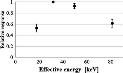
The GAFCHROMIC LD-V1 radiochromic film is widely used in dosimetry because it can provide high-resolution two-dimensional dose distributions without processing. This study aimed to evaluate the response characteristics at different effective energies, from the low-energy range of mammography to the high-energy range of computed tomography. Net pixel value (NPV)-absorbed dose calibration curves for the GAFCHROMIC LD-V1 were generated using x-rays with effective energies of 18, 30, 50, and 80 keV to reflect those used in different diagnostic radiographic modalities. The film response was analyzed using calibration curves at each energy level. The coefficients of determination for the calibration curves at 18, 30, 50, and 80 keV were 0.9992, 0.9997, 0.9999, and 0.9976, respectively. The pixel value change at 30 keV was the largest and most sensitive, while the smallest change in pixel value and lowest sensitivity were noted at 18 keV. Because the energy dependence of the GAFCHROMIC LD-V1 is significant below 18 keV and above 80 keV, it is necessary to establish an appropriate NPV-absorbed dose calibration curve for energies below 18 keV and consider the possibility of underestimating the dose at energies above 80 keV.
Keywords: GAFCHROMIC LD‐V1; diagnostic radiographic modalities; energy dependence.
© 2025 The Author(s). Journal of Applied Clinical Medical Physics published by Wiley Periodicals, LLC on behalf of The American Association of Physicists in Medicine.
Conflict of interest statement
The authors declare no conflicts of interest.
Figures


References
-
- Niroomand‐Rad A, Chiu‐Tsao ST, Grams MP, et al. Report of AAPM task group 235 radiochromic film dosimetry: an update to TG‐55. Med Phys. 2020;47(12):5986‐6025. - PubMed
-
- Gotanda T, Katsuda T, Gotanda R, et al. Evaluation of effective energy using radiochromic film and a step‐shaped aluminum filter. Australas Phys Eng Sci Med. 2011;34(2):213‐222. - PubMed
-
- Gotanda R, Katsuda T, Gotanda T, et al. Computed tomography phantom for radiochromic film dosimetry. Australas Phys Eng Sci Med. 2007;30(3):194‐199. - PubMed
-
- Gotanda R, Katsuda T, Gotanda T, Tabuchi A, Yatake H, Takeda Y. Dose distribution in pediatric CT head examination using a new phantom with radiochromic film. Australas Phys Eng Sci Med. 2008;31(4):339‐344. - PubMed
-
- Gotanda T, Katsuda T, Gotanda R, et al. Half‐value layer measurement: simple process method using radiochromic film. Australas Phys Eng Sci Med. 2009;32(3):150‐158. - PubMed
MeSH terms
Grants and funding
LinkOut - more resources
Full Text Sources
Medical
Research Materials
