. 2025 May 20;6(5):102092.
doi: 10.1016/j.xcrm.2025.102092. Epub 2025 May 8.

Cancer vaccine from intracellularly gelated tumor cells functionalized with CD47 blockage and damage-associated molecular pattern exposure

Affiliations

Cancer vaccine from intracellularly gelated tumor cells functionalized with CD47 blockage and damage-associated molecular pattern exposure

Cheng Gao et al. Cell Rep Med. .

Abstract

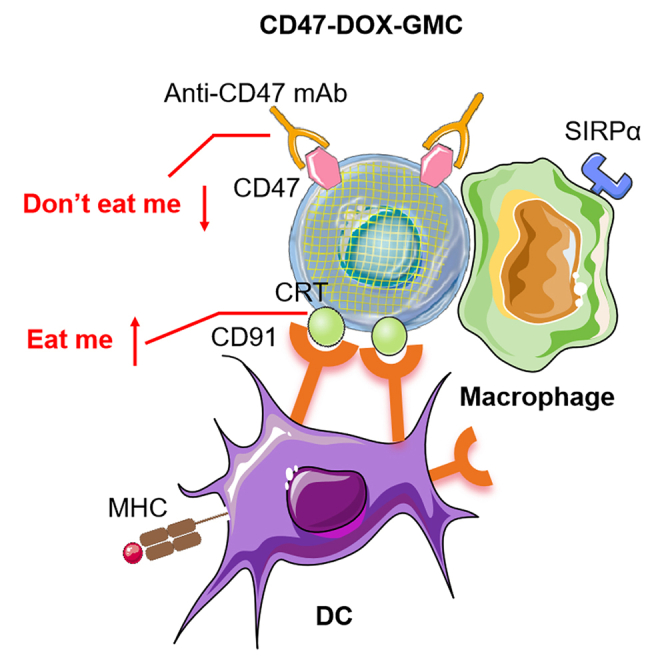
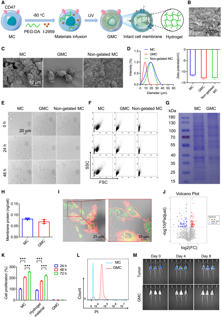
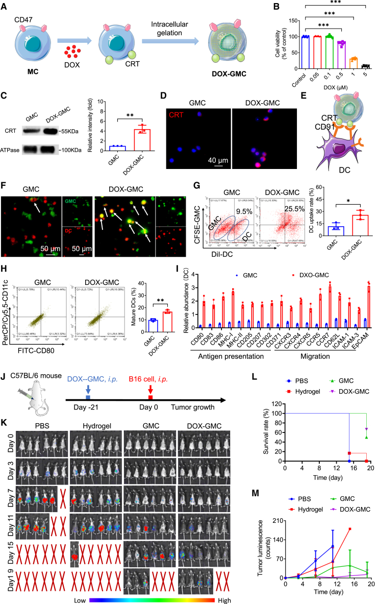
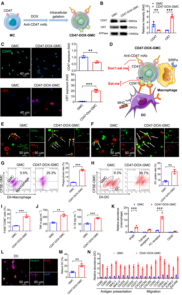
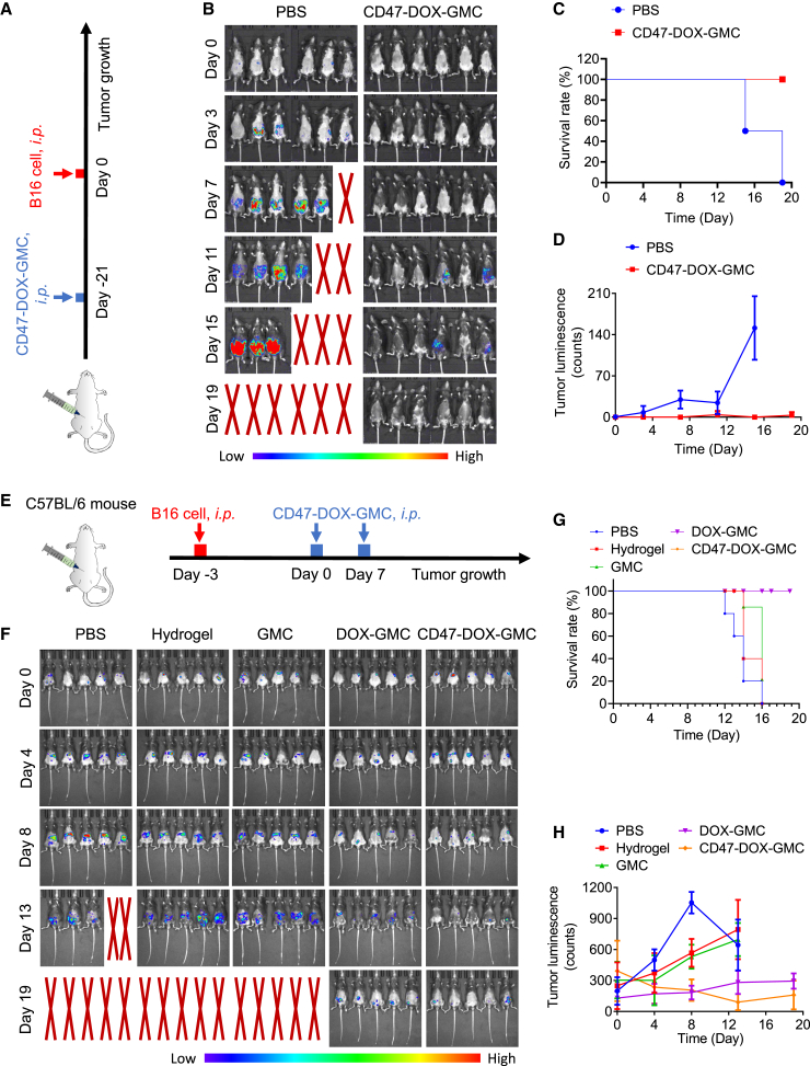
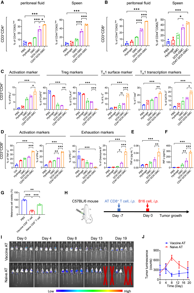
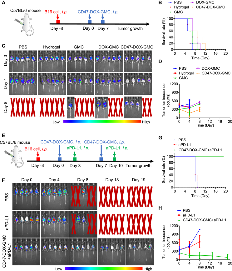
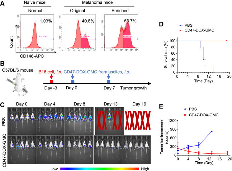
The effectiveness of whole tumor cell vaccines prepared by traditional inactivation methodology is often hindered by insufficient immunogenicity. Here, we report development of a cancer vaccine through the intracellular gelation of tumor cells, combined with CD47 blockade and damage-associated molecular pattern (DAMP) exposure, for effective tumor prevention and treatment. Intracellular hydrogelation preserves the morphology and antigenicity of tumor cells. CD47 blockade and DAMP exposure synergistically enhance the "eat me" signals and inhibit the "don't eat me" signals on tumor cells, significantly improving their immunogenicity. In the context of tumor prevention and treatment of pre-existing tumors, this vaccine polarizes CD4+ T cells toward a TH1 phenotype, reduces regulatory T cells and T cell exhaustion, and elicits a robust tumor-antigen-specific T cell response. When combined with an immune checkpoint inhibitor, this vaccine demonstrates enhanced efficacy in eradicating established tumors. The successful application of this vaccine using ascites and subcutaneous tumor cells supports the feasibility of developing personalized whole tumor cell vaccines for diverse tumor types.

Keywords: cancer treatment; hydrogel; intracellular assembly; surface engineering; tumor cell vaccine.


Conflict of interest statement

Declaration of interests The authors declare no competing interests.

Figures

None
Graphical abstract
Figure 1
Figure 1
GMC maintained cell morphology and intact antigen proteins but exhibited no cell viability (A) Schematic illustration on the construction of GMC. (B) SEM imaging on the PEG hydrogel. (C) SEM imaging on the MC, GMC, and non-gelated MC. (D) Mean diameter and zeta potential of MC, GMC, and non-gelated MC. (E and F) Confocal images (E) and FSC-SSC scatterplots (F) of MC, GMC, and non-gelated MC after placement in PBS for different times (0, 24, and 48 h). (G) Coomassie blue staining on the membrane proteins of MC and GMC. (H) BCA protein quantification on the membrane proteins of MC and GMC. (I) Confocal fluorescence imaging on the GMC labeled with red fluorescence membrane dye DiI and green fluorescence hydrogel material FITC-PEG-DA. (J) LC-MS/MS analysis on the whole proteins of MC and GMC. (K) Cell growth evaluation on free MC, MC treated with hydrogel materials, and GMC. (L) Flow cytometry analysis on PI-stained MC and GMC. (M) IVIS imaging on the C57BL/c mouse i.p. administered with MC and GMC (n = 3). The experiments were repeated three times (n = 3), and data were presented as mean ± SD. Statistical analyses for (D) and (H) are performed using one-way ANOVA. All other analyses were conducted using two-way ANOVA. ∗∗∗p ≤ 0.001.
Figure 2
Figure 2
Tumor cell ICD improved DC uptake and antigen presentation, and DOX-GMC vaccine prevented tumor growth (A) Illustration on the construction of DOX-GMC. (B) Cell viability of B16-F10 cells treated with different concentrations of DOX (0.05, 0.1, 0.5, 1, and 5 μM) for 24 h. (C) Western blot analysis on the CRT expression of GMC and DOX-GMC. (D) Fluorescence imaging on the red fluorescence probe-labeled CRT protein of GMC and DOX-GMC. (E) Schematic illustration on the enhanced recognition between DC and DOX-GMC through CD91-CRT interaction. (F) Confocal fluorescence imaging on the phagocytosis behavior of DC toward GMC and DOX-GMC after incubation for 6 h. Red: DiI-stained DC. Green: CFSE-stained GMC and DOX-GMC. (G) Quantification on the phagocytosis rate by flow cytometry. (H) The ratio of CD80+CD11c+ cell in DC after treatment with GMC and DOX-GMC for 6 h. (I) The relative abundances of antigen presentation-related proteins and migration-related proteins in DC incubated with GMC and DOX-GMC, determined by LC-MS/MS. (J) 6-week-old C57BL/6 male mice were i.p. inoculated with PBS, 10 mg/kg of hydrogel, GMC, and DOX-GMC at a dose of 106 per mouse (n = 6), and then challenged with B16-F10-Luc cells. (K) Tumor bioluminescence was monitored by using IVIS at different time points (day 0, 3, 7, 11, 15, and 19). (L and M) The recorded survival rate curve (N) and semi-quantification on the change of bioluminescence intensity in melanoma-bearing mice (M). The experiments except in vivo antitumor study were repeated three times (n = 3), and data were presented as mean ± SD. Statistical analysis for (B) was performed using t test. Statistical analysis for (J) was performed using two-way ANOVA. All other analyses were conducted using one-way ANOVA. ∗p ≤ 0.05, ∗∗p ≤ 0.01, and ∗∗∗p ≤ 0.001.
Figure 3
Figure 3
DAMP exposure and CD47 blockage strengthened the immunogenicity of CD47-DOX-GMC through induction of “eat me signal” and inhibition of “don’t eat me signal” (A) Diagram on the construction of CD47-DOX-GMC. (B) Western blot analysis on the CRT and CD47 expression of GMC and CD47-DOX-GMC. (C) Confocal fluorescence imaging on the green fluorescence probe-labeled CD47 protein and red fluorescence probe-labeled CRT protein of GMC and CD47-DOX-GMC. (D) Schematic illustration on the improved recognition of macrophage and DC toward CD47-DOX-GMC through induction of CD91-CRT interaction and blockage of SIRPα-CD47 interaction. (E) Confocal fluorescence imaging on the phagocytosis behavior of macrophage toward GMC and CD47-DOX-GMC after incubation for 6 h. Red: DiI-stained macrophage. Green: CFSE-stained GMC and CD47-DOX-GMC. (F) Confocal fluorescence imaging on the phagocytosis behavior of DC toward GMC and CD47-DOX-GMC after incubation for 6 h. Red: DiI-stained DC. Green: CFSE-stained GMC and CD47-DOX-GMC. (G and H) Quantification on the phagocytosis rate of macrophage (G) and DC (H) by flow cytometry. (I) The ratio of F4/80+CD86+ cell in macrophage after treatment with GMC and CD47-DOX-GMC for 6 h. (J) The levels of TNF-α and IL-1β released from macrophage treated with GMC and CD47-DOX-GMC for 6 h. (K) The number and relative abundance of upregulated immune response-related proteins in macrophages treated with GMC and CD47-DOX-GMC, respectively. (L) Confocal fluorescence imaging on the internalization and intracellular location of CD47-DOX-GMC in DC following 1 h incubation. (M) The ratio of CD80+CD11c+ cell in DC after treatment with GMC and CD47-DOX-GMC for 6 h. (N) The number and relative abundance of upregulated immune response-related proteins in DC treated with GMC and CD47-DOX-GMC, respectively. The experiments were repeated three times (n = 3), and data were presented as mean ± SD. Statistical analyses for (B), (K), and (N) were performed using two-way ANOVA. All other analyses were conducted using one-way ANOVA. ∗∗p ≤ 0.01 and ∗∗∗p ≤ 0.001.
Figure 4
Figure 4
CD47-DOX-GMC effectively prevented tumor and treated pre-existing tumor (A) 6-week-old C57BL/6 male mice were i.p. inoculated with PBS and CD47-DOX-GMC at a dose of 106 per mouse (n = 6), and then challenged with B16-F10-Luc cells. (B) Tumor bioluminescence was monitored by using IVIS at different time points (day 0, 3, 7, 11, 15, and 19). (C and D) The recorded survival rate curve (C) and semi-quantification on the change of bioluminescence intensity in melanoma-bearing mice (D). (E) 6-week-old C57BL/6 male mice were i.p. injected with B16-F10-Luc cells for 3 days, and then inoculated with PBS, 10 mg/kg of hydrogel, GMC, DOX-GMC, and CD47-DOX-GMC at a dose of 106 per mouse (n = 5), with two doses in total at day 0 and day 7. (F) Tumor bioluminescence was monitored by using IVIS at different time points (day 0, 4, 8, 13, and 19). (G and H) The recorded survival rate curve (G) and semi-quantification on the bioluminescence intensity in melanoma-bearing mice (H). The data in (D) and (H) were presented as mean ± SD.
Figure 5
Figure 5
CD47-DOX-GMC significantly activated tumor-associated lymphocytes and cytokines to achieve antitumor therapeutic effect (A and B) 6-week-old C57BL/6 male mice were i.p. injected with B16-F10-Luc cells for 3 days, and then inoculated with PBS, 10 mg/kg of hydrogel, GMC, DOX-GMC, and CD47-DOX-GMC at a dose of 106 per mouse (n = 3), with two doses in total at day 0 and day 7. The peritoneal fluid and spleen were collected at day 12, and T cell type and activation status were analyzed by flow cytometry. Percent of CD4+ and CD8+ T cells with effector memory (CD44+CD62Llow) phenotypes in ascites (A) and spleen (B) from treated mice. (C) Percent of CD4+ cells expressing activation marker IFN-γ, regulatory T cell markers FoxP3 and CTLA4, and TH1 surface markers and transcription markers in ascites from treated mice. (D) Percent of CD8+ cells expressing activation markers TNF-α and IFN-γ and exhaustion markers PD-1 and granzyme B in ascites from treated mice. (E and F) Cytokine analysis on ascites TNF-α (E) and IFN-γ (F) from treated mice. (G) Tumor cell death in co-cultures containing peritoneal CD8+ T cells from naive or vaccinated mice. (H) Tumor-naive 6-week-old C57BL/6 male mice were i.p. injected with peritoneal 2 × 10⁵ magnetically enriched CD8+ cell, and then challenged with B16-F10-Luc cells after 1 day (n = 5). (I) Tumor bioluminescence was monitored by using IVIS at different time points (day 0, 4, 8, 13, and 19). (J) Semi-quantification on the change of bioluminescence intensity in melanoma-bearing mice. The experiments except in vivo antitumor imaging were repeated three times (n = 3), and data were presented as mean ± SD. All statistical analyses were conducted using one-way ANOVA. ∗p ≤ 0.05, ∗∗p ≤ 0.01, and ∗∗∗p ≤ 0.001.
Figure 6
Figure 6
Immune checkpoint inhibitor facilitated the T cell-mediated tumor-killing effect induced by CD47-DOX-GMC on established tumor (A) 6-week-old C57BL/6 male mice were i.p. inoculated with B16-F10-Luc cells for 8 days to establish melanoma-bearing mice and i.p. treated with GMC, DOX-GMC, and CD47-DOX-GMC at a dose of 106 per mouse for two doses in total at day 0 and day 7 (n = 5). (B) Tumor bioluminescence was monitored by using IVIS at different time points (day 0, 4, and 8). (C and D) The recorded survival rate curve (C) and semi-quantification on the change of bioluminescence intensity in melanoma-bearing mice (D). (E) Melanoma mice were additionally i.p. administered with aPD-L1 after CD47-DOX-GMC treatment for 3 days (n = 5), with a dose of 4 mg/kg at day 3 and day 10, respectively. (F) Tumor bioluminescence was monitored by using IVIS at different time points (day 0, 4, 8, 13, and 19). (G and H) The recorded survival rate curve (G) and semi-quantification on the change of bioluminescence intensity in melanoma-bearing mice (H). The data in (D) and (H) were presented as mean ± SD.
Figure 7
Figure 7
Personalized CD47-DOX-GMC vaccine from ascites achieved effective antitumor efficacy (A) B16-F10 cells were collected from ascites of tumor-bearing mice and enriched by using filtration capture. (B) 6-week-old C57BL/6 male mice were i.p. administered with B16-F10-Luc cells for 3 days, and then inoculated with ascites-derived CD47-DOX-GMC at a dose of 106 per mouse (n = 5), with two doses in total at day 0 and day 7. (C) Tumor bioluminescence was monitored by using IVIS at different time points (day 0, 4, 8, 13, and 19). (D and E) The recorded survival rate curve (D) and semi-quantification on the change of bioluminescence intensity in melanoma-bearing mice (E). The data in (E) were presented as mean ± SD.

Similar articles

References

    1. Harari A., Graciotti M., Bassani-Sternberg M., Kandalaft L.E. Antitumour dendritic cell vaccination in a priming and boosting approach. Nat. Rev. Drug Discov. 2020;19:635–652. - PubMed
    1. Lorentzen C.L., Haanen J.B., Met Ö., Svane I.M. Clinical advances and ongoing trials of mRNA vaccines for cancer treatment. Lancet Oncol. 2022;23:e450–e458. - PMC - PubMed
    1. Chen L., Qin H., Zhao R., Zhao X., Lin L., Chen Y., Lin Y., Li Y., Qin Y., Li Y., et al. Bacterial cytoplasmic membranes synergistically enhance the antitumor activity of autologous cancer vaccines. Sci. Transl. Med. 2021;13 - PubMed
    1. Ma L., Diao L., Peng Z., Jia Y., Xie H., Li B., Ma J., Zhang M., Cheng L., Ding D., et al. Immunotherapy and Prevention of Cancer by Nanovaccines Loaded with Whole-Cell Components of Tumor Tissues or Cells. Adv. Mater. 2021;33 - PubMed
    1. Fang L., Zhao Z., Wang J., Zhang P., Ding Y., Jiang Y., Wang D., Li Y. Engineering autologous tumor cell vaccine to locally mobilize antitumor immunity in tumor surgical bed. Sci. Adv. 2020;6 - PMC - PubMed