Case Reports
. 2025 May 7;30(9):103283.
doi: 10.1016/j.jaccas.2025.103283. Epub 2025 Feb 26.

Lymphatic Abnormalities in Fontan: Case Report of Plastic Bronchitis

Affiliations
Case Reports

Lymphatic Abnormalities in Fontan: Case Report of Plastic Bronchitis

Daniela Torres-Gómez et al. JACC Case Rep. .

Abstract

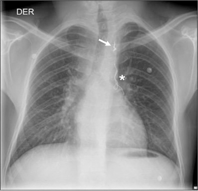
Plastic bronchitis is a rare but severe complication in Fontan patients. Central lymphatic evaluation may direct therapy for these complications. We present a 22-year-old mn post-Fontan who presented with bronchial casts. Initial medical management failed to reduce cast formation. Dynamic contrast-enhanced magnetic resonance lymphangiography was conducted for targeted management, revealing an aberrant lymphatic channel draining to the right pulmonary hilum. After 2 failed attempts, a percutaneous intervention with a proctoring aid resolved symptoms, improving the patient's condition. This case contributes to the literature on the role of central lymphatic imaging in diagnosing and treating lymphatic complications in Fontan patients. However, expertise is needed to successfully deliver targeted interventional lymphatic therapy. Understanding the management of Fontan patients requires recognizing lymphatic complications. Studying the central lymphatic system through dynamic contrast-enhanced magnetic resonance lymphangiography in patients with Fontan aids in diagnosing lymphatic abnormalities and targeting treatment.

Keywords: Fontan palliation; congenital heart disease; lymphatic abnormalities; magnetic resonance lymphangiogram; plastic bronchitis; therapeutic embolization.


Conflict of interest statement

Funding Support and Author Disclosures The authors have reported that they have no relationships relevant to the contents of this paper to disclose.

Figures

None
Graphical abstract
Visual Summary
Visual Summary
Patient Case Timeline: Disease Course, Interventions, and Outcomes A 22-year-old man status post-Fontan palliation who manifested coughing bronchial casts. Failure of medical management led to a dynamic contrast-enhanced magnetic resonance lymphangiography, which showed an aberrant lymphatic channel. Percutaneous intervention was achieved with symptom resolution.
Figure 1
Figure 1
Bronchial Casts (A and B) Expectoration reported by the patient, consistent with bronchial casts.
Figure 2
Figure 2
Transthoracic Echocardiogram (A) Four-chamber apical view depicting the morphology of tricuspid atresia with an ejection fraction of 35% to 40%. (B and E) Normal inferior vena cava flow with normal forward phasic flow and reduced respirophasic variability, suggesting dysfunction. (C and F) Fontan conduit with a 6-mm fenestration and a gradient of 6 mm Hg. (D and G) Patent superior vena cava to right pulmonary artery connection with antegrade and phasic flow and reduced respirophasic variability, suggesting dysfunction.
Figure 3
Figure 3
Dynamic Contrast-Enhanced Magnetic Resonance Lymphangiography (A) Coronal reconstruction in maximal intensity projection shows the thoracic duct (arrowhead) and a tortuous aberrant lymphatic channel originating from the left venous angle to the right pulmonary hilum (arrows). (B) Coronal reconstruction in maximal intensity projection shows chylolymphatic reflux in the supraclavicular, upper mediastinum, and hilar regions with pulmonary extension, right more than left.
Figure 4
Figure 4
Selected Lymphatic Embolization The lymphangiography images illustrate the lymphatic embolization procedure. (A) Localization of the tortuous aberrant lymphatic channel (arrow). (B) Successful occlusion of the aberrant lymphatic channel achieved through targeted embolization (arrow). A previous aortopulmonary embolization using a coil device, from the innominate vein to the coronary sinus, is observed (∗).
Figure 5
Figure 5
Postprocedure Chest Radiograph Anteroposterior view shows embolization coils projected over the superior aspect of the previously described aberrant lymphatic channel in the superior mediastinum (arrow). A previous aortopulmonary embolization using a coil device, from the innominate vein to the coronary sinus, is observed (∗). Also note the increased perihilar bronchovascular markings, more prominent on the right than left.

References

    1. Book W.M., Gerardin J., Saraf A., Marie Valente A., Rodriguez F. Clinical phenotypes of fontan failure: implications for management. Congenit Heart Dis. 2016;11(4):296–308. doi: 10.1111/chd.12368. - DOI - PubMed
    1. Benjamin J., O'Leary C., Hur S., Gurevich A., Klein W.M., Itkin M. Imaging and Interventions for lymphatic and lymphatic-related disorders. Radiology. 2023;307(3) doi: 10.1148/radiol.220231. - DOI - PubMed
    1. Ramirez-Suarez K.I., Tierradentro-Garcia L.O., Smith C.L., et al. Dynamic contrast-enhanced magnetic resonance lymphangiography. Pediatr Radiol. 2022;52(2):285–294. doi: 10.1007/s00247-021-05051-6. - DOI - PubMed
    1. Itkin M. Magnetic resonance lymphangiography and lymphatic embolization in the treatment of pulmonary complication of lymphatic malformation. Semin Intervent Radiol. 2017;34(3):294–300. doi: 10.1055/s-0037-1604301. - DOI - PMC - PubMed
    1. Kamsheh A.M., O'Connor M.J., Rossano J.W. Management of circulatory failure after Fontan surgery. Front Pediatr. 2022;10 doi: 10.3389/fped.2022.1020984. - DOI - PMC - PubMed

Publication types

LinkOut - more resources