Case Reports
. 2025 May;32(5):e70198.
doi: 10.1111/ene.70198.

Cerebral Amyloid Angiopathy-Related Inflammation in Iatrogenic Cerebral Amyloid Angiopathy

Affiliations
Case Reports

Cerebral Amyloid Angiopathy-Related Inflammation in Iatrogenic Cerebral Amyloid Angiopathy

Larysa Panteleienko et al. Eur J Neurol. 2025 May.

Abstract

Introduction: Cerebral amyloid angiopathy (CAA) related inflammation (CAA-ri) is considered to be a distinct syndrome caused by an inflammatory response to amyloid-β deposition in the walls of small leptomeningeal and cortical vessels in patients with sporadic CAA. However, recent data suggest that inflammation might contribute to a broader range of CAA subtypes.

Results: We describe a case of probable iatrogenic CAA (iCAA), which manifested with multiple intracerebral haemorrhages complicated by the development of clinical and radiological features of CAA-ri, which responded to steroids. Clinical, neuroimaging and CSF data suggested possible co-existing Alzheimer's pathology.

Discussion: CAA-ri may occur in association with iCAA, suggesting that a broader spectrum of patients might benefit from steroid treatment than previously assumed.

Keywords: amyloid‐beta; cadaveric dura mater; cerebral amyloid angiopathy‐related inflammation; iatrogenic cerebral amyloid angiopathy; prion.


Conflict of interest statement

The authors declare no conflicts of interest.

Figures
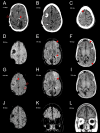
FIGURE 1
FIGURE 1
Representative CT and MRI images of the disease progression (with time indicators from hospitalisation due to the described case, days). At first presentation to the Emergency Department, this patient's CT head scan (A) showed no acute findings, but did demonstrate presence of an intraventricular catheter (red arrow), an old right parietal craniotomy (open red arrow), and scattered calcified lesions in both cerebral hemispheres with bilateral occipital white matter volume loss (consistent with her prior history of congenital toxoplasmosis). Two weeks later, when she presented again, her CT head showed an acute intracerebral haemorrhage involving the right middle frontal gyrus (B). Following a further clinical deterioration, her CT scan 3 days later revealed an acute convexity subarachnoid haemorrhage involving the right superior frontal sulcus (C). MRI brain (D), performed 5 days after the acute convexity subarachnoid haemorrhage, showed widespread disseminated cortical superficial siderosis involving both hemispheres and scattered foci of susceptibility artefact, representing lobar microhaemorrhages. T2‐weighted fluid‐attenuated inversion recovery (FLAIR) imaging showed sulcal hyperintensity and mild subcortical vasogenic oedema (arrows) involving the left frontal lobe and near the left temporal stem (E). Post‐gadolinium T1‐weighted imaging showed extensive and prominent leptomeningeal enhancement along the basal surface of the left frontal lobe and over the left parieto‐occipital region (F). Repeat MRI (17 days from admission) showed a further acute convexity subarachnoid haemorrhage overlying the left frontal convexity (open red arrows; G) and two acute intracerebral haemorrhages, involving the right superior frontal gyrus (arrow; G) and the left sub‐insular white matter (arrow; H). Although the previously observed leptomeningeal enhancement had improved (I), there was progression of the left frontal and temporal vasogenic oedema (H). Two months after treatment with steroids, the haematomas had matured, cortical superficial siderosis had progressed (J) and the vasogenic oedema, sulcal hyperintensities (K) and leptomeningeal enhancement (L) had resolved.

References

    1. Fandler‐Höfler S., Ambler G., Banerjee G., et al., “Temporal and Spatial Clustering of Intracerebral Hemorrhage in Cerebral Amyloid Angiopathy,” Neurology 103, no. 5 (2024): e209770, 10.1212/WNL.0000000000209770. - DOI - PMC - PubMed
    1. Sellimi A., Panteleienko L., Mallon D., et al., “Inflammation in Cerebral Amyloid Angiopathy‐Related Transient Focal Neurological Episodes,” Annals of Neurology 97 (2025): 475–482, 10.1002/ana.27164. - DOI - PMC - PubMed
    1. Banerjee G., Samra K., Adams M. E., et al., “Iatrogenic Cerebral Amyloid Angiopathy: An Emerging Clinical Phenomenon,” Journal of Neurology, Neurosurgery, and Psychiatry 93, no. 7 (2022): 693–700, 10.1136/jnnp-2022-328792. - DOI - PubMed
    1. Banerjee G., Collinge J., Fox N. C., et al., “Clinical Considerations in Early‐Onset Cerebral Amyloid Angiopathy,” Brain 146, no. 10 (2023): 3991–4014, 10.1093/brain/awad193. - DOI - PMC - PubMed
    1. Fandler‐Höfler S., Kaushik K., Storti B., et al., “Clinical‐Radiological Presentation and Natural History of Iatrogenic Cerebral Amyloid Angiopathy,” Journal of Neurology, Neurosurgery, and Psychiatry, ahead of print, February 12, 2025, 10.1136/jnnp-2024-335164. - DOI - PubMed

Publication types

MeSH terms

LinkOut - more resources