. 2025 May 9;16(1):4327.
doi: 10.1038/s41467-025-59503-w.

KAT5 regulates neurodevelopmental states associated with G0-like populations in glioblastoma

Affiliations

KAT5 regulates neurodevelopmental states associated with G0-like populations in glioblastoma

Anca B Mihalas et al. Nat Commun. .

Abstract

Quiescence cancer stem-like cells may play key roles in promoting tumor cell heterogeneity and recurrence for many tumors, including glioblastoma (GBM). Here we show that the protein acetyltransferase KAT5 is a key regulator of transcriptional, epigenetic, and proliferative heterogeneity impacting transitions into G0-like states in GBM. KAT5 activity suppresses the emergence of quiescent subpopulations with neurodevelopmental progenitor characteristics, while promoting GBM stem-like cell (GSC) self-renewal through coordinately regulating E2F- and MYC- transcriptional networks with protein translation. KAT5 inactivation significantly decreases tumor progression and invasive behavior while increasing survival after standard of care. Further, increasing MYC expression in human neural stem cells stimulates KAT5 activity and protein translation, as well as confers sensitivity to homoharringtonine, to similar levels to those found in GSCs and high-grade gliomas. These results suggest that the dynamic behavior of KAT5 plays key roles in G0 ingress/egress, adoption of quasi-neurodevelopmental states, and aggressive tumor growth in gliomas.


Conflict of interest statement

Competing interests: The authors declare no competing interests.

Figures
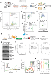
Fig. 1
Fig. 1. Identification of KAT5 as a G0-trap in GSC-0827 cells.
a Schematic of the G0-trap screen. For the screen, GSC-0827 cells containing mCherry-CDT1 (G0/G1) and p27-mVenus (G0) were transduced with a genome-wide CRISPR-Cas9 library, allowed to expand for 10 days, and sorted for double positive cells using the top 20% of p27-mVenus+ cells as a cut off. FACS machine cartoon taken from BioRender. Paddison, P. (2025) https://BioRender.com/o2c4y3l. b Results G0-trap screen from sgRNA-seq of p27hi vs. total cell population (n = 3; edgeR was used to assess p values and logFC cuttoffs) (Supplementary Data 1). Supporting QC data and gene set enrichment can be found in Supplementary Fig. 1. c FACS-based assessment of total RNA and DNA content, using pyronin Y and DAPI, respectively, in GSC-0827 cells via nucleofection of sgRNA:Cas9 RNPs. Additional retest assays are available in Supplementary Figs. 2, 3. d Quantification of (c) (≥2350 cells per condition from two independent treatments; KS test, p < .0001; median and quartiles shown). e FACS-based assessment of EdU incorporation after KAT5 KO using nucleofection of sgRNA:Cas9 RNPs (5 days post-nucleofection) in GSC-0827 cells. Quantification is shown in Supplementary Fig. 3a. f GSC-0827 cells were engineered to have a doxycycline controllable KAT5 open reading frame and knockout insertion-deletion mutations in the endogenous KAT5 gene. We found one such clone, #13 (C13), to have Dox-dependent KAT5 expression and requirement of Dox+ for continued growth. g Western blot analysis of KAT5 and histones H3 and H4 acetylation and methylation status after Dox withdrawal of 0, 4, 7, and 14 days in C13 cells. *indicates KAT5 targets. h FACS analysis of histone H4 acetylation and p27-mVenus levels after 0, 7, and 14 days of Dox withdrawal in C13 cells. i A growth curve with C13 cells grown in various concentrations of Dox (n = 3; mean ± SEM is shown, significance determined by unpaired, 2 tailed student’s t test). j FACS analysis of p27 induction in C13 cells grown in shown concentrations of Dox for 7 days. k Violin plot of H4-Ac levels in C13 cells grown in shown concentrations of Dox for 5 days. ≥5703 cells plotted for single trial. KS test was used to test significance, p <0.0001. Median and quartiles displayed. Source data with exact p values are provided with this paper as a Source Data file.
Fig. 2
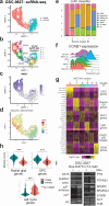
Fig. 2. Single cell gene expression analysis of GSC-0827 cells after KAT5 inhibition in vitro.
a UMAP projection of scRNA-seq data for sgCD8 (control) and sgKAT5 cells in GSC-0827 cells 5 days post nucleofection. Filtering scheme and data quality assessment for this data is available in Supplementary Fig. 4. b The UMAP projection from a showing de novo clusters generated. Cell cycle state predictions using the ccSeurat and ccAF classifiers and RNA velocity analysis for this data from are available in Supplementary Fig. 5. Associated data files include: cluster-based gene expression analysis (Supplementary Data 2) and gene set enrichment for top 200 expressed and top 200 depleted genes for each cluster (Supplementary Data 3, 4, respectively). For control cells, there is one G0-like cluster (cluster 4) which showed some weak expression for p53-associated target genes, suggestive of a DNA damage-induced G0-like state, a common feature of cultured cells (Arora et al., 2017; Spencer et al., 2013) also to some degree Neural G0 genes (see text). This state likely represents the p27hi Edu- population observed in vitro in Supplementary Fig. 1b, which are capable of re-entering the cell cycle. c MKI67/Ki67 gene expression, which is only expressed in cycling cells, among cells and clusters from (b). d Gene Set Variation Analysis (GSVA) associated with clusters and cell cycle phases showing MCM pathway required for initiation of DNA replication in early S-phase. Cell cycle pathways are shown in Supplementary Fig. 5. e Cell cycle phase predictions using the ccAF classifier for scRNA-seq data from (a). f Analysis of CCNB1 gene expression in ccAF predicted cell cycle phases: Neural G0 contains fewest cells expressing CCNB1, while G2/M the most. g Heatmap of representative genes upregulated in scRNA-seq clusters from (b). h Violin plots of gene expression module scores for each cell from scRNA-seq data of sgCD8A and sgKAT5KO GSC-0827 cells. oRG: outer radial glia. OPC oligodendrocyte precursor cells. Each data point = single cell. KS test was used to test significance (p < 0.0001)(1323 CD8 KO control cells; 840 KAT5 KO cells). Genes contained in each module are available in Supplementary Data 5. i Western blot validation studies of gene expression changes associated with loss of KAT5 activity. Protein extracts from GSC-0827 C13 cells were used from Dox+ or Dox- (7 days) conditions. Western blots were performed at least twice with similar results (except nAURKA = 1, nS100B = 1, nKAT5 = 4). Source data with exact p values are provided with this paper as a Source Data file.
Fig. 3
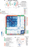
Fig. 3. KAT5-dependent cell state changes across 9 different GBM isolates.
Single cell gene expression was used to assess changes in cell states in response cells after KAT5 inhibition in vitro. GSCs were collected for analysis post nucleofection of sgCD8 (control) and sgKAT5 RNPs after 5 days of growth in NSC self-renewal media. a Scheme for comparing KAT5 versus CD8 KO populations in scRNA-seq data. For each isolate, we co-embedded scRNA-seq data from KAT5 KO and control CD8 KO cells into a single merged scRNA-seq object. Then, de novo clustering was applied to each GSC line, and each cluster was examined for admixtures of KAT5 and CD8 KO cells. For assessment of gene expression changes, for clusters mainly composed of control CD8 KO cells (CD8 KO:KAT5 KO cell ratios of ≳3) or KAT5 KO cells (CD8 KO: KAT5 KO cell ratios of ≲.33), cluster marker gene enrichment (or “method 2”) was used. For clusters composed of mixed populations of CD8 and KAT5 KO cells (cell ratios between 3 and. 33), DESeq2 was used to compare gene expression changes between CD8 KO to KAT5 KO cells within that cluster (or “method 1”). For both methods cutoffs were log2 fold change of 0.321 and adjusted pvalue for 0.05. The example in (a) highlights three clusters from each category. UMAPs for all 9 GSC isolates can be found in Supplementary Fig. 6. The full results from this analysis can be found in Supplementary Data S6. b Comparisons of KAT5 KO dependent changes in cell states for 9 GSC isolate. The genes found significantly changed for each GSC cluster by either method 1 or 2 were compared using the Fisher’s exact hypergeometric enrichment test and the -log10 p value and the overlapping marker genes were recorded for each pairwise comparison. Call out clusters from a are shown as filled circle, square, and triangle. *denotes novel KAT5 KO clusters from Fig. 2. c Gene set enrichment analysis for gene enriched in CD8 KO. Comparisons of 272 genes enriched in CD8 KO clusters with S, S/G2, and G2/M phase genes form the ccAF cell cycle classifier. d Comparisons of 718 gene enriched in KAT5 KO clusters with Neural G0 genes and outer radial glial genes (from human fetal brain development). Genes were required to overlap with ≥50% of clusters to be considered for (c, d).
Fig. 4
Fig. 4. KAT5 KO triggers gene expression changes in GSCs consistent with predicted quiescent states in patient-derived xenograft tumors.
a, b Projections of scRNA-seq data for GSC-0827 and GSC-464T tumor references, respectively. Data was visualized using uniform manifold approximation and projection (UMAP) for dimensional reduction of data and generation of de novo cell-based clusters (Becht et al. 2018). Overview of experiment, filter cutoffs, and QC analysis are available in Supplementary Fig. 7. Supporting data includes: top enriched genes for each cluster; gene expression modules; and individual and gene set expression profiles (Supplementary Data 7-10; Supplementary Figs. 8–11). c–f Cells from scRNA-seq analysis performed on GSC-0827 and GSC-464T CD8 and KAT5 KO cells. The tumor references from (a, b) were used for mapping cells from scRNA-seq analysis performed on GSC-0827 and GSC-464T CD8 and KAT5 KO cells passed quality control metrics. g, h Cell cycle predictions using ccAFv2 computational classifier for (a, b), respectively. i, j Cyclin B1 gene expression for each predicted phase of the cell cycle from (g, h), respectively. k, l Relative proportions of mapped single cells appearing in clusters from (c–f) for GSC−0827 and GSC-464T cells, respectively.
Fig. 5
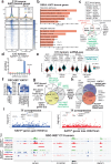
Fig. 5. Analysis of KAT5 target genes, gene expression changes, and epigenetic patterning associated with KAT5on and KAT5off states in GSC-0827 tumors.
a KAT5 binding associated with transcription start sites in GSC-0827 tumors. An antibody recognizing the V5 epitope tag was used to perform CUT&TAG on KAT5-V5 ectopically expressed in C13 Dox + /KAT5on tumors. Dox-/KAT5off tumors (6 days Dox0-) were used as a control. K-mean clustering for KAT5on was used to define separate cluster classes shown (Supplementary Data 11). b Gene set enrichment analysis for KAT5 bound genes from clusters 2, 3, and 4 from (a). c Comparisons of KAT5 bound genes with common essential genes (depmap.org) and essential transcription factors in GSC-0827s. d Fold change of cells in each scRNA-seq cluster for KAT5off tumors. e Violin plots of gene expression module scores for each cell from scRNA-seq data of Dox+ vs. Dox- GSC-0827 C13 tumors. Each data point = single cell. KS test was used to test significance (p < 0.0001)(4951 Dox+ cells; 5507 Dox- cells). Genes associated with each model are available in Supplementary Fig. 12 and Supplementary Data 5. f ChromHMM analysis of genomic regions in KAT5on C13 tumor cells showing 8 possible chromatin states (i.e., emission states) for H3K4me2, H3K27ac, and H3K27me3, the associated number of genes, and emission state region genomic annotations. Genome% = intergenic space; TES = transcription end sites; TSS = transcription start sites. The darker blue color corresponds to a greater probability of observing the mark in the state. The full data set is available in Supplementary Data 12. g, h Overlap of genes associated with emission state E4 from (f), which display both activating and repressive chromatin marks, and those with significant changes H3K27ac and H3K27me3 after loss of KAT5 activity in GSC-0827 tumors. DiffBind and DESeq2 were used to score significant changes in chromatin marks (Supplementary Data 13,14, 15) from KAT5on and KAT5off C13 tumor samples (n = 2). Note: DiffBind could not be performed on H3K4me2 marks for C13 tumor samples because one replicate failed to produce sufficient quality data. i Enrichment for genes associated with expression of human transcription factors (TFs). Left panel: genes enriched for H3K27ac regions after loss of KAT5 activity. Right panel: genes depleted for H3K27me3 regions after loss of KAT5 activity. The analysis was performed using the Enrichr pipeline (Kelshov et al., 2016) using the hypergeometric test by comparing the top 300 genes associated with specific human transcription factors in the ARCHS4 database (Lachmann, et al. 2018). j Examples of multivalent genes in KAT5on C13 tumors that upon Dox withdrawal gain H3K27ac and lose H3K27me3 marks (i.e., overlapping genes from h). Additional examples including GNG7, SLIT1, and SOX8 are shown in Supplementary Fig. 12.
Fig. 6
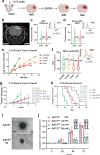
Fig. 6. Modulation of KAT5 activity in GSC-derived tumors and during GSC invasion assays.
a Scheme for using C13 GSC-0827 cells for creating PDX tumors in NSG mice. Created in BioRender. Paddison, P. (2025) https://BioRender.com/tehoi00. b Representative MRI image of mouse head with Dox-KAT5 tumor in cortex at 36 days post-injection. c Analysis of p27 levels after Dox withdrawal in C13 tumor containing mouse. KS test was used to test significance (p < 0.0001). d Analysis of EdU incorporation (6 h) after 7 days of Dox withdrawal in C13 tumor containing mouse. KS test was used to test significance (p < 0.0001). e Tumor growth as assessed by volume using MRI in C13-induced PDX tumors after switching drinking water to concentration of Dox indicated (µg/mL). Linear regression analysis was used to assess significance (p val overall = 0.001; p val2000 vs. 200 < 0.0001; p val2000 vs. 20 = 0.0049; p val2000 vs. 0 = 0.0183). f EdU incorporation for 20 h at 14 days after Dox concentration from (e). KS test was used to test significance (p < 0.0001). g C13 xenografts were treated with standard of care (SOC) in the presence (Dox + ) and/or absence (Dox–) of KAT5. Tumor growth was assessed by volume using MRI. The plot represents a linear regression analysis with the first MRI time point at enrollment of mice in experimental cohorts. The asterisk denote statistically significant differences between pairs of slopes for the indicated experimental conditions by color (nDox + = 8, nDox- = 8, nDox + /SOC = 9, nDox-/SOC = 5). Plots show mean ± SEMs. with simple linear regression curve comparisons for significance. h Kaplan-Meier survival probability plot for tumor bearing mice from (g). Survival probability differences between paired comparisons was assessed by log rank (Mantel-Cox) test. The asterisk denote statistically significant differences between pairs of comparisons for the indicated experimental conditions by color. Average survival days gained: Dox- = 17.75; SOC/Dox + = 17.97; and SOC/Dox- = 39.55. i, j KAT5on vs. KAT5off C13 invasion assays. C13 cells were grown as spheres for 3 days in either 1μg/ml Doxycycline for KAT5on or no Doxycycline for KAT5off conditions and transferred to Matrigel covered wells for the specified number of days (3 d, 7 d, or 9 d). Phase-contrast images were captured every 24 h for 72 h. The area covered by invading cells was measured using FIJI. i Representative phase contrast microscopy images of invasion assay. Spheroids were embedded into Matrigel. Scale bar is 200 μm. j Quantification of (e). nKAT5on = 3, nKAT5off 3d = 3, nKAT5off 7d = 4, nKAT5off 9d = 5. Mean ± SEM shown (significance was tested using repeated measurements 2-way ANOVA with Giesser-Greenhouse correction and Sidak multiple corrections test; p ≤ 0.01). Additional isolates are assayed in Supplementary Fig. 13. Source data with exact p values are provided with this paper as a Source Data file.
Fig. 7
Fig. 7. Assessment of KAT5 activity and protein translation rates in primary glioma tumor samples.
a Examination of protein synthesis as measured by L-azidohomoalaine (AHA) incorporation in parental 827 cells and for KAT5 inhibited C13 cells after 7 days of Dox withdrawal. KS test was used to test significance. Note: AHA was used for these experiments because OPP (below) cannot be used in puromycin resistant cells. KS test was used to test significance. *p val < 0.0001 (n ≥ 16970). b Examination of protein synthesis via AHA incorporation in C13 cells grown in shown concentrations of Dox for 5 days. KS test was used to test significance. *p val < 0.0001 (n ≥ 5703). c Examination of total protein content in C13 cells using ponceau staining of total protein extract from 200,000 C13 cells Dox withdrawal of 0, 4, 7, and 14 days. Protein measurement was performed three times with similar results. d The scheme used for examining protein synthesis using O-propargyl-puromycin (OPP) incorporation in primary tumor cells followed by FACS-based assessment of OPP, histone H4 acetylation (i.e., KAT5 activity), CD45 + , and viability. Samples were dissociated, OPP-labeled, viably frozen, thawed, and flow analyzed as a cohort. Tumor cells are CD45-. FACS machine cartoon created in BioRender. Paddison, P. (2025) https:// BioRender.com/o2c4y3l. e Plots of OPP versus pan-H4-Ac in LGG (UW36) and HGG (UW40). f, g Violin plots of OPP and pan-H4-Ac assay results, respectively, from two LGG (IDH1/2mut) (blue), one HGG (IDH1/2 mut) (pink) and one HGG (IDH1/2 wt) (red). Each flow event = single cell. KS test was used to assess significance (p < 0.0001)(n ≥ 2751); LGG vs either HGG WT or IDH1/2mut. h, i Violin plots of OPP and pan-H4-Ac assay results for 3 LGG (IDH1/2mut) (blue), two HGG (IDH1/2 mut) (pink) and 5 HGG (IDH1/2 wt) (red). FACS values are normalized in order for to compare across cohort groups. Each flow event = single cell. KS test was used to assess significance (p < 0.0001)(n ≥ 59074); LGG vs either HGG WT or IDH1/2mut.Supplementary Fig. 15 shows similar data for other individual tumors for LGG, HGG, and IDH1/2 mut HGG tumors. Supplementary Data 18 provides descriptions of each tumor sample used. j Combined of OPP versus pan-H4-Ac for LGG (Pearson r = 0.77), HGG (IDH1/2mut) tumors (Pearson r = 0.93), and HGG (Pearson r = 0.68), respectively; p < 0.0001 for each.
Fig. 8
Fig. 8. Assessment of KAT5 activity, AHA incorporation, and homoharringtonine sensitivity.
a Flow-based assessment of H4-Ac and AHA incorporation in NSCs, transformed NSCs and GSC-0827 cells. b Homoharringtonine (HHT) sensitivity of NSCs, transformed NSCs, and GSCs. The approximate IC50 (100 nM) for NSC-CB660 cells was used for this assay. Cells were treated for 4 days 100 nM HHT (the ~IC50 of NSC-CB660 cells) after which viability was measured (n ≥ 3). Means and ± SEMs are shown, unpaired, 2 tailed t-tests were used for significance. Supplementary Fig. 16 shows MYC and MYCN expression as well as H4-Ac and AHA incorporation for GSC-464T and GSC-1406. c Model of KAT5 function in HGG and LGG tumors arising from this work. LGGs have lower levels of KAT5 activity and cell cycle genes but appear to maintain similar levels of MYC, MYCN, and KAT5 transcripts as HGGs (Supplementary Fig. 16). If true, one possibility is that the presence of excessive cellular 2-hydroxyglutarate, produced by oncogenic IDH1/2 mutant enzymes, keeps KAT5 activity. Created in BioRender. Paddison, P. (2025) https://BioRender.com/6xzam1p. Source data and exact p values are provided with this paper as a Source Data file.

References

    1. Patel, A. P. et al. Single-cell RNA-seq highlights intratumoral heterogeneity in primary glioblastoma. Science344, 1396–1401 (2014). - PMC - PubMed
    1. Neftel, C. et al. An integrative model of cellular states, plasticity, and genetics for glioblastoma. Cell178, 835–849.e821 (2019). - PMC - PubMed
    1. Hanahan, D. Hallmarks of cancer: New dimensions. Cancer Discov.12, 31–46 (2022). - PubMed
    1. Lathia, J. D., Mack, S. C., Mulkearns-Hubert, E. E., Valentim, C. L. & Rich, J. N. Cancer stem cells in glioblastoma. Genes Dev.29, 1203–1217 (2015). - PMC - PubMed
    1. O’Connor, S. A. et al. Neural G0: a quiescent-like state found in neuroepithelial-derived cells and glioma. Mol. Syst. Biol.17, e9522 (2021). - PMC - PubMed

MeSH terms

Substances