. 2025 May 9;17(1):38.
doi: 10.1038/s41368-025-00375-7.

Profiling and functional characterization of long noncoding RNAs during human tooth development

Affiliations

Profiling and functional characterization of long noncoding RNAs during human tooth development

Xiuge Gu et al. Int J Oral Sci. .

Abstract

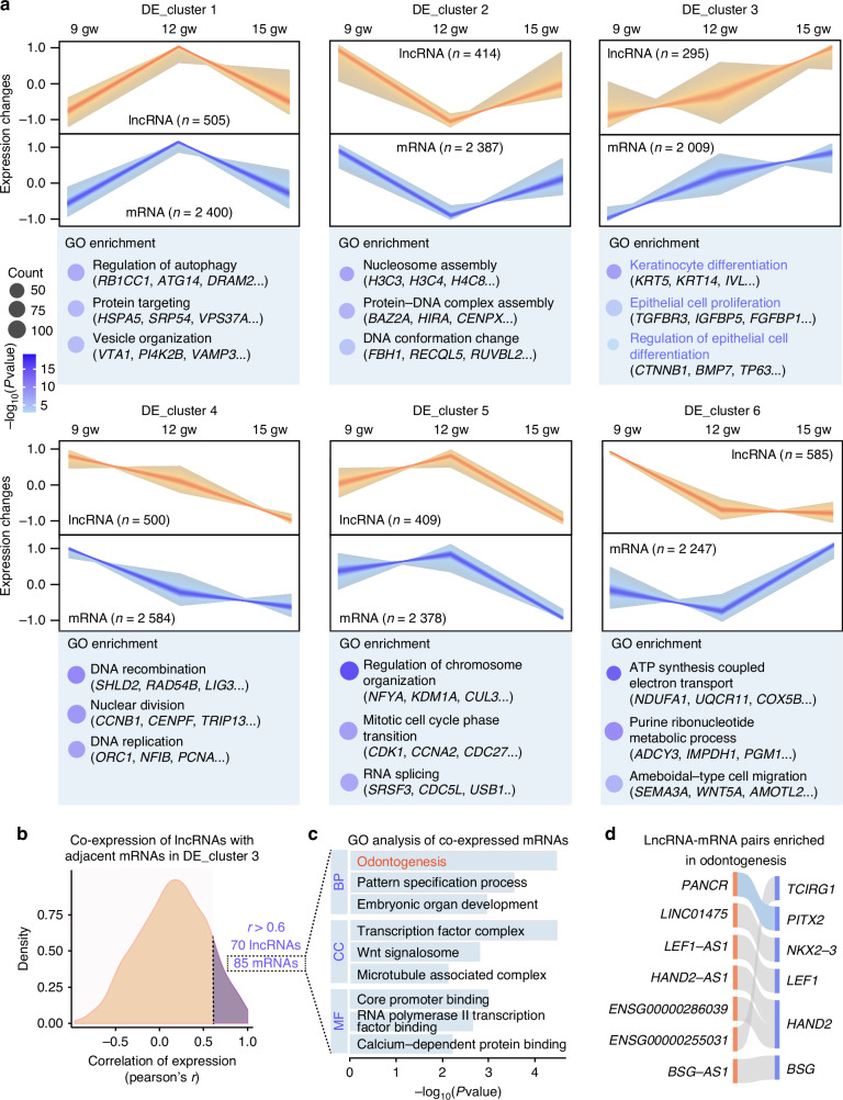
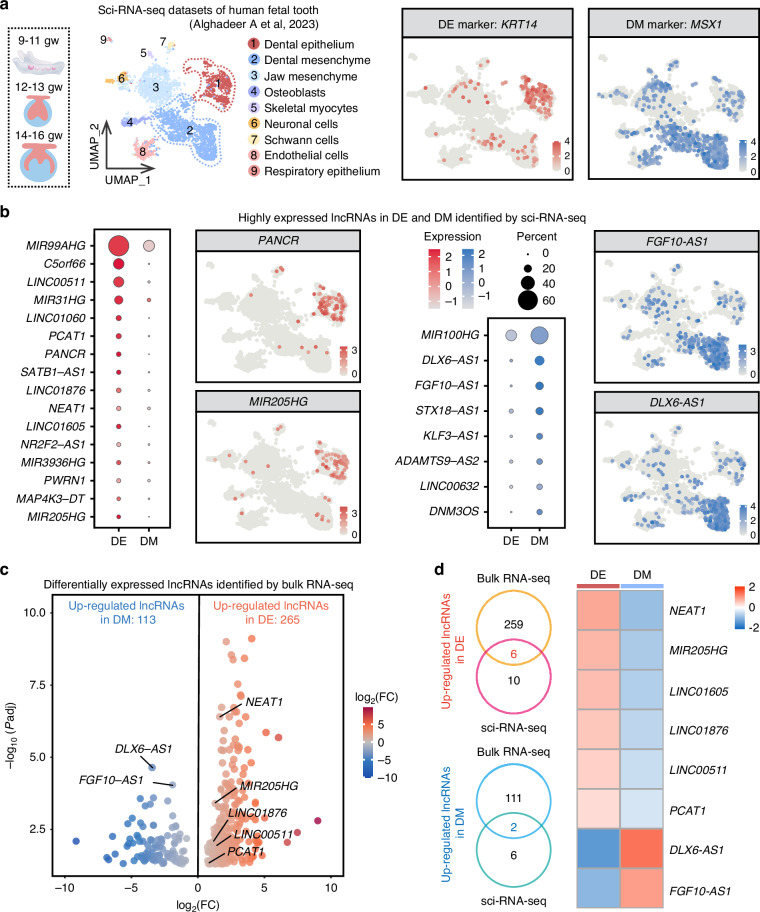
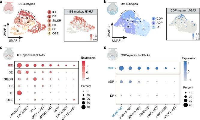
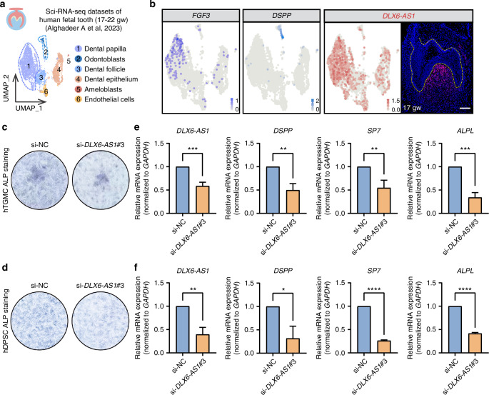
The regulatory processes in developmental biology research are significantly influenced by long non-coding RNAs (lncRNAs). However, the dynamics of lncRNA expression during human tooth development remain poorly understood. In this research, we examined the lncRNAs present in the dental epithelium (DE) and dental mesenchyme (DM) at the late bud, cap, and early bell stages of human fetal tooth development through bulk RNA sequencing. Developmental regulators co-expressed with neighboring lncRNAs were significantly enriched in odontogenesis. Specific lncRNAs expressed in the DE and DM, such as PANCR, MIR205HG, DLX6-AS1, and DNM3OS, were identified through a combination of bulk RNA sequencing and single-cell analysis. Further subcluster analysis revealed lncRNAs specifically expressed in important regions of the tooth germ, such as the inner enamel epithelium and coronal dental papilla (CDP). Functionally, we demonstrated that CDP-specific DLX6-AS1 enhanced odontoblastic differentiation in human tooth germ mesenchymal cells and dental pulp stem cells. These findings suggest that lncRNAs could serve as valuable cell markers for tooth development and potential therapeutic targets for tooth regeneration.


Conflict of interest statement

Competing interests: The authors declare no competing interests. Ethics approval: The present study is approved by the Ethics Commission of Beijing Friendship Hospital, Capital Medical University (CMU) (2021-P2-198-02), the Beijing Laboratory of Oral Health (BLOH), CMU (Z2022SY064), and the Beijing Obstetrics and Gynecology Hospital (BOGH), CMU (2023-KY-082-01). Human fetal tissues were initially collected from BOGH and CMU, and were obtained under informed consent from donors. All human fetal tissues came from aborted fetuses for family planning purposes.

Figures

Fig. 1
Fig. 1
Identification of lncRNAs in developing fetal teeth. a Schematic diagram of bulk RNA-seq samples from dental epithelium (DE) and dental mesenchyme (DM) of developing fetal teeth at the late bud, cap, and early bell stages. n = 3 per group. gw gestational week. b Boxplots showing the expression (FPKM) of KRT5 and VIM in DE and DM across the three developmental stages, through bulk RNA-seq. c Pie graph showing the quantity of robustly expressed mRNAs and lncRNAs. The number of genes expressed in DE and DM across the three developmental stages is also shown. d Cumulative expression plots comparing the number of mRNAs (left) and lncRNAs (right), that constitute the total sum of gene expression (FPKM), respectively. Percentage of total lncRNA expression (FPKM) accounted for by the union of the top 12 most highly expressed lncRNAs in DE (e) and DM (f)
Fig. 2
Fig. 2
Dynamic expression patterns and functional insights of lncRNAs and mRNAs in DE. a Fuzzy c-means clustering identified six distinct temporal expression patterns of lncRNAs and mRNAs in DE. GO enrichment analysis and representative genes were identified for each mRNA cluster. b Density distributions of the Pearson correlation coefficients between mRNAs and their adjacent lncRNAs in DE_cluster 3. This analysis revealed a positive correlation (r > 0.6) between 85 mRNAs and 70 lncRNAs. c GO enrichment analysis of the 85 co-expressed mRNAs shown in (b). d Alluvial plot depicting lncRNA-mRNA pairs enriched in odontogenesis, as shown in (c)
Fig. 3
Fig. 3
Dynamic expression patterns and functional insights of lncRNAs and mRNAs in DM. a Fuzzy c-means clustering identified six distinct temporal expression patterns of lncRNAs and mRNAs in DM. GO enrichment analysis and representative genes were identified for each mRNA cluster. b Density distributions of the Pearson correlation coefficients between mRNAs and their adjacent lncRNAs in DM_cluster 6. This analysis revealed a positive correlation (r > 0.6) between 335 mRNAs and 263 lncRNAs. c GO enrichment analysis of the 335 co-expressed mRNAs shown in (b). d Alluvial plot depicting lncRNA-mRNA pairs enriched in odontogenesis of dentin-containing tooth, as shown in (c)
Fig. 4
Fig. 4
Integration of sci-RNA-seq and bulk RNA-seq reveals cell-specific lncRNAs in developing fetal teeth. a UMAP visualization of nine clusters integrated from previously published sci-RNA-seq datasets of developing fetal teeth at the cap stage (9–11 and 12–13 gw) and early bell stage (14–16 gw). DE and DM clusters annotated with KRT14 and MSX1. b Dot plots of highly expressed lncRNAs in DE (red) and DM (blue) from sci-RNA-seq. Feature plot highlighting high expression of PANCR and MIR205HG in DE, and FGF10-AS1 and DLX6-AS1 in DM. c Volcano plot of differentially expressed lncRNAs in DE and DM identified by bulk RNA-seq. d Venn diagrams and heatmap of up-regulated lncRNAs in DE and DM showing overlap between bulk RNA-seq and sci-RNA-seq
Fig. 5
Fig. 5
IEE-specific and CDP-specific lncRNAs identified by sci-RNA-seq. a UMAP plot of six single-cell clusters in DE from sci-RNA-seq datasets of developing fetal teeth at 9–16 gw. IEE cluster annotated with RYR2. IEE inner enamel epithelium, DE dental epithelium, SI&SR stratum intermedium and stellate reticulum, EK enamel knot, OE oral epithelium, OEE outer enamel epithelium. b UMAP representation of three single-cell clusters in DM from sci-RNA-seq datasets of developing fetal teeth at 9–16 gw. CDP cluster annotated with FGF3. CDP coronal dental papilla, ADP apical dental papilla, DF dental follicle. Dot plots showing IEE-specific lncRNAs (c) and CDP-specific lncRNAs (d)
Fig. 6
Fig. 6
CDP-specific DLX6-AS1 enhances odontoblastic differentiation of hTGMCs and hDPSCs. a UMAP plot of six clusters from previously published sci-RNA-seq datasets of developing fetal teeth at the late bell stage (17–19 and 20–22 gw). b UMAP plot of DP marker FGF3, odontoblast marker DSPP, and lncRNA DLX6-AS1. RNAscope ISH staining of DLX6-AS1 in human fetal molars at 17 gw. Scale bar, 100 μm. ALP staining after 7-day odontogenic induction in hTGMCs (c) and hDPSCs (d) with DLX6-AS1 knockdown. Relative expression of DLX6-AS1, DSPP, SP7, and ALPL after 7-day odontogenic induction in hTGMCs (e) and hDPSCs (f) with DLX6-AS1 knockdown was measured by qRT-PCR

Similar articles

References

    1. Tucker, A. S., Matthews, K. L. & Sharpe, P. T. Transformation of tooth type induced by inhibition of BMP signaling. Science282, 1136–1138 (1998). - PubMed
    1. Yu, T. & Klein, O. D. Molecular and cellular mechanisms of tooth development, homeostasis and repair. Development147, dev184754 (2020). - PMC - PubMed
    1. Li, J., Parada, C. & Chai, Y. Cellular and molecular mechanisms of tooth root development. Development144, 374–384 (2017). - PMC - PubMed
    1. Pispa, J. & Thesleff, I. Mechanisms of ectodermal organogenesis. Dev. Biol.262, 195–205 (2003). - PubMed
    1. Herman, A. B., Tsitsipatis, D. & Gorospe, M. Integrated lncRNA function upon genomic and epigenomic regulation. Mol. Cell82, 2252–2266 (2022). - PMC - PubMed

Publication types

Substances

LinkOut - more resources