. 2025 May 9;11(1):34.
doi: 10.1038/s41514-025-00219-y.

Wearable sleep recording augmented by artificial intelligence for Alzheimer's disease screening

Affiliations

Wearable sleep recording augmented by artificial intelligence for Alzheimer's disease screening

Elisabeth R M Heremans et al. NPJ Aging. .

Abstract

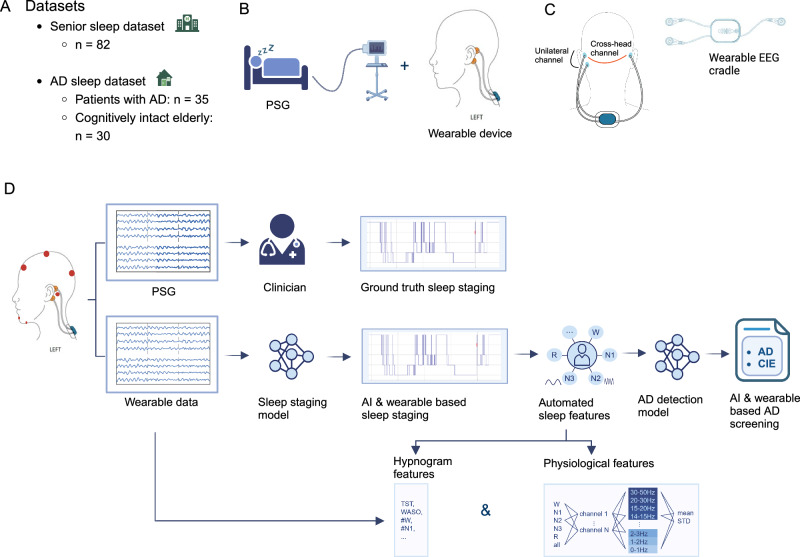
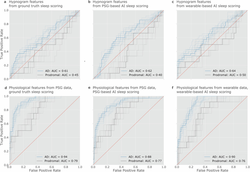
The recent emergence of wearable devices will enable large scale remote brain monitoring. This study investigated whether multimodal wearable sleep recordings could help screening for Alzheimer's disease (AD). Measurements were acquired simultaneously from polysomnography and a wearable device, measuring electroencephalography (EEG) and accelerometry (ACM) in 67 elderly without cognitive symptoms and 35 AD patients. Sleep staging was performed using an AI model (SeqSleepNet), followed by feature extraction from hypnograms and physiological signals. Using these features, a multi-layer perceptron was trained for AD detection, with elastic net identifying key features. The wearable AD detection model achieved an accuracy of 0.90 (0.76 for prodromal AD). Single-channel EEG and ACM physiological features captured sufficient information for AD detection and outperformed the hypnogram features, highlighting these physiological features as promising discriminative markers for AD. We conclude that wearable sleep monitoring augmented by AI shows promise towards non-invasive screening for AD in the older population.


Conflict of interest statement

Competing interests: The authors declare no competing interests. Ethical approval: Studies were conducted in accordance with the Declaration of Helsinki and approved by the Ethical Committee of University Hospitals Leuven. Informed consent was obtained from all participants or their caregiver. The clinical trial numbers were NCT04755504 (S64190/B3222020000148), NCT03617497 (S61745) respectively.

Figures

Fig. 1
Fig. 1. Overview of the methodology.
A The datasets used for this study. The Senior Sleep Dataset (n = 82) and Alzheimer’s Sleep Dataset (n = 65) consist of in-hospital and home-based acquired sleep studies, respectively. B Technical set-up. PSG was simultaneous acquired with a wearable EEG device. C Detailed technical set-up of the wearable device. The device is placed in the neck. Two electrodes are attached behind one ear, and one electrode behind the opposite ear, creating a cross-head and ipsilateral EEG channel. The wearable EEG device also has an ACM in it. D A schematic representation of the methodology. The annotated PSG by the clinician was used as a ground truth. An automated sleep staging AI model was developed on the wearable data. Hypnogram and physiological features (EEG and accelerometry signals) from this AI-model were used for the AD detection MLP model in order to distinguish patients with AD from CIE. Parts of the figure were modified from Byteflies. Abbreviations: ACM accelerometry, AD Alzheimer’s disease, AI artificial intelligence, all all sleep stages, CIE cognitively intact elderly, PSG polysomnography, STD standard deviation. Abbreviations for hypnogram features are explained in Supplementary.
Fig. 2
Fig. 2. The performance in detecting patients with AD and the subset with prodromal AD.
The transparent lines represent the ROC curves of all ten cross-validation runs for each prediction task, while the non-transparent lines represent the ROC curves obtained using the mean predictions with these ten repeats. Their AUC is reported in the legend. As the prodromal AD patients are a subgroup of AD, the curves labeled with “AD” show the performance in discriminating CIE patients from the whole AD cohort, consisting of patients in the prodromal and dementia disease stage. The curves labeled with “prodromal” show the performance in discriminating the prodromal AD patients from the CIE group. The upper row shows the performances using hypnogram features, calculated based on a the manually scored PSG, which is the ground truth, (b) the AI-scored PSG, (c) the AI-scored wearable data. The lower row shows the performances using the physiological features, calculated based on both the raw data and sleep stage labels. In (d), the PSG data was used with manual labels, in (e), the PSG data was used with AI-scored labels, and in (f), the wearable data was used with AI-scored labels. The ROC curves show that the physiological features by far outperform the hypnogram features. For the physiological features, the features derived with AI scoring (based on both PSG and wearable) are almost on par with the features based on ground truth scoring. AD Alzheimer’s disease, AI artificial intelligence, AUC area under the curve, CIE cognitively intact elderly, PSG polysomnography, ROC receiver operating characteristic.
Fig. 3
Fig. 3. ROC curves showing the AD detection results for the physiological features without sleep staging.
These results show the AD detection performance when using as features the frequency bands for the different channels (both mean and STD) but aggregating them over the whole night without sleep staging. The transparent lines represent the ROC curves of all ten classifiers trained for each prediction task, while the non-transparent lines represent the ROC curves obtained using the mean predictions with ten repeats. Their AUC is reported in the legend. The curves show the performances using (a), the raw PSG data, and (b), the raw wearable data. AD Alzheimer’s disease, AUC area under the curve, OSA obstructive sleep apnea, PSG polysomnography, ROC receiver operating characteristic.
Fig. 4
Fig. 4. Correlation between MMSE and sleep features based on wearable data.
The hypnogram features are shown in the upper part of the figure, and a few selected physiological features are shown in the lower part. The first column shows the ground truth sleep features based on the wearable data, but scored with the ground truth manual PSG scoring, and the second column shows sleep features based on the wearable data scored by AI. Pearson’s correlation test was performed, with the subjects as samples. The MMSE score was not recorded in the patients from the Senior Sleep Dataset, so the correlations are only computed for the 65 patients of the Alzheimer’s Sleep Dataset (see Table 2). Correlations are shown through the colors, with p values below 0.05 reported as numbers to indicate the significance of the correlations. The wearable-based features based on AI scoring and based on ground truth scoring agree on most significant correlations. ACM accelerometry, AI artificial intelligence, All all sleep stages, EEG electroencephalography, MMSE Mini-Mental State Examination, STD standard deviation. Abbreviations for hypnogram features are explained in Supplementary Table 1.
Fig. 5
Fig. 5. Most selected wearable-based and PSG-based physiological features for the AD detection model.
The features that were selected more than 85% of the time by the a wearable-based and b PSG-based AD detection model. In both cases, the features were calculated using ground truth sleep scoring (for the features calculated using AI scoring, see Supplementary Fig. 5). Features are visualized using three rings showing to which (1) sleep stage, (2) channel, and (3) frequency range each feature corresponds. The frequency range is only shown for the EEG and EOG channels due to the limited interpretability of the EMG and ACM frequencies. The discrete color bar shows the interpretation of the frequency range. The surrounding black ring shows which features were computed using STD, with the remaining calculated using the mean. The width of each segment indicates how many features of that type were selected more than 85% of the times. This width should not be mistaken for overall importance of a channel or sleep stage: information may be more spread out over multiple frequency bands for some channels or sleep stages than for others. Rather, this figure shows which channels are important in which sleep stages, and which frequencies were decisive in those channels. ACM accelerometry, AD Alzheimer’s disease, AI artificial intelligence, All features computed over the whole recording, EEG electroencephalography, EOG electrooculography, EMG electromyography, PSG polysomnography, STD standard deviation.

References

    1. Scheltens, P. et al. Alzheimer’s disease. Lancet397, 1577–1590 (2021). - PMC - PubMed
    1. Georges J., Miller O. & Bintener, C. Dementia in Europe Yearbook 2019: Estimating The Prevalence Of Dementia In Europe, accessed 17 Apr 2024. https://www.researchgate.net/publication/339401240_Estimating_the_preval... (Alzheimer Europe, 2020).
    1. Lucey, B. P. et al. Reduced non-rapid eye movement sleep is associated with tau pathology in early Alzheimer’s disease. Sci. Transl. Med.11, eaau6550 (2019). - PMC - PubMed
    1. Winer, J. R. et al. Sleep as a potential biomarker of Tau and β-amyloid burden in the human brain. J. Neurosci.39, 6315–6324 (2019). - PMC - PubMed
    1. Mander, B. A., Winer, J. R., Jagust, W. J. & Walker, M. P. Sleep: a novel mechanistic pathway, biomarker, and treatment target in the pathology of Alzheimer’s disease?. Trends Neurosci.39, 552–566 (2016). - PMC - PubMed

LinkOut - more resources