. 2025 Nov;21(11):1783-1793.
doi: 10.1038/s41589-025-01907-2. Epub 2025 May 9.

Enzymatic combinatorial synthesis of E-64 and related cysteine protease inhibitors

Affiliations

Enzymatic combinatorial synthesis of E-64 and related cysteine protease inhibitors

Mengting Liu et al. Nat Chem Biol. 2025 Nov.

Abstract

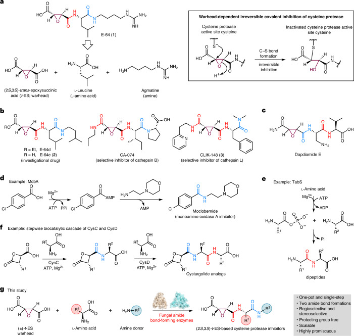
E-64 is an irreversible cysteine protease inhibitor prominently used in chemical biology and drug discovery. Here we uncover a nonribosomal peptide synthetase-independent biosynthetic pathway for E-64, which is widely conserved in fungi. The pathway starts with epoxidation of fumaric acid to the warhead (2S,3S)-trans-epoxysuccinic acid with an Fe(II)/α-ketoglutarate-dependent oxygenase, followed by successive condensation with an L-amino acid by an adenosine triphosphate grasp enzyme and with an amine by the fungal example of amide bond synthetase. Both amide bond-forming enzymes display notable biocatalytic potential, including scalability, stereoselectivity toward the warhead and broader substrate scopes in forming the amide bonds. Biocatalytic cascade with these amide bond-forming enzymes generated a library of cysteine protease inhibitors, leading to more potent cathepsin inhibitors. Additionally, one-pot reactions enabled the preparative synthesis of clinically relevant inhibitors. Our work highlights the importance of biosynthetic investigation for enzyme discovery and the potential of amide bond-forming enzymes in synthesizing small-molecule libraries.


Conflict of interest statement

Competing interests: The authors declare no competing interests.

Figures

Fig. 1
Fig. 1. Amide bond-forming enzyme(s) are hypothesized to be involved in the biosynthesis of E-64.
a, Structure, mode of action and retrobiosynthesis of 1. E-64 contains the t-ES warhead that is the site of covalent inhibition of cysteine proteases. E-64 is proposed to form from the condensation of (2S,3S)-t-ES, l-Leu and agmatine. b, Structures of synthetic cysteine protease inhibitors based on 1. c, Structure of dapdiamide E. d, Mechanism and application of bacterial ABSs such as McbA from the marinacarboline biosynthetic pathway. e, Mechanism and applications of bacterial ATP-grasp enzymes such as TabS from the tabtoxin biosynthetic pathway. f, Stepwise combination of two bacterial amide bond-forming enzymes CysC and CysD to synthesize cystargolide analogs. g, In this work, we discovered and biochemically characterized the fungal ATP-grasp enzyme and ABS from the biosynthesis of 1 and used the enzymes in the synthesis of diverse E-64 analogs.
Fig. 2
Fig. 2. Biosynthetic pathway for E-64 and identifying standalone amide-forming enzymes.
a, BGCs of 1 from A.flavus and A.oryzae and other homologous BGCs. The percentage amino acid sequence identity to each corresponding Cp1 enzyme is shown. b, LC–QTOF analysis of metabolites produced by different gene combinations from cp1 and cp2 clusters in the heterologous host A.nidulans is shown in (i)–(v). Selected ion chromatography traces presented on the same scale are shown and the colors of the traces match the indicated mass and compounds. The y axis represents ion counts. *Not isolated. c, Cp1A catalyzed the epoxidation on fumaric acid to t-ES. Assays were carried out at 30 °C for 3 h in 100 μl of 50 mM sodium phosphate buffer with 0.2 mM FeSO4, 2 mM αKG, 2 mM ascorbate, 1 mM fumaric acid and 10 μM Cp1A or MfaA. The products were derivatized with 3-NPH to increase MS sensitivity. Selected ion monitoring of 3-NPH-t-ES ([M + H]+ = 403) is shown. The y axis represents ion counts and the chromatograms are presented on the same scale. d, Enzyme assays with Cp1B and Cp1D. Reactions were performed at 30 °C for 16 h in 100 μl of 50 mM sodium phosphate buffer (pH 8.0). Reaction components for each reaction were as follows: (i) 25 μM Cp1B, 5 mM (±)-t-ES, 2.5 mM l-Ile, 10 mM ATP and 10 mM MgCl2; (ii) 10 μM Cp1D, 2 mM 14, 2.5 mM putrescine, 10 mM ATP and 10 mM MgCl2; (iii) 25 μM Cp1B, 25 μM Cp1D, 5 mM (±)-t-ES, 2.5 mM l-Ile, 5 mM putrescine, 10 mM ATP and 10 mM MgCl2. Traces represent selected ion monitoring of 4 ([M + H]+ = 316) and 14 ([M + H]+ = 246). The y axis represents ion counts and the chromatograms are presented on the same scale. e, Biosynthetic pathway of 1 and the related compounds from the cp1 pathway.
Fig. 3
Fig. 3. Stereoselective (2S,3S)-t-ES-l-amino acid synthesis by Cp1B and Cp2B.
a, Crystal structure of Cp1B in a complex with adenosine and MES. Domains A (residues 5–200 and 444–502), B (residues 201–323) and C (residues 365–443) are shown as deep salmon, wheat and cyan, respectively. The helical connection (residues 324–364) between domains B and C is shown in gray. The P-loop and A-loop are highlighted in purple and blue, respectively. Adenosine is colored in light green and MES is colored in cyan. mFo − DFc polder omit maps for adenosine and MES are shown in gray mesh and contoured at 3.0σ. b, Enlarged view of the substrate-binding site. Conserved residues with hGSH synthetases are labeled in red, which are proposed to be involved in phosphorylation of substrate carboxylate. Residues surrounding MES that are possibly involved in substrate binding are shown in blue. c, Amino acid nucleophile (proteinogenic amino acids and a1a32) scope for Cp1B and Cp2B. The heat map shows percentage yields in analytical-scale reactions catalyzed by Cp1B and Cp2B, estimated from the standard curves generated at λ = 204 nm of the purified compounds. The analytical-scale reactions were performed in 100 μl of 50 mM sodium phosphate buffer (pH 8.0) at 30 °C for 16 h. Each reaction contained 25 μM enzyme, 5 mM (±)-t-ES, 2.5 mM amino acid, 10 mM MgCl2 and 10 mM ATP. Isolated percentage yields from the preparative-scale reactions catalyzed using either Cp1B or Cp2B are shown under the structures. Preparative-scale reactions were performed in 20 ml of 50 mM sodium phosphate buffer (pH 8.0) at 30 °C for 16 h. Each reaction contained 2.5 μM Cp1B or Cp2B, 5 mM (±)-t-ES, 2.5 mM amino acid, 10 mM MgCl2 and 10 mM ATP. Notes on percentage yields: (i) isolated percentage yields with Cp1B; (ii) isolated percentage yields with Cp2B; (iii) analytical percentage yield estimated from the standard curves generated at λ = 204 nm of purified 14 is shown because t-ES-Met was not isolated from the preparative-scale reaction in this study.
Fig. 4
Fig. 4. Substrate scope of Cp1D toward amine nucleophiles.
Amine substrates (b1b41) that were accepted by Cp1D as nucleophiles to form the corresponding diamides (15-b1 to 15-b41) using 15 as the electrophile. Analytical-scale reactions to estimate percentage yields were performed in 100 μl of 50 mM sodium phosphate buffer (pH 8.0) at 30 °C for 16 h. Each reaction contained 25 μM Cp1D, 2 mM 15, 5 mM amine, 10 mM ATP and 10 mM MgCl2. The reactions were analyzed by LC–MS. Preparative-scale reactions to determine structures and isolated percentage yields were performed in 15 ml of 50 mM sodium phosphate buffer (pH 8.0) at 30 °C for 16 h. Each reaction contained 2.5 μM Cp1D, 2 mM 15, 5 mM amine, 10 mM ATP and 10 mM MgCl2. Notes on analytical and isolated percentage yields: (i) analytical percentage yield was not determined as the product peak overlapped with 15 in the LC–MS chromatogram; (ii) isolated yields from preparative-scale reactions; (iii) estimated percentage yields of the amide products from analytical-scale reactions. The analytical percentage yields were estimated from the standard curves generated at λ = 204 nm of purified 15-b15 (for 15-b1 to 15-b17, 15-b28, 15-b39 and 15-b40), 15-b24 (for 15-b21 to 15-b23), 15-b27 (for 15-b25 to 15-b26) or 15-b37 (for 15-b34 to 15-b36).
Fig. 5
Fig. 5. Biocatalytic platform for screening and synthesis of t-ES-based cysteine protease inhibitors.
a, General workflow. Created with BioRender.com. b, Cathepsin B inhibitory activity of the crude reaction containing (2S,3S)-t-ES-AA-agmatine (b41) where AA is an amino acid (proteinogenic amino acids and a1a32). c, Cathepsin B inhibitory activity of the crude reaction containing (2S,3S)-t-ES-a10-B where B is an amine (b1b41). The average inhibition activity is presented as a percentage of the positive control (n = 2). d, Selected data output for fluorescence-based cathepsin B inhibition assay using a fluorogenic substrate Z-Phe-Arg-AMC. e, Biocatalytic one-pot synthesis of 2 and the identified cathepsin B inhibitors. The percentage isolated yields are shown for all synthesized compounds. IC50 values of the selected inhibitors against cathepsin B are also shown as the mean ± s.d. (n = 3). Reaction conditions for b,c: 25 μM Cp1B, 25 μM Cp1D, 2 mM (±)-t-ES, 1 mM l-amino acid, 1 mM amine, 10 mM ATP and 10 mM MgCl2 in 100 μl of 50 mM sodium phosphate buffer (pH 8.0) at 30 °C for 16 h. Reaction conditions for e: 5 mM (±)-t-ES, 2.5 mM l-amino acid, 2.5 mM amine donor, 10 mM ATP, 10 mM MgCl2, 2.5 μM Cp1B and 2.5 μM Cp1D in 50 mM sodium phosphate (pH 8.0) at 30 °C for 16 h. f, Chemoenzymatic synthesis of CLIK-148 (3) in preparative-scale reaction. After the biocatalytic synthesis of (2S,3S)-t-ES-Phe-b28 using l-Phe and dimethylamine (b28), the crude mixture was directly used for the subsequent chemical condensation. The chemoenzymatic approach can also be applied to synthesize the cathepsin C-selective inhibitors starting with the biocatalytic synthesis of (2S,3S)-t-ES-a3-b44.
Extended Data Fig. 1
Extended Data Fig. 1. Significance and biosynthetic machinery of amide functionality.
a, Selected top-selling drugs containing amide bond. b, Representative amide-containing natural products used in clinic and agriculture. c, The mechanisms of the biocatalytically competent amide bond synthetase McbA and the ATP-grasp enzyme TabS. d, Examples of non-ribosomal strateiges for amide synthesis in fungi. The amide bond formations in isopenicillin N (IPN) is catalyzed by three modules NRPS PcbAB. NRPS-independent siderophore synthetase (NIS) AnkE is proposed to catalyze the amide bond in NK13650B. The pair of CoA ligase PclA and N-acyltransferase PenDE forms the amide bond in the production of penicillin G and 2-aminoadipic acid (2-AAA). AnkA catalyzed the tRNA-dependent amidation to form cyclo-Tyr-Arg. Domain abbreviations: A: adenylation; T: peptidyl-carrier protein; C: condensation; E: epimerization; TE: thioesterase. While a few ATP-grasp enzymes were proposed to involve the biosynthesis of the fungal peptides (also see Extended Data Fig. 2), ABSs have not been identified in the fungal natural product biosynthesis.
Extended Data Fig. 2
Extended Data Fig. 2. Limited examples of fungal ATP-grasp enzyme involved in natural product biosynthesis.
a, FsqD from fumisoquin biosynthesis was proposed to activate L-tyrosine to form tyrosyl phosphate. The function of FsqD has not been biochemically characterized. b, AnkG was proposed to be an ATP-grasp enzyme that catalyzes the amide bond formation between L-aspartic acid and NK13650D to form NK13650C based on the in vivo experiments. c, Sequence similarity network analysis of Cp1B homologs from the UniProt database. Note that 5,000 maximum number target sequences from a blastp search with Cp1B as a query (expect threshold value: 5) were retrieved and subjected to SSN construction, with an alignment score threshold of 7. Cp1B and Cp2B in this study and the proposed ATP-grasp enzyme AnkG are highlighted. While Cp1B and Cp2B were located at a separate clade from AnkG, the SSN and the amino acid sequence identity between them ( ~ 30%) suggest these enzymes are distinct but distantly related. While no characterized enzymes were found in this SSN, the SSN showed that putative Cp1B-like ATP-grasp enzymes are conserved in not only many fungi (Ascomycota and Basidiomycota) but also in a few bacteria. Domain abbreviations: A: adenylation; T: peptidyl-carrier protein; R: reductase domain; P: pyridoxal phosphate binding domain.
Extended Data Fig. 3
Extended Data Fig. 3. E-64-like biosynthetic gene clusters are widely conserved in fungi.
a, Selected clusters are shown in a dendrogram (based on identity to query sequences) from cblaster search. A darker tint of blue indicates a higher percentage identity of the query in the output cluster. The three gene cassette (cpA, cpB, and cpD) is highly conserved in more than > 40 different fungal genera such as Aspergillus spp, Penicillium spp, Metarhizium spp, Trichoderma spp, and Mycena spp (Basidiomycota). Two copies of E-64 like cluster are also present in Mycena galopus. b. Selected E-64 homologous biosynthetic gene clusters in clinker visualization, including reported E-64 analog producing fungi (Aspergillus oryzae, Penicillium citrinum, and Colletotrichium spp) and fungi not known to produce E-64 (Trichoderma atroviride, Metarhizium anisopliae, and Mycena galopus ATCC 62051). Nearly all the genes in those clusters are conserved except for PLP-dependent decarboxylase (the homolog of Cp1C).
Extended Data Fig. 4
Extended Data Fig. 4. LC/MS analysis of extracts from the heterologous expression of cp1 and cp2 in A. nidulans.
LC/MS analyses include cp1ABCD (i), cp1BCD (ii), cp1ACD (iii), cp1ABC (iv), cp1ABD (v), and cp2ABCD (vi). Selected ion chromatograms correspond to the [M + H]+ for 1 ([M + H]+ = 358), 4 ([M + H]+ = 316), 5 ([M + H]+ = 330), 6 ([M + H]+ = 358), 7 ([M + H]+ = 372), 8 ([M + H]+ = 364), 9 ([M + H]+ = 380), 10 ([M + H]+ = 406), 11 ([M + H]+ = 422), 12 ([M + H]+ = 318), and 13 ([M + H]+ = 300). Y-axis represents ion counts and the chromatograms are presented on the same scale. Heterologous expression of three gene cassette cp1ABD is sufficient for the biosynthesis of 1 and the analogs in A. nidulans. As polyamines are abundant primary metabolites in fungi, the PLP-dependent decarboxylase Cp1C is not essential for the biosynthesis of 1 and the analogs in heterologous host A. nidulans. Interestingly, the heterologous expression of cp1BCD led to the formation of malic acid (12) and fumaric acid (13) derivatives, suggesting the role of Cp1A as an epoxidase. This result further supported the promiscuous substrate specificities of both Cp1B and Cp1D. The structures of all compounds except 11 were determined by NMR.
Extended Data Fig. 5
Extended Data Fig. 5. Absolute configuration of t-ES and substrate for Cp1A.
a, Enzymatic synthesis of (2S,3S)-t-ES from Cp1A or MfaA. Briefly, the reaction was performed in 50 mM sodium phosphate buffer (pH 8.0) containing 0.2 mM FeSO4, 2 mM αKG, 2 mM ascorbate, 1 mM of substrate, and 10 μM of Cp1A or MfaA at 30 °C for 16 h. The protein was removed by Amicon concentrators (Millipore). Subsequently 10 μM Cp1B, 2.5 mM l-isoleucine, 10 mM ATP, 10 mM MgCl2 were added followed by incubation at 30 °C for 16 h. The enzymatic synthesis of (2S,3S)-14 allowed determination of the absolute configuration of the epoxide to be (2S, 3S), based on retention times of standards. HPLC analysis was performed with a CHIRALPAK® IA-3 column (150 ×4.6 mm, 3 μm) at room temperature (flow rate 1 mL/min, 40% MeCN–H2O with 0.1% trifluoroacetic acid). Y-axis represents UV absorption (λ=204 nm) and the chromatograms are not presented on the same scale. b, LC/MS analysis of reaction of Cp1A with 12 or 13. 100 μL reactions were performed at 30 °C for 3 h, in 50 mM sodium phosphate buffer (pH 8.0) containing 0.2 mM FeSO4, 2 mM αKG, 2 mM ascorbate, 1 mM of substrate 12 or 13, and 10 μM of Cp1A. 4 was not observed in enzymatic assay of Cp1A with 12 (ii) and 13 (iii) in the presence of αKG, ascorbate, and Fe2+. The traces show selected ion monitoring of 4 ([M + H]+ = 316). Y-axis represents ion counts and the chromatograms are presented on the same scale. c, Enzymatic reaction of Cp1A with succinic acid. The same reaction condition with Extended Data Fig. 5b was used except for succinic acid being used as the substrate. After overnight incubation at 30 °C, the product was derivatized with 3-NPH. Selected ion monitoring of 3-NPH-t-ES ([M + H]+ = 403) is shown. Y-axis represents ion counts and the chromatograms are presented on the same scale. Note that 3-NPH-fumaric acid was also observed when succinic acid was used as the substrate.
Extended Data Fig. 6
Extended Data Fig. 6. Cp1B is an ATP-grasp enzyme.
a, Relative activity of ADP formation upon incubation of Cp1B with dicarboxylic acid substrates. Reactions were performed in 200 μL of 100 mM Tris-HCl (pH 8.0) containing 0.25 μM Cp1B, 10 mM ATP, 12 mM MgCl2, 300 μM NADH, 500 μM phosphoenolpyruvic acid (PEP), 41 units/mL pyruvate kinase (PK, Sigma), 59 units/mL lactate dehydrogenase (LDH, Sigma), 10 mM KCl with 1 mM acid donors and 5 mM l-Phe. The relative phosphorylation activities of Cp1B towards each substrate were derived by the consumption of NADH at the time point where each reaction mixture was incubated at 30 °C for 30 min. Values and error bars represent the average and s.d. of three independent replicates (black filled circles), respectively (n = 3). b, Apparent Michaelis-Menten plots for the Cp1B catalyzed phosphorylation of (2S,3S)-t-ES. The values represent means ± s.d., and error bars indicate s.d. of three independent replicates (n = 3). The reaction mixtures (100 μL) contained 1.0 μM Cp1B, 10 mM ATP, 12 mM MgCl2, 300 μM NADH, 500 μM phosphoenolpyruvic acid (PEP), 41 units/mL pyruvate kinase (PK, Sigma), 59 units/mL lactate dehydrogenase (LDH, Sigma), 10 mM KCl and 100 mM Tris-HCl (pH 8.0) with various concentration (0.04 mM to 1 mM) of (2S,3S)-t-ES and 5 mM l-Phe. The reaction mixture was incubated at 30 °C, and the consumption of NADH at 10 min was used to derive the reaction velocity for enzyme kinetics. Kinetic constants were derived from velocity versus substrate concentration data using a nonlinear regression fitting method with GraphPad Prism 9. c, Structure-based multiple sequence alignment of Cp1B with other characterized ATP-grasp enzymes. d, Activity of Cp1B mutants quantified by the formation of 14. Reactions are performed at 30 °C for 20 min in 100 μL of 50 mM sodium phosphate buffer (pH 8.0). Reaction components are 25 μM Cp1B, 5 mM (±)-t-ES, 2.5 mM l-Ile, 10 mM ATP, and 10 mM MgCl2. Values and error bars represent the average and s.d. of three independent replicates (black filled circles), respectively (n = 3). Source data
Extended Data Fig. 7
Extended Data Fig. 7. The crystal structure of Cp1B likely adopts a closed active site form.
Comparisons of overall structures of (a) hGSH synthetase with the open active site form (3KAK), γ-glutamylcysteine is shown in magenta; (b) hGSH synthetase with closed active site form (3KAL). Mg2+ ions are shown as magenta spheres. ADP is shown in green and hGSH is shown in cyan; and (c) Cp1B in complex with adenosine shown in green and MES shown in cyan. Surface representations of the crystal structures are shown below. All structures have three characteristic domains typical of ATP-grasp enzymes: Domain A (deep salmon), Domain B (wheat), and lid domain (Domain C, cyan). P-loop (Gly-rich loop) and A-loop (Ala-rich loop) are shown in purple and blue, respectively. In the open form, the P-loop and A-loop are disordered in contrast to those in the closed form and in the Cp1B structure. Consequently, in the open form (3KAK), the nucleotide binding site open. In contrast, the lid domain with P-loop and A-loop enclose the active site in the closed form (3KAL) and partially in the Cp1B structure. These structural comparisons therefore suggested that the crystal structure of Cp1B adopts a closed active site, possibly as a result of MES binding in the active site.
Extended Data Fig. 8
Extended Data Fig. 8. Substrate scope of Cp1D/Cp2D towards N-succinyl-AA.
a, Substrate scope assay for Cp1D catalyzed amidation of N-succinyl-l-AA with isopentylamine. Cp1D was found to accept N-succinyl-l-AA of which the AA are hydrophobic amino acids (L, I, V, M, F, Y, and W). Assays were performed in 100 μL of 50 mM sodium phosphate buffer (pH 8.0) with 25 μM enzyme, 2 mM N-succinyl-l-AA, 5 mM isoamylamine, 10 mM MgCl2 and 10 mM ATP. Reactions were analyzed by LC/MS after incubation at 30 °C for 16 h. Analytical % conversion of N-succinyl-l-AA to the corresponding amide product was estimated from HPLC peak area ratios between product and starting material at λ = 204 nm (% Conversion = (peak area of product / (peak area of substrate + peak area of product)) × 100%). ND: not detected. b, Hydroxamate-based colorimetric assay was performed to assess adenylation specificity towards N-succinyl-l-AA for Cp1D/Cp2D. The reaction was performed in 150 μL of Tris buffer (pH 8.0) containing 20 μM of Cp1D or Cp2D, 15 mM of ATP, 5 mM of N-succinyl-l-AA, 200 mM hydroxylamine, and 10 mM MgCl2. After incubation for 8 h at 30 °C, the reaction was quenched by addition of equivalent volume of stopping solution (10% (w/v) FeCl3 and 3.3% (w/v) trichloroacetic acid dissolved in 0.7 M HCl). The precipitated enzyme was removed by centrifugation and the supernatant was measured for absorbance at 540 nm by a TECAN M200 plate reader. The absorbance at 540 nm was used to calculate the relative activity, and the absorbance of N-succinyl-l-Leu and N-succinyl-l-Tyr after the subtraction of that from each negative control (without Cp1D or Cp2D) were set as 100% activity for Cp1D and Cp2D, respectively. Values and error bars represent the average and s.d. of three independent replicates (white circles), respectively (n = 3). The assays confirmed that both Cp1D and Cp2D prefer hydrophobic l-amino acids in N-succinyl-l-AA, while Cp2D has a stronger preference for aromatic amino acids. Source data
Extended Data Fig. 9
Extended Data Fig. 9. Structures of the active site of papain (no inhibitor bound, panels a-b) and papain bound to E64 analogs from X-ray diffraction (panels c-k).
For the unliganded active site, atomic coordinates are superimposed on an Fo-Fc map at 3σ (green mesh) following complete modeling and refinement (a), and the 2Fo-Fc composite omit map (indigo mesh) generated following refinement at 1.5σ (b). For E-64 (1)-bound active site, atomic coordinates are superimposed on a ligand-omit Fo-Fc map at 3σ, revealing positive density that 1 (translucent magenta model) was modeled to fit (c), and the fully refined structure with the ligand modeled is superimposed on a 2Fo-Fc composite omit map at 1.5σ carved a 1.6 Angstrom radius from all ligand atoms (d). The same is shown for the papain-E-64c active site (e-f) the papain-(2S,3S)-t-ES-a9-b7 active site (g-h), and the papain-E-64d active site (i-j). The same papain-E-64d structure is additionally superimposed against a 2Fo-Fc feature-enhanced map (cyan mesh) carved a 1.6 Angstrom radius from all E-64d atoms (k). Insets for each structure highlight a hydrophobic pocket adjacent to the active site occupied by hydrophobic side-chains of each ligand (top inset), and the solvent-facing region adjacent to the active site occupied by each inhibitor’s tail (bottom inset). Yellow dashed lines indicate potential hydrogen-bonding interactions.

References

    1. Hoffmann, M. et al. SARS-CoV-2 cell entry depends on ACE2 and TMPRSS2 and is blocked by a clinically proven protease inhibitor. Cell181, 271–280 (2020). - PMC - PubMed
    1. Satoyoshi, E. Therapeutic trials on progressive muscular dystrophy. Intern. Med.31, 841–846 (1992). - PubMed
    1. Hook, G., Yu, J., Toneff, T., Kindy, M. & Hook, V. Brain pyroglutamate amyloid-β is produced by cathepsin B and is reduced by the cysteine protease inhibitor E64d, representing a potential Alzheimer’s disease therapeutic. J. Alzheimers Dis.41, 129–149 (2014). - PMC - PubMed
    1. Tamai, M. et al. In vitro and in vivo inhibition of cysteine proteinases by EST, a new analog of E-64. J. Pharmacobiodyn.9, 672–677 (1986). - PubMed
    1. Barrett, A. J. et al. l-trans-Epoxysuccinyl-leucylamido(4-guanidino)butane (E-64) and its analogues as inhibitors of cysteine proteinases including cathepsins B, H and L. Biochem. J.201, 189–198 (1982). - PMC - PubMed

MeSH terms

LinkOut - more resources