. 2025 May 9;26(1):465.
doi: 10.1186/s12864-025-11658-y.

Multi-omics integrated analysis reveals the molecular mechanism of tail fat deposition differences in sheep with different tail types

Affiliations

Multi-omics integrated analysis reveals the molecular mechanism of tail fat deposition differences in sheep with different tail types

Wannian Wang et al. BMC Genomics. .

Abstract

Background: The accumulation of tail fat in sheep is a manifestation of adaptive evolution to the environment. Sheep with different tail types show significant differences in physiological functions and tail fat deposition. Although these differences reflect the developmental mechanism of tail fat under different gene regulation, the situation of sheep tail fat tissue at the single cell level has not been explored, and its molecular mechanism still needs to be further elucidated.

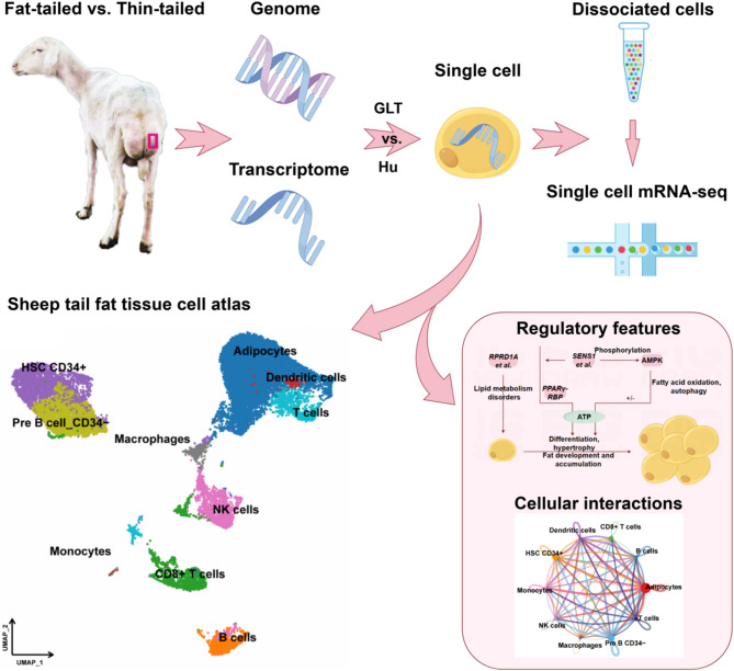
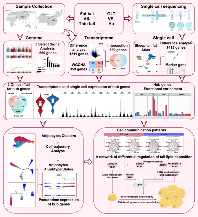
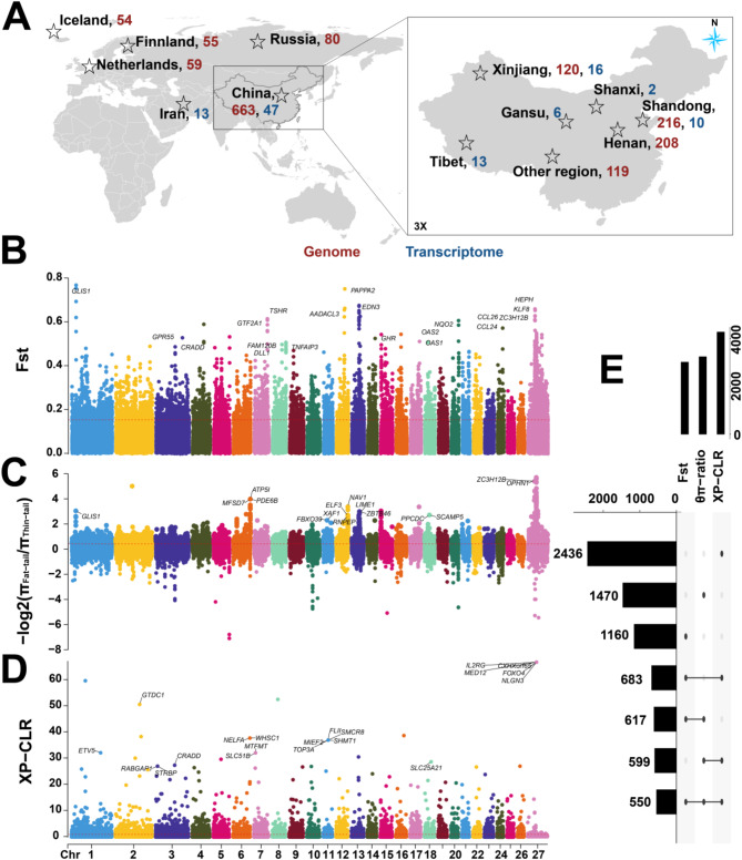
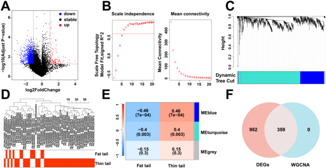
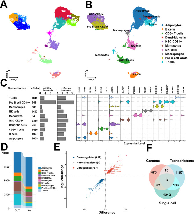
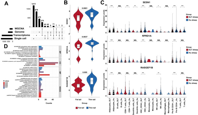
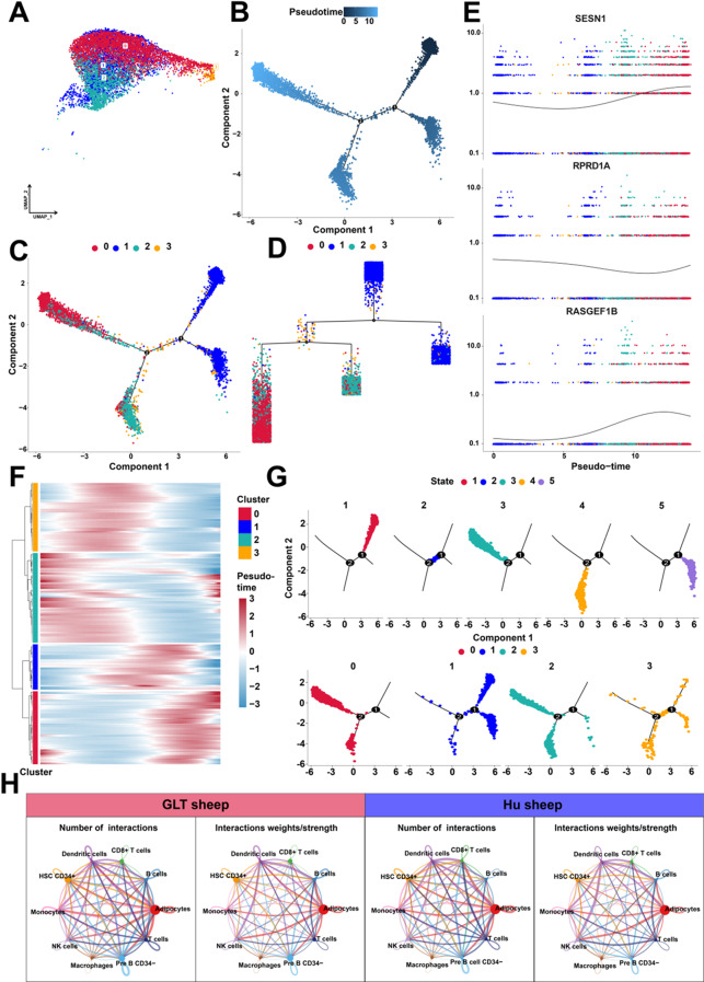
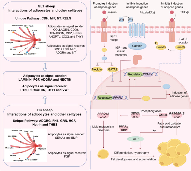
Results: Here, we characterized the genomic features of sheep with different tail types, detected the transcriptomic differences in tail adipose tissue between fat-tailed and thin-tailed sheep, established a single-cell atlas of sheep tail adipose tissue, and screened potential molecular markers (SESN1, RPRD1A and RASGEF1B) that regulate differences in sheep tail fat deposition through multi-omics integrated analysis. We found that the differential mechanism of sheep tail fat deposition not only involves adipocyte differentiation and proliferation, but is also closely related to cell-specific communication networks (When adipocytes act as signal outputters, LAMININ and other signal pathways are strongly expressed in guangling large tailed sheep and hu sheep), including interactions with immune cells and tissue remodeling to drive the typing of tail fat. In addition, we revealed the differentiation trajectory of sheep tail adipocytes through pseudo-time analysis and constructed the cell communication network of sheep tail adipose tissue.

Conclusions: Our results provide insights into the molecular mechanisms of tail fat deposition in sheep with different tail types, and provide a deeper explanation for the development and functional regulation of adipocytes.

Keywords: Molecular mechanism; Multi-omics; Sheep; Single cell; Tail fat deposition.


Conflict of interest statement

Declarations. Ethics approval and consent to participate: The experimental animals used in this study were all raised at the Animal Station of Shanxi Agricultural University. The ownership of the animals belongs to the co-author of this study (Liying Qiao), who has informed and consented to the conduct of this experiment. All experimental protocols were reviewed and approved by the Experimental Animal Ethics Committee of Shanxi Agricultural University (Ethics Committee Approval Reference Number: SXAU-EAW-2022 S.UV.010009). All authors approved the conduct of the experiment and the publication of the manuscript. In this study, in compliance with the requirements of the Animal Science and Veterinary College IACUC, sheep were humanely euthanized by exsanguination after deep anesthesia, and sodium pentobarbital (≥ 90 mg/KG) was injected intravenously to relieve the animals suffering. Consent for publication: Not applicable. Competing interests: The authors declare no competing interests.

Figures

Fig. 1
Fig. 1
Research ideas of this study
Fig. 2
Fig. 2
Schematic view of the procedures for data collection and analyses in the present study
Fig. 3
Fig. 3
Genome-wide selection signals in fat-tailed and thin-tailed sheep. (A) Geographical distribution of sampling for whole genome and transcriptome sequencing. A total of 663 fat-tailed sheep and 248 thin-tailed sheep whole genome data were collected, and transcriptome data of 13 fat-tailed sheep and 47 thin-tailed sheep were collected. (B) Whole-genome selective signals between fat-tailed sheep and thin-tailed sheep based on the pairwise FST selection test. (C) Whole-genome candidate selective regions between fat-tailed sheep and thin-tailed sheep by the log2(π ratio) selection test. (D) Genome-wide selective signals between fat-tailed sheep and thin-tailed sheep by the XP-CLR test. The horizontal red dashed line corresponds to the genome-wide significance threshold (top 5%: FST = 0.1608, log2(π ratio) = 0.5215, and XP-CLR = 0.8870). (E) The upset plot shows the number of genes and their intersection obtained from the three genome-wide selection signal analyses when the significance threshold is top 5%
Fig. 4
Fig. 4
Screening for differentially expressed genes regulating sheep tail fat deposition using multiple methods. (A) DEGs regulating tail fat deposition in sheep; red represents upregulation, blue represents downregulation. (B) Soft threshold power versus scale-free topological model fit index and average connectivity, with 9 selected as the appropriate soft power. (C) Gene clustering based on dissimilarity metrics. (D) Clustering of samples and indicator traits. (E) Association analysis between gene modules and different traits, the blue module is the most specific module for the fat-tail trait. (F) Venn plot showing the intersection of DEGs with blue module genes
Fig. 5
Fig. 5
Single-cell transcriptome landscape of tail adipose tissue in fat-tailed and thin-tailed sheep. (A) UMAP analysis of 19,471 single cells from GLT and Hu tail adipose tissue. In the UMAP map, 16 cell type clusters are marked with different colors. (B) Single-cell atlas of GLT and Hu tail adipose tissue identified a total of 10 cell types based on the expression of marker gene signatures. (C) From left to right: cell type information; number of cells of each type; number of unique molecular identifiers (UMIs) in each cell type; number of genes detected in each cell type and violin plots of marker gene expression in each cell type. (D) Comparison of the number of cells in each cell type in GLT and Hu tail adipose tissue. (E) DEGs in tail adipocytes of GLT and Hu. (F) Venn diagram showing the intersection of DEGs between fat-tailed and thin-tailed sheep at the genome, transcriptome, and single-cell levels
Fig. 6
Fig. 6
Expression and functional enrichment of hub genes regulating sheep fat tail formation. (A) Upset plot shows the intersection of DEGs in multi-omics analysis. (B) The expression of genes that regulate differential fat deposition in sheep tail at the transcriptome level, P < 0.05 indicates significant difference. (C) The expression of genes that regulate differential fat deposition in sheep tail at the single-cell level, * indicates P < 0.05, ** indicates P < 0.01, *** indicates P < 0.001. (D) Functional enrichment of genes with significant differences in two or more omics
Fig. 7
Fig. 7
Adipocytes exhibit temporal heterogeneity in their differentiation trajectories. (A) 9,059 adipocytes were subjected to UMAP analysis, and 4 clusters were marked with different colors. (B) Pseudo-time processes in adipocytes. (C-D) Adipocytes differentiation trajectory in a pseudo-time process. (E) Expression of genes regulating sheep tail fat deposition in pseudo-time trajectory. (F) Heatmap showing dynamic expression changes of genes in cell clusters. (G) Pseudo-time trajectories of adipocytes classified by state and cell clusters. (H) Communication network between GLT and Hu adipocytes
Fig. 8
Fig. 8
Potential regulatory network for tail fat deposition in sheep

Similar articles

References

    1. Xu YX, Wang B, Jing JN, Ma R, Luo YH, Li X, et al. Whole-body adipose tissue multi-omic analyses in sheep reveal molecular mechanisms underlying local adaptation to extreme environments. Commun Biol. 2023;6(1):159. 10.1038/s42003-023-04523-9. - PMC - PubMed
    1. Mereu P, Pirastru M, Sanna D, Bassu G, Naitana S, Leoni GG. Phenotype transition from wild mouflon to domestic sheep. Genet Sel Evol. 2024;56(1):1. 10.1186/s12711-023-00871-6. - PMC - PubMed
    1. Dong K, Yang M, Han J, Ma Q, Han J, Song Z, et al. Genomic analysis of worldwide sheep breeds reveals PDGFD as a major target of fat-tail selection in sheep. BMC Genomics. 2020;21(1):800. 10.1186/s12864-020-07210-9. - PMC - PubMed
    1. Liu T, Feng H, Yousuf S, Xie L, Miao X. Differential regulation of mRNAs and LncRNAs related to lipid metabolism in Duolang and small tail Han sheep. Sci Rep. 2022;12(1):11157. 10.1038/s41598-022-15318-z. - PMC - PubMed
    1. Hao K, Liu X, Chen Y, Zeng W, Chen L, Wang J, et al. PPARγ regulates lipid metabolism and viability of sheep trophoblast cells. Reprod Domest Anim. 2023;58(11):1559–68. 10.1111/rda.14471. - PubMed

LinkOut - more resources