Review
. 2025 May 9;26(1):454.
doi: 10.1186/s12891-025-08669-x.

Appropriate treatment for nail breakage following femur intertrochanteric fractures without additional reduction: case series and literature review

Affiliations
Review

Appropriate treatment for nail breakage following femur intertrochanteric fractures without additional reduction: case series and literature review

Hong Man Cho et al. BMC Musculoskelet Disord. .

Abstract

Background: Intramedullary nail breakage is an uncommon complication in patients with femoral intertrochanteric fractures treated with proximal femoral nail antirotation. Salvage surgery for nail breakages associated with delayed union or nonunion is challenging, particularly when breakage occurs within an acceptable reduction range, complicating implant selection. This study evaluated outcomes in patients with proximal femoral nail antirotation breakage, acceptable reduction, and fixation treated with the long proximal femoral nail antirotation change with distal screw dynamization and lateral cortical notching procedure.

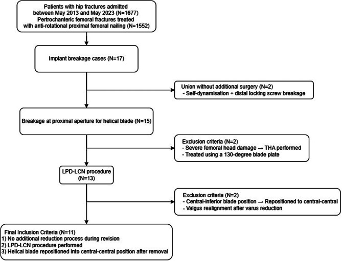
Methods: Eleven patients who underwent the long proximal femoral nail antirotation change with distal screw dynamization and lateral cortical notching procedure between May 2013 and May 2023 with no additional fracture reduction required during salvage surgery and with a helical blade screw resinserted at the same position after removal were observed for > 1 year.

Results: The average time to full weight-bearing was 8.44 ± 0.82 weeks (mean ± standard deviation) and to return to normal activities was 18.05 ± 0.89 weeks. The Harris Hip Score was 78.64 ± 1.03 at 12 months; all patients resumed independent walking at the final follow-up. The average fracture union time was 21.49 ± 1.75 weeks. In seven cases with a fracture gap at the time of breakage, significant change was observed in the tip-apex distance (P = 0.016) or lateral extension of the proximal femoral nail antirotation blade (P = 0.005) between the immediate post-surgery and final follow-up results, with the gap healing radiographically at 21.50 ± 1.65 weeks.

Conclusion: Patients with high functional demands, good femoral head bone stock, no acetabular disease, and isolated nail breakage due to nonunion or delayed union in the intertrochanteric area treated with proximal femoral nail antirotation are suitable candidates for osteosynthesis. When reduction is within an acceptable range and the lag screw is centrally placed, long proximal femoral nail antirotation with lateral notching and dynamization offers a minimally invasive approach that reduces soft tissue injury and can yield successful outcomes.

Clinical trial number: Not applicable.

Keywords: Bone screws; Femur head; Fracture fixation; Internal hip fractures.


Conflict of interest statement

Declarations. Ethics approval and consent to participate: The protocol of this study was approved by Gwangju Veterans Hospital Institutional Review Board (no. 2024-2-1). All data were anonymized, and the requirement for informed consent was waived by the Institutional Review Board due to the retrospective nature of the study design. This research was conducted in accordance with the Declaration of Helsinki. Consent for publication: Not applicable. Competing interests: The authors declare no competing interests.

Figures

Fig. 1
Fig. 1
Flow chart to identify patients who met the inclusion criteria for this study
Fig. 2
Fig. 2
Schematic Diagram of surgical procedure shows the 11.0 mm drill bit was then slowly advanced through the existing opening in the lateral cortex for proximal femoral nail antirotation blade insertion, ensuring that it passed through without resistance until fully inserted. (A) The insertion of an 11.0 mm drill bit is hindered by the varus deformity of the (A) fracture and the lateral cortex, where the old blade was removed, for proximal femoral nail antirotation (PFNA) blade insertion. (B) The lower limb is tractioned on the fracture table to ensure that the 11.0 mm drill bit is inserted without resistance. After reaming, the blade is inserted
Fig. 3
Fig. 3
Schematic Diagram of surgical procedure (long proximal femoral nail antirotation with lateral notching and dynamization). (A) Arbeitsgemeinschaft für Osteosynthesefragen/Orthopedic Trauma Association 31-A2 trochanteric fracture treated with proximal femoral nail antirotation, showing acceptable fracture reduction, helical blade position, and fracture gap. (B) Illustration shows nail breakage at the proximal aperture of the proximal femoral nail antirotation blade with trochanteric fracture nonunion due to fracture gap. (C) In cases where resistance was encountered, it was due to varus deformity caused by nail breakage. To address this, traction of the lower limb using the fracture bed was performed until resistance was eliminated, allowing the restoration of the same cephalo-diaphyseal angle as the old proximal femoral nail antirotation (D) long proximal femoral nail antirotation with lateral notching and distal screw dynamization and helical blade re-insertion at the same position of femoral head (center-center). (E) compressed fracture site and distal screw dynamization. (F) fracture is healed completely without a fracture gap
Fig. 4
Fig. 4
Radiographic measurements performed in this study. (A) (B) The tip-apex distance is measured as the sum of the distance, in millimeters, from the tip of the lag screw to the apex of the femoral head in the anteroposterior and lateral view of the radiographs. The lateral extension distance (for screw back-up) was measured between the lateral end of the screw and lateral edge of the intramedullary nail. The gap of the cortical bone (G) was measured as the maximum value of the gap observed in the anteroposterior or lateral view
Fig. 5
Fig. 5
Pelvis anteroposterior radiographs of an intertrochanteric fracture in a male patient in his 70s. (a) Preoperative radiographs show an intertrochanteric fracture (Arbeitsgemeinschaft für Osteosynthesefragen/Orthopedic Trauma Association 31-A2.1). (b) Immediate postoperative radiograph after fixation using proximal femoral nail antirotation showing a fracture gap. (c) The nail broke at the proximal aperture 6.9 months later due to nonunion. Fracture gap is persistent although compared to the immediate post-operation radiograph. (d) Using a chisel we performed a small osteotomy just below the sleeve of the helical blade on the lateral side. (e) The long proximal femoral nail antirotation with lateral notching and dynamization was performed after proximal femoral nail antirotation removal. (f) The radiograph taken 19.8 months after salvage surgery shows no gap, indicating that there is good bone contact, and the fracture has healed

Similar articles

References

    1. Karapinar L, Kumbaraci M, Kaya A, Imerci A, Incesu M. Proximal femoral nail anti-rotation (PFNA) to treat peritrochanteric fractures in elderly patients. Eur J Orthop Surg Traumatol. 2012;22:237–43.
    1. Lopez-Hualda A, García-Cabrera EM, Lobato-Perez M, Martinez-Martin J, Rossettini G, Leigheb M, et al. Mechanical complications of proximal femur fractures treated with intramedullary nailing: A retrospective study. Med (Kaunas Lithuania). 2024;60:718. - PMC - PubMed
    1. Xue D, Yu J, Zheng Q, Feng G, Li W, Pan Z, et al. The treatment strategies of intertrochanteric fractures nonunion: an experience of 23 nonunion patients. Injury. 2017;48:708–14. - PubMed
    1. Butler BA, Selley RS, Summers HD, Stover MD. Preventing wedge deformities when treating intertrochanteric femur fractures with intramedullary devices: a technical tip. J Orthop Trauma. 2018;32:e112–6. - PubMed
    1. Petrie J, Sassoon A, Haidukewych GJ. When femoral fracture fixation fails: salvage options. Bone Joint J. 2013;95–B(Suppl A):7–10. - PubMed

LinkOut - more resources