. 2025 May 9;32(1):47.
doi: 10.1186/s12929-025-01143-9.

Keratin 6A promotes skin inflammation through JAK1-STAT3 activation in keratinocytes

Affiliations

Keratin 6A promotes skin inflammation through JAK1-STAT3 activation in keratinocytes

Mengting Chen et al. J Biomed Sci. .

Abstract

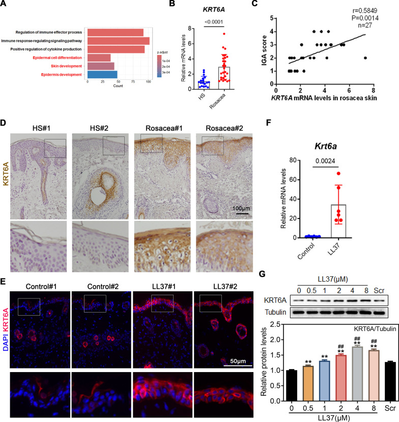
Background: Skin barrier dysfunction and immune activation are hallmarks of inflammatory skin diseases such as rosacea and psoriasis, suggesting shared pathogenic mechanisms. While barrier disruption may trigger or exacerbate skin inflammation, the precise underlying mechanisms remain unclear. Notably, epidermal barrier compromise leads to a marked increase in barrier alarmin expression. Among these, keratin 6A (KRT6A) plays a role in maintaining skin barrier integrity.

Methods: We treated mouse skin and human keratinocytes, with and without KRT6A expression, with LL37/TNF-α and assessed the severity of inflammation. The specific mechanism by which KRT6A promotes skin inflammation was investigated using mass spectrometry and immunoprecipitation assays.

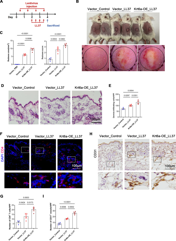
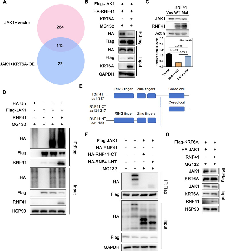
Results: KRT6A expression was elevated in lesional skin from patients and mouse models of rosacea and psoriasis. In mice with LL37-induced rosacea-like and imiquimod (IMQ)-induced psoriasis-like skin inflammation, KRT6A knockdown alleviated inflammation, whereas KRT6A overexpression exacerbated inflammatory responses. Mechanistically, KRT6A activated signal transducer and activator of transcription 3 (STAT3) and enhanced proinflammatory cytokine expression in keratinocytes by reducing Janus kinase 1 (JAK1) ubiquitination. This occurred through inhibition of ring finger protein 41 (RNF41)-mediated JAK1 binding.

Conclusions: Our findings indicate that KRT6A expression increases following epidermal barrier disruption and contributes to exacerbated skin inflammation in disease conditions. Targeting KRT6A may represent a novel therapeutic approach for inflammatory skin diseases associated with epidermal dysfunction.

Keywords: Inflammation; JAK1-STAT3; Keratin 6; Psoriasis; Rosacea.


Conflict of interest statement

Declarations. Ethics approval and consent to participate: This study was approved by the ethical committee of the Xiangya Hospital of Central South University (IRB number 202203076), and all subjects obtained written informed consent. All animals were purchased from Slack Company (Shanghai, China) and comply with the National Research Council’s Guide for the Care and Use of Laboratory Animals (IRB No. 2022020368). Consent for publication: Not applicable. Competing interests: The authors declare no competing interests.

Figures

Fig. 1
Fig. 1
KRT6A is up-regulated in rosacea. A GO analysis showed that differentially expressed genes in rosacea lesions were associated with immune response and skin development. B The mRNA expression levels of KRT6A in the skin of HS (n = 19) and patients with rosacea (n = 46). C Correlation analysis between KRT6A expression and IGA score in rosacea lesions. D Representative immunohistochemistry (IHC) images showing the expression of KRT6A in rosacea lesions and healthy controls. E Representative immunofluorescence (IF) images showing the expression of KRT6A in LL37-induced rosacea-like mice. F RT-qPCR analysis showing the mRNA expression of KRT6A in the skin of LL37-induced rosacea-like mice and controls. G Representative western blot images showing the expression of KRT6A in LL37-treated keratinocytes. Quantification of relative protein expression is shown in the bottom panel. Scr: scrambled 37 amino acid polypeptide sequences. *P < 0.05; **P < 0.01, compare with the 0 group; #P < 0.05; ##P < 0.01, compare with the Scr group. All experiments were performed in 3 independent biological replicates. Data represents the mean ± SEM. Two-tailed unpaired Student’s t-test (B, F, G) was used
Fig. 2
Fig. 2
KRT6A knockdown relieves the development of rosacea. A Schematic diagram of AAV-shKRT6A-injected mice treated with LL37 or PBS. B The back skins of the control group and AAV shRNA-mediated knockdown of the Krt6a group treated with or without LL37 (n = 6/group). C The severity of the rosacea-like phenotype was evaluated on account of the redness area and score. D HE staining of lesional skin sections from (B). E Quantitative result of HE staining for dermal cellular infiltrates is shown. Data represent the mean ± SEM. F Immunostaining of CD4 in skin sections. G Quantitative result of CD4+ T cells is shown. Data represent the mean ± SEM. H Immunostaining of CD31 in skin sections. G Quantitative result of CD31+ vessels is shown. Data represent the mean ± SEM. 1-way ANOVA with Bonferroni’s post hoc test (C, E, G, I) was used
Fig. 3
Fig. 3
KRT6A aggravates skin inflammation in rosacea. A Schematic diagram of lentivirus-injected mice treated with LL37 or PBS. B The back skin of mice in control group and KRT6A-overexpressed group injected with LL37 or PBS (n = 4/group). C The severity of the rosacea-like phenotype was evaluated on account of the redness area and score. D HE staining of lesional skin sections from (B). E Quantitative result of HE staining for dermal cellular infiltrates is shown. Data represent the mean ± SEM. F Immunostaining of CD4 in skin sections. G Quantitative result of CD4+ T cells is shown. Data represent the mean ± SEM. H Immunostaining of CD31 in skin sections. I Quantitative result of CD31+ vessels is shown. Data represent the mean ± SEM. 1-way ANOVA with Bonferroni’s post hoc test (C, E, G, I) was used
Fig. 4
Fig. 4
KRT6A affects STAT3 activation and downstream cytokine expression in keratinocytes. A Immunoblotting of p-STAT3, STAT3, p-STAT1, STAT1, p-p65, P65, p-p38, P38, p-AKT, AKT, p-ERK or ERK in cell lysates from HaCaT cells infected with siKRT6A or Scr and stimulated with TNF-α for 2 h. Quantification of relative protein expression is shown in the right panel. B The STAT3 activation in KRT6A-overexpressed HaCaT cells. Quantification of relative protein expression is shown in the right panel. C Expression of STAT3 and pSTAT3 in cytoplasm or nucleus from HaCaT cells infected with siKRT6A or Scr and stimulated with TNF-α for 2 h. D Expression (left) and relative fluorescence intensity (right) of pSTAT3 in KRT6A-knockdown HaCaT cells treated with or without TNF-α for 2 h. E The mRNA levels of downstream cytokines in KRT6A-knockdown HaCaT cells. F Immunoblotting of p-STAT3 and STAT3 in skin lesions from LL37-induced mice treated with AAV-scr or Krt6a shRNA. Quantification of relative protein expression is shown in the right panel. All experiments were performed in 3 independent biological replicates. Data represents the mean ± SEM. *P < 0.05; **P < 0.01, ns for not significant. Two-tailed unpaired Student’s t-test (E) or 1-way ANOVA with Bonferroni’s post hoc test (A, B, D, F) was used
Fig. 5
Fig. 5
KRT6A increases JAK1 expression by targeting the ubiquitination. The heat map (A), volcano graph (B) and GO analysis (C) of differentially expressed protein in KRT6A-knockdown HaCaT cells. D Representative IHC images showing the expression of JAK1 in rosacea lesions and healthy controls in the upper panel; Representative IHC images showing the expression of JAK1 in LL37-induced rosacea-like mice in the bottom panel. E The JAK1 expression in KRT6A-knockdown HaCaT cells. Quantification of relative protein expression is shown in the bottom panel. F The STAT3 activation in KRT6A-knockdown HaCaT cells combined with or without JAK1 overexpression. G The STAT3 activation in KRT6A-overexpressed HaCaT cells combined with or without JAK1 knockdown. H The mRNA levels of downstream cytokine in KRT6A-knockdown HaCaT cells combined with or without JAK1 overexpression. I The JAK1 expression in KRT6A-knockdown HaCaT cells treated with or without MG132. Quantification of relative protein expression is shown in the bottom panel. J The JAK1 expression treated with CHX in control or KRT6A-knockdown HaCaT cells. Quantification of relative protein expression is shown in the right panel. K The JAK1 expression treated with CHX in control or KRT6A-overexpressed HaCaT cells. Quantification of relative protein expression is shown in the right panel. L The ubiquitination of JAK1 with or without KRT6A overexpression. All experiments were performed in 3 independent biological replicates. Data represents the mean ± SEM. *P < 0.05; **P < 0.01, ***P < 0.001, ****P < 0.0001. Two-tailed unpaired Student’s t-test (E) or 1-way ANOVA with Bonferroni’s post hoc test (I, J, K) was used
Fig. 6
Fig. 6
KRT6A inhibits the interaction between RNF41 and JAK1. A The Venn diagram of proteins interacted with JAK1 detected by IP-MS after transferring of Flag-JAK1 plasmid with or without KRT6A plasmid into HEK293T cells. B Flag-JAK1 and HA-RNF41 plasmids were transfected into HEK293T cells with or without KRT6A plasmid and the interaction between JAK1 and RNF41 was detected. C The JAK1 expression was detected in RNF41-overexpressed and enzymatic-null RNF41-expressed HEK293T cells. D The ubiquitination of JAK1 with or without RNF41 overexpression. E Schematic representation of RNF41 and its truncated forms. F Flag-JAK1, full-length HA-RNF41, or truncated mutants of RNF41 were coexpressed in HEK293T cells for co-IP assay with anti-Flag beads. G Flag-KRT6A and HA-JAK1 plasmids were transfected into HEK293T cells with or without RNF41 plasmid for co-IP assay with anti-Flag beads. All experiments were performed in three independent biological replicates

References

    1. Medgyesi B, Dajnoki Z, Béke G, Gáspár K, Szabó IL, Janka EA, et al. Rosacea is characterized by a profoundly diminished skin barrier. J Invest Dermatol. 2020;140(10):1938-50.e5. - PubMed
    1. Montero-Vilchez T, Segura-Fernández-Nogueras MV, Pérez-Rodríguez I, Soler-Gongora M, Martinez-Lopez A, Fernández-González A. Skin barrier function in psoriasis and atopic dermatitis: transepidermal water loss and temperature as useful tools to assess disease severity. J Clin Med. 2021. 10.3390/jcm10020359. - PMC - PubMed
    1. Gether L, Overgaard LK, Egeberg A. Incidence and prevalence of rosacea: a systematic review and meta-analysis. Br J Dermatol. 2018;179(2):282–9. - PubMed
    1. Parisi R, Iskandar IYK, Kontopantelis E, Augustin M, Griffiths CEM, Ashcroft DM. National, regional, and worldwide epidemiology of psoriasis: systematic analysis and modelling study. BMJ (Clinical Research ed). 2020;369: m1590. - PMC - PubMed
    1. Darlenski R, Kazandjieva J, Tsankov N, Fluhr JW. Acute irritant threshold correlates with barrier function, skin hydration and contact hypersensitivity in atopic dermatitis and rosacea. Exp Dermatol. 2013;22(11):752–3. - PubMed