. 2025 May;32(5):e70146.
doi: 10.1111/ene.70146.

Progressive Thalamo-Cortical Disconnection in Amyotrophic Lateral Sclerosis Genotypes: Structural Degeneration and Network Dysfunction of Thalamus-Relayed Circuits

Affiliations

Progressive Thalamo-Cortical Disconnection in Amyotrophic Lateral Sclerosis Genotypes: Structural Degeneration and Network Dysfunction of Thalamus-Relayed Circuits

Marlene Tahedl et al. Eur J Neurol. 2025 May.

Abstract

Background: The thalamus is a key subcortical hub of numerous corticobasal and corticocortical circuits mediating a wealth of cognitive, behavioural, sensory and motor processes. While thalamic pathology is increasingly recognised in amyotrophic lateral sclerosis, its degeneration is often assessed in isolation instead of adopting a network-wise perspective and assessing the integrity of its rich cortical projections.

Methods: A prospective imaging study was conducted in a cohort of genetically stratified patients to assess the structural and functional integrity of thalamo-cortical circuits and volumetric alterations longitudinally.

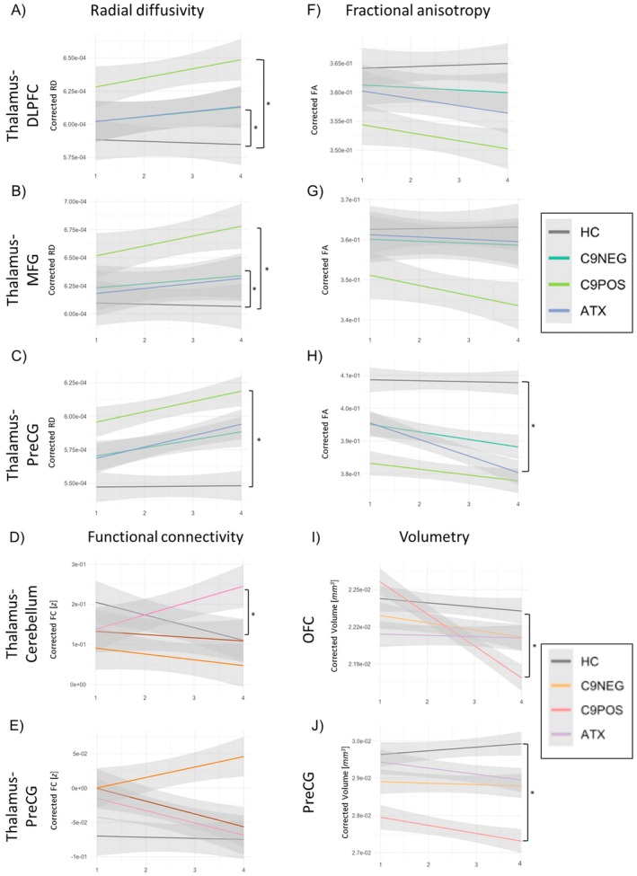
Results: The white matter integrity of thalamic projections to the anterior cingulate cortex, cerebellum, dorsolateral prefrontal cortex (DLPFC), Heschl's gyrus, medial frontal gyrus (MFG), orbitofrontal cortex, parietal cortex, postcentral gyrus and precentral gyrus (PreCG) is affected at baseline in ALS, which is more marked in C9orf72 hexanucleotide repeat carriers. Precentral gyrus and cerebellar grey matter volumes are also reduced, particularly in C9orf72. Longitudinal analyses capture progressive disconnection between the thalamus and frontal regions (DLPFC and MFG) in both C9orf72 positive and sporadic patients and progressive thalamo-PreCG disconnection in the sporadic C9orf72 negative cohort. Functional connectivity analyses revealed increasing thalamo-cerebellar connectivity in sporadic ALS and increasing thalamo-DLPFC connectivity in intermediate-length CAG repeat expansion carriers in ATXN2 over time.

Discussion: Our data provide evidence of extensive thalamo-cortical connectivity alterations in ALS. Corticobasal circuits mediating extrapyramidal, somatosensory, cognitive and behavioural functions are increasingly affected as the disease progresses. The degeneration of thalamic projections support the conceptualisation of ALS as a 'network disease' and the notion of 'what wires together degenerates together'.

Keywords: amyotrophic lateral sclerosis; magnetic resonance imaging; motor neuron disease; neuroimaging.


Conflict of interest statement

The authors declare no conflicts of interest.

Figures

FIGURE 1
FIGURE 1
Thalamo‐cortical tractography. Representative tractography outputs between the thalamus and 11 cortical regions of interest. Sagittal, coronal and axial views are presented from left to right for each cortical projection. ACC, anterior cingulate cortex; DLPFC, dorsolateral prefrontal cortex; Heschl, Heschl's gyrus; Mammillary, mammillary bodies; MFG, medial frontal gyrus; OFC, orbitofrontal cortex; Parietal, parietal cortex; PostCG, postcentral gyrus; PreCG, precentral gyrus; V1, primary visual cortex.
FIGURE 2
FIGURE 2
Cross‐sectional structural connectivity (SC) profiles. Radial diffusivity (RD) distributions are shown on the left (A‐D), fractional anisotropy (FA) profiles on the right (E‐H). * indicates adjusted p ≤ 0.05 in post hoc pairwise comparisons using Tukey's HSD testing. ATX, Patients with ALS carrying an intermediate length CAG repeat expansion in ATXN2, C9NEG = Sporadic patients with ALS testing negative for ALS associated genetic variants as well as C9orf72 hexanucleotide repeat expansions; C9POS, Patients with ALS carrying GGGGCC hexanucleotide repeat expansions in C9orf72; DLPFC, dorsolateral prefrontal cortex; HC, healthy controls; PostCG, postcentral gyrus; PreCG, precentral gyrus.
FIGURE 3
FIGURE 3
Cross‐sectional functional connectivity (FC) and volumetry. Baseline functional connectivity profiles are shown on the left (A‐C) and baseline volume distributions on the right (D‐F) * indicates adjusted p‐values ≤ 0.05 in post‐hoc pairwise comparisons using Tukey’s HSD testing. Abbreviations: ATX, Patients with ALS carrying an intermediate length CAG repeat expansion in ATXN2; C9NEG, Sporadic patients with ALS testing negative for ALS associated genetic variants as well as C9orf72 hexanucleotide repeat expansions; C9POS, Patients with ALS carrying GGGGCC hexanucleotide repeat expansions in C9orf72; HC, Healthy controls; PreCG, precentral gyrus.
FIGURE 4
FIGURE 4
Longitudinal findings. Progressive radial diffusivity (A‐C) , fractional anisotropy (F‐H), functional connectivity (D,E) and volumetric alterations (I,J) in the four study groups. * indicates p‐values ≤ 0.05, comparing the interaction effect Genotype X Time versus HC x Time in a linear mixed effects model. Abbreviations: ATX, Patients with ALS carrying an intermediate length CAG repeat expansion in ATXN2; C9NEG, Sporadic patients with ALS testing negative for ALS associated genetic variants as well as C9orf72 hexanucleotide repeat expansions; C9POS, Patients with ALS carrying GGGGCC hexanucleotide repeat expansions in C9orf72; DLPFC, dorsolateral prefrontal cortex; HC, Healthy controls; MFG, medial frontal gyrus, OFC, orbitofrontal cortex; PreCG, precentral gyrus.

Similar articles

Cited by

  • New developments in imaging in ALS.
    Kleinerova J, Querin G, Pradat PF, Siah WF, Bede P. Kleinerova J, et al. J Neurol. 2025 May 12;272(6):392. doi: 10.1007/s00415-025-13143-8. J Neurol. 2025. PMID: 40353906 Free PMC article. Review.

References

    1. Westeneng H. J., Verstraete E., Walhout R., et al., “Subcortical Structures in Amyotrophic Lateral Sclerosis,” Neurobiology of Aging 36 (2015): 1075–1082. - PubMed
    1. Bede P., Omer T., Finegan E., et al., “Connectivity‐Based Characterisation of Subcortical Grey Matter Pathology in Frontotemporal Dementia and ALS: A Multimodal Neuroimaging Study,” Brain Imaging and Behavior 12 (2018): 1696–1707. - PubMed
    1. Bocchetta M., Todd E. G., Tse N. Y., et al., “Thalamic and Cerebellar Regional Involvement Across the ALS‐FTD Spectrum and the Effect of C9orf72,” Brain Sciences 12, no. 3 (2022): 336, 10.3390/brainsci12030336. - DOI - PMC - PubMed
    1. Cistaro A., Pagani M., Montuschi A., et al., “The Metabolic Signature of C9ORF72‐Related ALS: FDG PET Comparison With Nonmutated Patients,” European Journal of Nuclear Medicine and Molecular Imaging 41 (2014): 844–852. - PMC - PubMed
    1. Sharma K. R., Saigal G., Maudsley A. A., and Govind V., “1H MRS of Basal Ganglia and Thalamus in Amyotrophic Lateral Sclerosis,” NMR in Biomedicine 24 (2011): 1270–1276. - PMC - PubMed

MeSH terms