. 2025 May 27;44(5):115629.
doi: 10.1016/j.celrep.2025.115629. Epub 2025 May 9.

Wolbachia-mediated reduction in the glutamate receptor mGluR promotes female promiscuity and bacterial spread

Affiliations

Wolbachia-mediated reduction in the glutamate receptor mGluR promotes female promiscuity and bacterial spread

Brandt Warecki et al. Cell Rep. .

Abstract

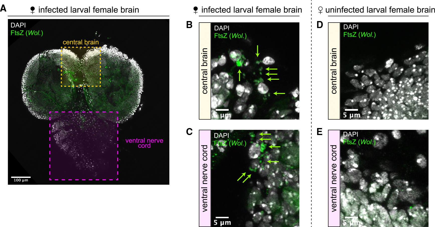
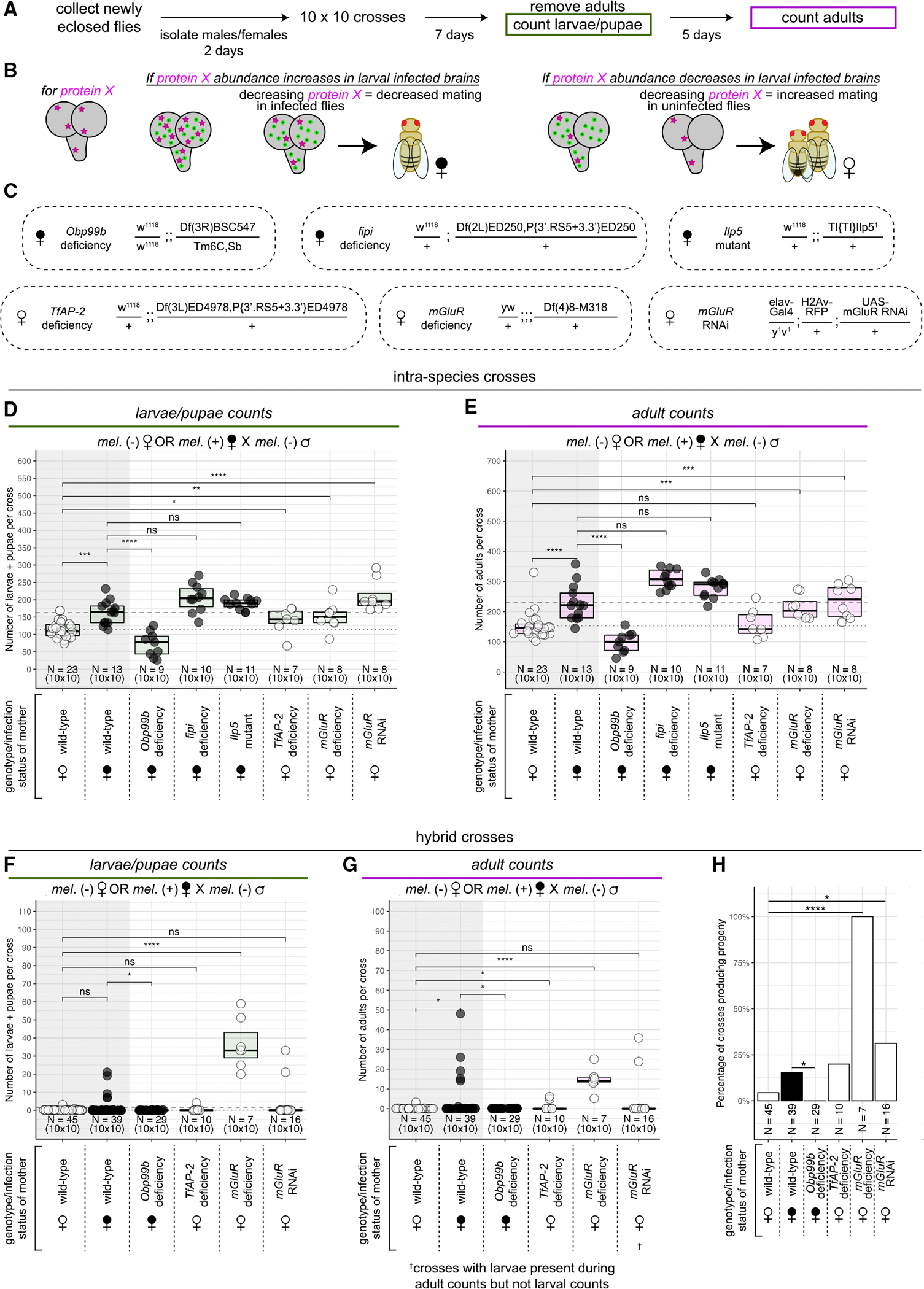
The molecular mechanisms by which parasites mediate host behavioral changes remain largely unexplored. Here, we examine Drosophila melanogaster infected with Wolbachia, a symbiont transmitted through the maternal germline, and find Wolbachia infection increases female receptivity to male courtship and hybrid mating. Wolbachia colonize regions of the brain that control sense perception and behavior. Quantitative global proteomics identify 177 differentially abundant proteins in infected female larval brains. Genetic alteration of the levels of three of these proteins in adults, the metabotropic glutamate receptor mGluR, the transcription factor TfAP-2, and the odorant binding protein Obp99b, each mimic the effect of Wolbachia on female receptivity. Furthermore, >700 Wolbachia proteins are detected in infected brains. Through abundance and molecular modeling analyses, we distinguish several Wolbachia-produced proteins as potential effectors. These results identify potential networks of host and Wolbachia proteins that modify behavior to promote mating success and aid the spread of Wolbachia.

Keywords: CP: Microbiology; CP: Neuroscience; Drosophila; Wolbachia; mGluR; mating; neurotransmission; proteomics; sense perception.


Conflict of interest statement

Declaration of interests The authors declare no competing interests.

Figures

Figure 1.
Figure 1.. Wolbachia increases the receptivity of female D. melanogaster to mating
(A) Ten 2-day-old females were crossed with ten 2-day-old males for 7 days at which point adults were removed and crawling third instar larvae and pupae were counted. After a further 5 days, emerged adults were counted. (B and C) The total number of larvae + pupae (B) and the total number of adults (C) collected from crosses with uninfected (white symbols and dots) and infected (black symbols and dots) D. melanogaster females crossed to uninfected D. melanogaster males. Each dot represents the progeny from one 10 × 10 cross. Data are represented as median +/− upper and lower quartiles. **p < 0.01, ***p < 0.001 by two-sided Mann-Whitney tests. (D) Seven to ten 2-day-old female flies were placed in a mating chamber with seven to ten 2-day-old male flies and filmed for 1 h (E) Comparison between the measured times to copulation for uninfected (white dots) and infected (black dots) D. melanogaster females presented with uninfected D. melanogaster males. Data are represented as median +/− upper and lower quartiles. Each dot represents one successful copulation. nsp > 0.05 by one-sided Mann-Whitney test. (F) Comparison between the measured copulation rates for uninfected (white dots) and infected (black dots) D. melanogaster females presented with uninfected D. melanogaster males. Data are represented as median +/− upper and lower quartiles. Each dot represents the percentage of females that copulated during one mating assay. *p < 0.05 by one-sided Mann-Whitney test. See also Figure S1. (G) Comparison between the percentage of total females that copulated over time for uninfected (white dots) and infected (black dots) D. melanogaster females presented with uninfected D. melanogaster males. (H and I) The numbers of larvae + pupae (H) and adults (I) collected from crosses with uninfected (white dots) and infected (black dots) D. melanogaster females crossed to uninfected (white symbols) and infected (black symbols) D. simulans males. Each dot represents the progeny from one 10 × 10 cross. Data are represented as median +/− upper and lower quartiles. nsp > 0.05, *p <0.05, **p < 0.01, ***p < 0.001 by two-sided Mann-Whitney tests.
Figure 2.
Figure 2.. Wolbachia colonize the central brain and ventral nerve cord regions of female D. melanogaster larval brains
(A) Female larval brain stained with the anti-bacterial antibody FtsZ (green) and DAPI (gray). Scale bar, 100 μm. (B and C) FtsZ-stained Wolbachia puncta (green arrows) are routinely observed in the central brain (B) and ventral nerve cord (C) regions of infected brains. Scale bars, 5 μm. (D and E) No FtsZ-stained puncta are observed in the central brain (D) or ventral nerve cord (E) regions of uninfected brains. Scale bars, 5 μm.
Figure 3.
Figure 3.. Wolbachia colonize regions of the female adult brain involved in sensory perception and higher order decision making
(A and B) Low-magnification view of infected (A) and uninfected (B) female adult brains stained with antibodies against FtsZ (green) to label Wolbachia and bruchpilot/nc82 (magenta) to label neuropil. Labels correspond to the relative areas examined in (C)–(L). al, antennal lobe; eb, ellipsoid body (not visible from this view); sog, suboesophageal ganglion; mb, mushroom body (not fully visible this view); fsb, fan-shaped body (not visible from this view). Scale bars, 100 μm. (C–L) High-magnification images of the antennal lobes (C and D), ellipsoid bodies (E and F), suboesophageal ganglia (G and H), mushroom bodies (I and J), and fan-shaped bodies (K and L) of infected (C, E, G, I, and K) and uninfected (D, F, H, J, and L) flies. Dotted outlines indicate brain regions. FtsZ-stained puncta (green arrows) correspond to Wolbachia adjacent to DAPI-stained host cell bodies (gray). Yellow dashed boxes indicate the zoomed-in regions. Scale bars, 20 and 5 μm (for brain region and zoomed-in regions, respectively). See also Figure S2.
Figure 4.
Figure 4.. Wolbachia infection is correlated with up- and downregulation of host proteins involved in sense perception and behavior
(A) Third instar brain tissue (brain lobes and ventral nerve cords) was used for mass spectrometry. The infection status of the samples was determined by performing PCR with primers targeting Wolbachia on five additional brains dissected simultaneously as samples. (B) Volcano plot of expression differences of D. melanogaster proteins in Wolbachia-infected and uninfected brains. Each dot represents one protein. Red dots are significantly upregulated in Wolbachia-infected brains. Blue dots are proteins significantly downregulated in Wolbachia-infected brain. (C) STRING-predicted protein-protein interaction network for differentially regulated proteins in Wolbachia-infected brains. Red and blue dots represent proteins up- and downregulated, respectively, in Wolbachia-infected brains. See also Figure S3. (D) Candidate proteins that may mediate increased female receptivity upon Wolbachia infection. Blue highlighted proteins were identified as belonging to the Monarch-defined “abnormal mating.” (E) Enrichment and false discovery rates (the likelihood of a predicted enrichment being untrue) for enriched Monarch-defined phenotypes among the candidate targets depicted in (D).
Figure 5.
Figure 5.. Genetic reduction of mGluR, TfAP-2, and Obp99b phenocopy the presence/absence of Wolbachia on female receptivity to mating
(A) Ten 2-day-old females were crossed with ten 2-day-old males for 7 days at which point adults were removed and crawling third instar larvae and pupae were counted. After a further 5 days, emerged adults were counted (same protocol as in Figure 1). (B) Diagram of the genetic screen. If an involved protein’s abundance is increased in larval infected brains, decreasing the level of the protein in infected flies should decrease mating. If an involved protein’s abundance is decreased in larval-infected brains, decreasing the level of the protein in uninfected flies should increase mating. (C) The genotypes of female flies used in the genetic screen determining which candidate differentially abundant host proteins may be involved in Wolbachia-correlated increase in female receptivity. (D and E) The numbers of larvae + pupae (D) and adults (E) collected from intra-species crosses with uninfected (white dots) and infected (black dots) D. melanogaster females crossed to uninfected D. melanogaster males. Each dot represents the progeny from one 10 × 10 cross. Data are represented as median +/− upper and lower quartiles. p values displayed were calculated by one-sided Mann-Whitney tests. nsp > 0.05, *p < 0.05, **p < 0.01, ***p < 0.001, ****p < 0.0001. (F and G) The numbers of larvae + pupae (F) and adults (G) collected from hybrid crosses with uninfected (white dots) and infected (black dots) D. melanogaster females crossed to uninfected D. simulans males. Each dot represents the progeny from one 10 × 10 cross. Data are represented as median +/− upper and lower quartiles. p values displayed were calculated by one-sided Mann-Whitney tests. nsp > 0.05, *p < 0.05, ****p < 0.0001. (H) The percentage of hybrid crosses that produced progeny for each given female genotype and infection status. *p < 0.05, ****p < 0.0001 by Fisher’s test.
Figure 6.
Figure 6.. Wolbachia-produced proteins are detected in infected female brains
(A) Detected average abundances for the components of the type IV secretion system encoded by the Wolbachia genome. Mean Wolbachia protein abundance in brain = 2.22 × 10⁶. (B) Diagram describing how candidate Wolbachia effectors were selected. WD0438 fits both selection criteria. (C) Candidate Wolbachia-produced effectors detected in infected female brains. Each dot represents an individual abundance value detected for a given protein. Data are represented as median +/− upper and lower quartiles. Proteins in the green region are in the top 10% of Wolbachia proteins detected based on abundance. Proteins highlighted in magenta were previously identified as causing growth defects when expressed in yeast. See also Figure S4. (D) Yeast were generated with either an empty vector or a Wolbachia gene of interest under control of the GAL1 promoter. Yeast were grown on either a 2% glucose control medium or 2% galactose medium to express the Wolbachia gene and growth was monitored with a spotting assay. (E) Spotting assays showing a strong growth defect in yeast grown on 2% galactose expressing the Wolbachia proteins WD1127 or WD0295. Expression of WD0523 or WD1220 did not induce a pronounced growth defect.
Figure 7.
Figure 7.. Model for how Wolbachia in the brain increases female receptivity to promote its spread
(A) In uninfected developing brains, mGluR, Obp99b, and TfAP-2 are present in wild-type levels, leading to normal sense perception and mating decisions in adult female flies. This results in the production of uninfected progeny. (B) In Wolbachia-infected developing brains, mGluR and TfAP-2 levels are decreased and Obp99b levels are increased. Decreased mGluR may lead to increased excitability of glutamatergic neurons. Increased Obp99b may lead to increased odor/pheromone transport. Decreased TfAP-2 may lead to altered central complex development. In the adult brain, Wolbachia colonizes areas important for sense perception and decision making. Collectively, this leads to altered sense perception and mating decisions in adult females and results in increased mating and an increased amount of infected progeny.

Similar articles

References

    1. Moore J (2002). Parasites and the Behavior of Animals (Oxford University Press; ).
    1. Rohrscheib CE, and Brownlie JC (2013). Microorganisms that Manipulate Complex Animal Behaviours by Affecting the Host’s Nervous System. Springer Sci. Rev. 1, 133–140. 10.1007/s40362-013-0013-8. - DOI
    1. Gowda V, Dinesh S, and Sharma S (2023). Manipulative neuroparasites: uncovering the intricacies of neurological host control. Arch. Microbiol. 205, 314. 10.1007/s00203-023-03637-2. - DOI - PubMed
    1. Boillat M, Hammoudi PM, Dogga SK, Pagès S, Goubran M, Rodriguez I, and Soldati-Favre D (2020). Neuroinflammation-Associated Aspecific Manipulation of Mouse Predator Fear by Toxoplasma gondii. Cell Rep. 30, 320–334.e6. 10.1016/j.celrep.2019.12.019. - DOI - PMC - PubMed
    1. Elya C, Lok TC, Spencer QE, McCausland H, Martinez CC, and Eisen M (2018). Robust manipulation of the behavior of Drosophila melanogaster by a fungal pathogen in the laboratory. Elife 7, e34414. 10.7554/eLife.34414. - DOI - PMC - PubMed

Substances

LinkOut - more resources