Comparative Study
. 2025 May 10;16(1):4362.
doi: 10.1038/s41467-025-59370-5.

T cell memory response to MPXV infection exhibits greater effector function and migratory potential compared to MVA-BN vaccination

Collaborators, Affiliations
Comparative Study

T cell memory response to MPXV infection exhibits greater effector function and migratory potential compared to MVA-BN vaccination

Ji-Li Chen et al. Nat Commun. .

Abstract

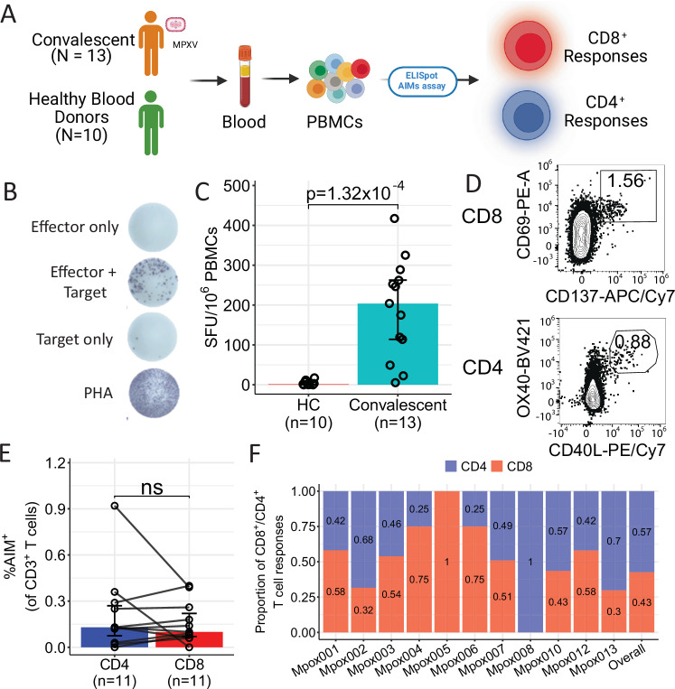
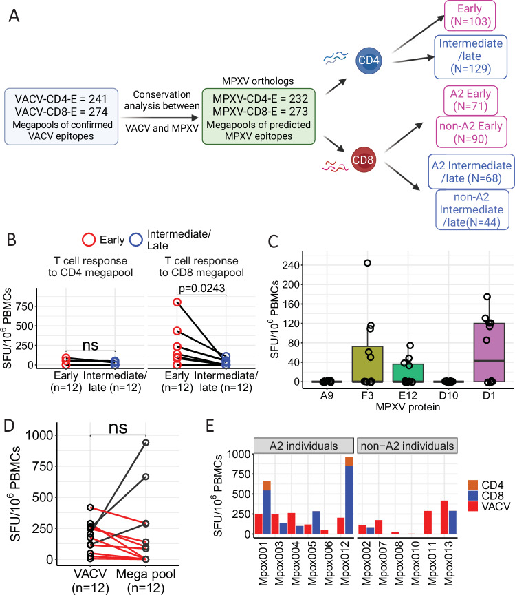
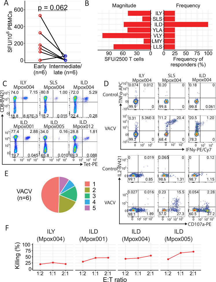
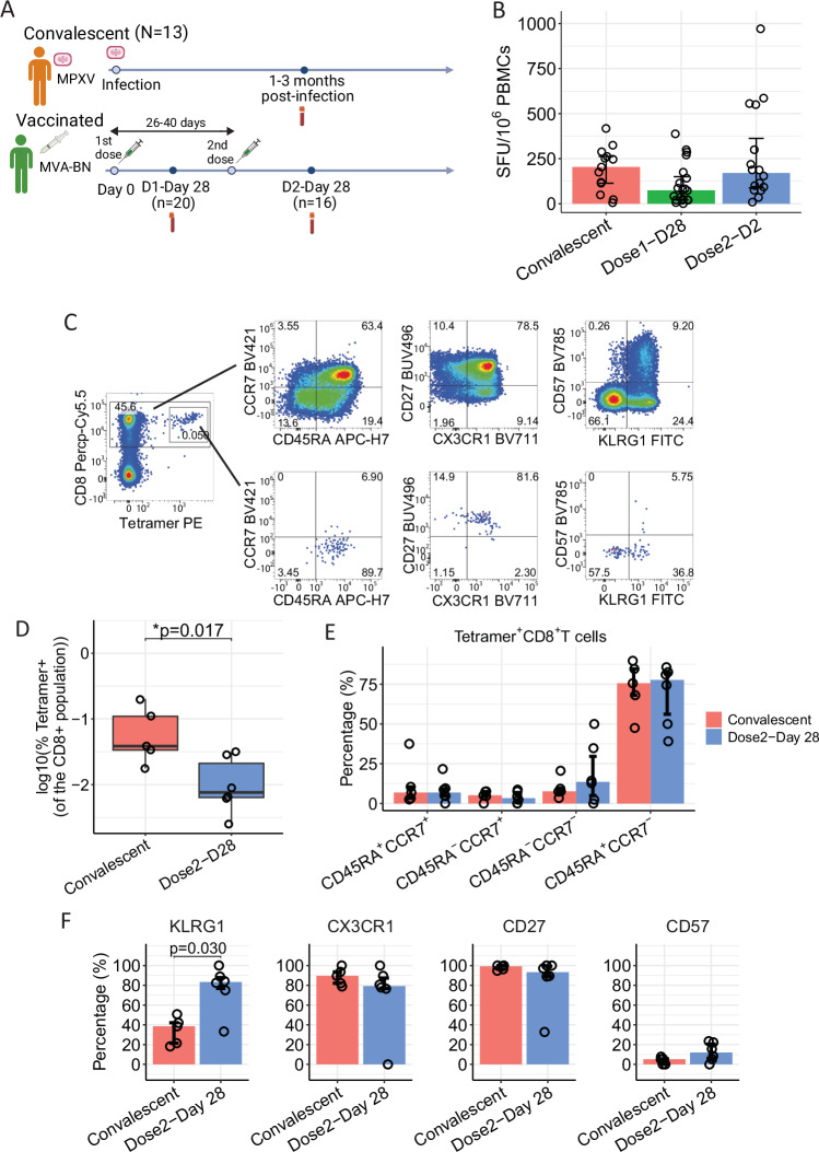
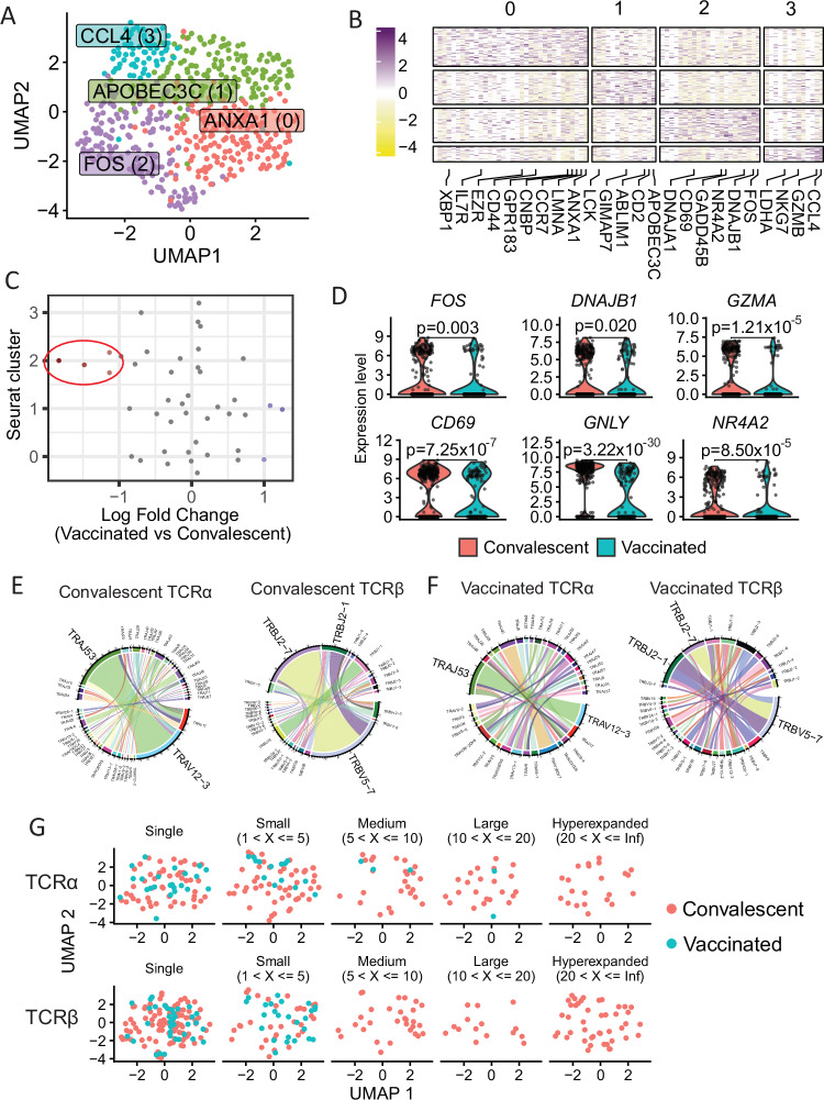
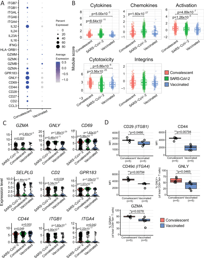
In 2022, a global mpox outbreak occurred, and remains a concern today. The T cell memory response to MPXV (monkeypox virus) infection has not been fully investigated. In this study, we evaluate this response in convalescent and MVA-BN (Modified Vaccinia Ankara - Bavarian Nordic) vaccinated individuals using VACV-infected cells. Strong CD8+ and CD4+ T cell responses are observed, and T cell responses are biased towards viral early expressed proteins. We identify seven immunodominant HLA-A*02:01 restricted MPXV-specific epitopes and focus our detailed phenotypic and scRNAseq analysis on the immunodominant HLA-A*02:01-G5R18-26-specific CD8+ T cell response. While tetramer+CD8+ T cells share similar differentiation and activation phenotypes, T cells from convalescent individuals show greater cytotoxicity, migratory potential to site of infection and TCR clonal expansion. Our data suggest that effective functional profiles of MPXV-specific memory T cells induced by Mpox infection may have an implication on the long-term protective responses to future infection.


Conflict of interest statement

Competing interests: The authors declare no competing interests.

Figures

Fig. 1
Fig. 1. MPXV-specific memory T cell response in mpox convalescent individuals and healthy controls (HC).
A Study design (Created in BioRender. Antoun, E. (2025) https://BioRender.com/a25q267). B Representative IFN-γ ELISpot response after stimulation with VACV. C Summary of VACV-induced memory T cell response in mpox convalescent (N = 13) and healthy control (HC, N = 10) participants. Data are presented as median ± IQR. D Representative flow cytometry plots measuring expression of activation-induced markers on VACV-reactive CD8+ and CD4+ T cells from mpox convalescent participants. E Overall percentage of VACV-reactive CD8+ and CD4+ T cells in total T cells. Data are presented as median ± IQR (N = 11). F Individual and overall relative proportion of VACV-reactive CD8+ (red) and CD4+ (blue) T cells, N = 11. The Mann–Whitney U-test was used for the analysis and two-tailed P values were calculated. ns not significant; IQR interquartile range. Source data are provided as a Source Data file.
Fig. 2
Fig. 2. Memory T cell responses in MPXV-convalescent individuals against epitope mega pools of early and intermediate/late antigens.
A Schematic graph of MPXV epitope mega pool design (see the “Methods” section for more details, Created in BioRender. Antoun, E. (2025) https://BioRender.com/a25q267). B T cell responses against MPXV CD4+/CD8+ early proteins (red) or intermediate/late proteins (blue), N = 12. C Overall T cell responses against overlapping peptide pools covering the full length of five selected MPXV antigens, N = 12. Boxplots represent the 25th and 75th percentiles with the median marked with whiskers at ±1.5*IQR. D, E Comparison of overall T cell response against VACV and that against MPXV mega pools (N = 12). A paired Wilcoxon signed-rank test for B and D was used and two-tailed P values were calculated. ns not significant, SFU spot forming units, MPXV Mpox virus, VACV vaccinia virus. Source data are provided as a Source Data file.
Fig. 3
Fig. 3. Identification and functional evaluation of immunodominant A2-restricted MPXV-specific T cells.
A Comparison of HLA-A*02:01-restricted T cell responses against MPXV-CD8-early (red) and MPXV-CD8-intermediate/late (blue) peptide pools using ex vivo IFNγ ELISpot, N = 6 participants. A paired Wilcox-signed rank test was carried out to compare between groups and two-sided p-values calculated. B Identification of dominant HLA-A*02:01-restricted MPXV-specific T cell epitopes using short-term T cells lines from N = 4 donors. C Specific T cells recognizing the three epitopes ILYDNVVTL (ILY), SLSNLDFRL (SLS), and ILDDNLYKV (ILD) were confirmed by tetramers. D Representative cytokine responses of different HLA-A*02:01-restricted MPXV-specific T cell lines from Mpox004. E Proportion of T cells against VACV expressing 1, 2, 3, 4, or all 5 of the following functional markers: IFNγ, TNFα, MIP1β, IL2 and CD107a expression, N = 6. F Percentage of specific lysis of VACV-infected HLA-A*02:01+ B cell lymphoblastoid cell lines (BCLs) by different epitope-specific CD8+ bulk lines. VACV vaccinia virus, SFU spot forming units. Source data are provided as a Source Data file.
Fig. 4
Fig. 4. Frequency and phenotype of ex vivo MPXV-specific T cell response in convalescent and vaccinated donors.
A Study design for MPXV-convalescent and vaccinated individuals (created in BioRender. Antoun, E. (2025) https://BioRender.com/a25q267). B Summary of VACV-induced memory T cell response in mpox convalescent (N = 13) and in individuals 28 days after 1 vaccination (N = 20) and 28 days after 2 vaccinations (N = 16). C Representative plot of tetramer+CD8+ and overall CD8+ T cells. D Comparison of frequency of HLA-A*02:01 ILD-specific Tetramer+ T cells between convalescent (N = 5) and vaccinated donors (N = 6). Boxplots represent the 25th and 75th percentiles with the median marked with whiskers at ±1.5*IQR. E Percentage of different memory and differentiation phenotype marker expression on CD8+tetramer+ T cells from convalescent participants (red, N = 5) and samples 28 days after 2 vaccinations (blue, N = 6). F Expression of memory and differentiation markers on CD45RA+CCR7-CD8+tetramer+ T cells from convalescent participants (red, N = 5) and samples 28 days after dose 2 of vaccine (blue, N = 6). Data are presented as median±IQR. The Mann–Whitney U-test was used and two-tailed P values were calculated. SFU spot forming units. Source data are provided as a Source Data file.
Fig. 5
Fig. 5. scRNAseq transcriptomic and TCR repertoire analysis of ex vivo MPXV-specific CD8+ T cells from vaccinated (n = 6) and convalescent (n = 4) individuals.
Tetramer+CD8+ T cells were used for single-cell transcriptomic and TCR repertoire analysis. A UMAP plot of tetramer+CD8+ T cells integrated based on patient of origin, identifying 4 clusters of cells. B Heatmap of the differentially expressed marker genes of the 4 cell clusters. C Beeswarm plot showing the log-fold distribution of cell abundance changes between convalescent and vaccinated samples. Neighbourhoods overlapping the same cell population are grouped together, neighbourhoods exhibiting differential abundance are coloured in red and are circled. D Violin plots of cluster 2 marker genes between vaccinated and convalescent samples. E and F Circos plots showing the TRAV-TRAJ and TRBV-TRBJ gene pairing for convalescent (E) and vaccinated (F) samples. G UMAP plot of the tetramer+CD8+ T cells, split by whether their TCR alpha (top) and beta (bottom) clonotypes are expanded. Differential expression between clusters was carried out using the MAST test. The Wilcoxon signed-rank test was used to test differences between convalescent and vaccinated samples and two-tailed P values were calculated. ****p < 0.0001, ***p < 0.001, **p < 0.01, *p < 0.05.
Fig. 6
Fig. 6. scRNAseq transcriptomic comparison between MPXV-specific and SARS-CoV-2-specific CD8+ T cells.
A Dot plot of the gene expression of cells from convalescent and vaccinated individuals for cytokines, cytotoxicity molecules, integrins and activation markers. B Comparison of module scores for cytokines, integrins, cytotoxicity genes, activation-related genes and chemokines between MPXV-specific (401 and 160 cells from convalescence and vaccination, respectively) and SARS-CoV-2-specific CD8+ T cells (912 cells). C Violin plots depicting the expression of select genes between MPXV-specific and SARS-CoV-2-specific CD8+ T cells. D Comparison of cytotoxicity molecules and integrins expressed on ILD-tetramer+CD8+ T cells between convalescent (N = 5) and vaccinated (N = 5) individuals by flow cytometry. The Wilcoxon signed-rank test was used in (B) and (C). In D, the Kolmogorov–Simonov test was used to test for normality and an unpaired t-test was used for CD29 and the Mann–Whitney U-test was used for the rest. Boxplots in B and D represent the 25th and 75th percentiles with the median marked with whiskers at ±1.5*IQR. Two-tailed p values were calculated. ****p < 0.0001, ***p < 0.001, **p < 0.01, *p < 0.05. Source data are provided as a Source Data file.

Similar articles

References

    1. Wenham, C. & Eccleston-Turner, M. Monkeypox as a PHEIC: implications for global health governance. Lancet400, 2169–2171 (2022). - PMC - PubMed
    1. McCollum, A. M. & Damon, I. K. Human monkeypox. Clin. Infect. Dis.58, 260–267 (2014). - PMC - PubMed
    1. WHO. Fifth Meeting of the International Health Regulations (2005) (IHR) (Emergency Committee on the Multi-country Outbreak of mpox (monkeypox), 2005).
    1. WHO. 2022–23 Mpox (Monkeypox) Outbreak: Global Trends (WHO, 2024).
    1. Vakaniaki, E. H. et al. Sustained human outbreak of a new MPXV Clade I lineage in eastern Democratic Republic of the Congo. Nat. Med.30, 2791–2795 (2024). - PMC - PubMed

Publication types