. 2025 May 10;16(1):4366.
doi: 10.1038/s41467-025-59736-9.

Phenotypic drug susceptibility testing for Mycobacterium tuberculosis variant bovis BCG in 12 hours

Affiliations

Phenotypic drug susceptibility testing for Mycobacterium tuberculosis variant bovis BCG in 12 hours

Buu Minh Tran et al. Nat Commun. .

Abstract

Drug-resistant tuberculosis (DR-TB) kills ~200,000 people every year. A contributing factor is the slow turnaround time (TAT) associated with drug susceptibility diagnostics. The prevailing gold standard for phenotypic drug susceptibility testing (pDST) takes at least two weeks. Here we show that growth-based pDST for slow-growing mycobacteria can be conducted in 12 h. We use Mycobacterium tuberculosis variant bovis Bacillus Calmette-Guérin (BCG) and Mycobacterium smegmatis as the mycobacterial pathogen models and expose them to antibiotics used in (multidrug-resistant) tuberculosis (TB) treatment regimens - i.e., rifampicin (RIF), isoniazid (INH), ethambutol (EMB), linezolid (LZD), streptomycin (STR), bedaquiline (BDQ), and levofloxacin (LFX). The bacterial growth in a microfluidic chip is tracked by time-lapse phase-contrast microscopy. A deep neural network-based segmentation algorithm is used to quantify the growth rate and to determine how the strains responded to drug treatments. Most importantly, a panel of susceptible and resistant M. bovis BCG are tested at critical concentrations for INH, RIF, STR, and LFX. The susceptible strains could be identified in less than 12 h. These findings are comparable to what we expect for pathogenic M. tuberculosis as they share 99.96% genetic identity.


Conflict of interest statement

Competing interests: J.E. has patented the method (US10,041,104B) and founded Astrego Diagnostics, but he has no current association with that company. No current company is associated with this work, but there may be in the future. All other authors declare no competing interests.

Figures
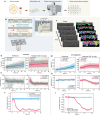
Fig. 1
Fig. 1. Schematic representation of the fast pDST workflow for M. bovis BCG and M. smegmatis.
a Main steps in the workflow (Created in BioRender. Lab, E. (2025) https://BioRender.com/e13c692). b Illustration of the microchamber cell traps. c Analysis of time-lapse stacks using Omnipose for cell segmentation. di Step-by-step growth rate calculation and normalization for M. bovis BCG (df) and M. smegmatis (gi). d. Total areas of M. bovis BCG [Reference (left) and Treatment population (right; isoniazid INH 0.5 mg/L)]. Each line is data from an individual microchamber. The dashed line indicates the drug addition time. e M. bovis BCG growth rates. f M. bovis BCG growth rate normalization (pDST profile) at INH 0.5 mg/L (mean ± SEM). gi Corresponding area growth (g), growth rates of M. smegmatis (rifampicin RIF 10 mg/L) (h), and its pDST profile at RIF 10 mg/L (i).
Fig. 2
Fig. 2. Training new models for mycobacterial cell segmentation.
a Phase contrast image and the corresponding cell membrane image stained by 3HC-3-Tre dye. b, c Segmentation performance of different training parameters using data excluding (b) and including (c) high-density cell microchambers. Mycobact_2* in (b) is the Mycobact_2 model performed using images from one camera. Inset in (c) is a zoom-in of the x and y-axis. di Representative micrographs of mycobact_2 model for cells in various conditions–(d) M. smegmatis on agarose gel, (e) and (f) M. smegmatis in microchamber, (g) M. bovis BCG in microchamber, (h) and (i) Relatively and highly dense M. smegmatis clumps. Ground truth is labeled from phase images. Flow field is an intermediate output from the neural network. Average precision at an IoU threshold of 0.5 (AP@0.5) for the entire image is reported on the top left corner of the outline - scale bar 5 µm.
Fig. 3
Fig. 3. Fast detection of response to drug treatment.
pDST assays detecting the fast response of susceptible M. bovis BCG Pasteur to (a) rifampicin (RIF), (b) isoniazid (INH), (c) ethambutol (EMB), and (d) linezolid (LZD); of susceptible M. smegmatis NCTC 8159 to (e) RIF, (f) INH, (g) EMB, and (h) LZD; and susceptible M. smegmatis mc2 155 to (i) RIF, (j) INH, (k) EMB, and (l) LZD. Data in each graph is from one biological replication. Multiple biological replication data are shown in Supplementary Fig. 2 and Supplementary Fig. 3.
Fig. 4
Fig. 4. Fast detection of resistant strains.
pDST profiles of lab-evolved resistant strains derived from M. smegmatis NCTC 8159 including (a) M. smegmatis RIF E1, (b) M. smegmatis RIF E2, (c) M. smegmatis RIF E3 in rifampicin treatment, (d) M. smegmatis INH E1, (e) M. smegmatis INH E2, (f) M. smegmatis INH E3 in isoniazid treatment, and (g) M. smegmatis LZD E1, (h) M. smegmatis LZD E2, (i) M. smegmatis LZD E3 in linezolid treatment. Data in each graph is from one biological replication. Multiple biological replication data are shown in Supplementary Fig. 8.
Fig. 5
Fig. 5. pDST of susceptible and resistant M. bovis BCG Russia at critical concentrations (CCs).
pDST profiles of M. bovis BCG Russia WT (a) and M. bovis BCG Russia INHR (b) in INH 0.5 mg/L; M. bovis BCG Russia WT (c) and M. bovis BCG Russia RIFR (d) in RIF 1 mg/L; M. bovis BCG Russia WT (e) and M. bovis BCG Russia STRR (f) in STR 1 mg/L; and M. bovis BCG Russia WT (g) and M. bovis BCG Russia FQR (h) in LFX 1 mg/L. Data in each graph is from one biological replication. Multiple biological replication data are shown in Supplementary Fig. 10.

References

    1. Barberis, I., Bragazzi, N. L., Galluzzo, L. & Martini, M. The history of tuberculosis: from the first historical records to the isolation of Koch’s bacillus. J. Prev. Med. Hyg.58, E9–E12 (2017). - PMC - PubMed
    1. Geneva: World Health Organization. Global Tuberculosis Report. https://www.who.int/teams/global-tuberculosis-programme/tb-reports/globa... (2024).
    1. Murray, C. J. L. et al. Global burden of bacterial antimicrobial resistance in 2019: a systematic analysis. Lancet399, 629–655 (2022). - PMC - PubMed
    1. Dheda, K. et al. Global control of tuberculosis: from extensively drug-resistant to untreatable tuberculosis. Lancet Respir. Med. 2, 321–338 (2014). - PMC - PubMed
    1. Pedersen, O. S. et al. Global treatment outcomes of extensively drug-resistant tuberculosis in adults: a systematic review and meta-analysis. J. Infect.87, 177–189 (2023). - PubMed

MeSH terms

LinkOut - more resources