. 2025 May 10;16(1):4349.
doi: 10.1038/s41467-025-59510-x.

RNA transcripts serve as a template for double-strand break repair in human cells

Affiliations

RNA transcripts serve as a template for double-strand break repair in human cells

Manisha Jalan et al. Nat Commun. .

Abstract

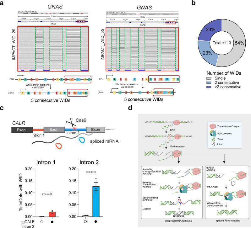
Double-strand breaks (DSBs) are toxic lesions that lead to genome instability. While canonical DSB repair pathways typically operate independently of RNA, growing evidence suggests that RNA:DNA hybrids and nearby transcripts can influence repair outcomes. However, whether transcript RNA can directly serve as a template for DSB repair in human cells remains unclear. In this study, we develop fluorescence and sequencing-based assays to show that RNA-containing oligonucleotides and messenger RNA can serve as templates during DSB repair. We conduct a CRISPR/Cas9-based genetic screen to identify factors that promote RNA-templated DSB repair (RT-DSBR). Of the candidate polymerases, we identify DNA polymerase zeta (Polζ) as a potential reverse transcriptase that facilitates RT-DSBR. Furthermore, analysis of cancer genome sequencing data reveals whole intron deletions - a distinct genomic signature of RT-DSBR that occurs when spliced mRNA guides repair. Altogether, our findings highlight RT-DSBR as an alternative pathway for repairing DSBs in transcribed genes, with potential mutagenic consequences.


Conflict of interest statement

Competing interests: A.S. is a co-founder, consultant, and shareholder for REPARE Therapeutics. S.N.P is a consultant for AstraZeneca, Varian Medical Systems, and Philips. J.S.R.-F. reports current employment at AstraZeneca and stocks in AstraZeneca, Repare Therapeutics, Paige.AI; J.S.R.-F. previously held a fiduciary role in Grupo Oncoclinicas and consulted with Goldman Sachs Merchant Banking, Bain Capital, Repare Therapeutics, Paige.AI, Volition Rx and MultiplexDx. P.C.B. sits on the Scientific Advisory Boards of Intersect Diagnostics Inc., BioSymetrics Inc., and Sage Bionetworks. The remaining authors declare no competing interests.

Figures

Fig. 1
Fig. 1. Human cells use RNA to template DSB repair.
a Schematic of the BFP-to-GFP assay designed to generate a green fluorescent signal via RNA templated DSB repair (RT-DSBR). This assay exploits the single amino acid change that differentiates Blue Fluorescent Protein (BFP) from Green Fluorescent Protein (GFP), switching the fluorescence from blue to green. A DSB is introduced at an integrated BFP locus using CRISPR/Cas9, and cells repair the break with a single-stranded DNA donor (DNAGFP) containing the GFP codon, switching from BFP to GFP fluorescence. To detect RT-DSBR activity, we used DNA/RNA chimeric donors in which the sequence required to swap the codon was encoded by ribonucleotides instead of deoxyribonucleotides. b Right: schematic of the 120 bp chimeric donors used in the BFP-to-GFP assay with green segments representing stretches of ribonucleotides. Left: GFP signal quantification was performed by flow cytometry with different donors (n = 3–6 biological replicates) and compared to a non-donor control. c Schematic of the AAVS1-seq assay. A targeted DSB is introduced at the AAVS1 genomic locus using CRISPR/Cas9, and the donor DNA or DNA/RNA chimeras containing a 3 bp insertion are transfected into the cells. Successful repair using the donor leads to the incorporation of the mutational signature, which is detected by PCR amplification and Next Generation Sequencing. d Right: a schematic of the 60 bp donor templates used in the AAVS1-seq assay, with red segments representing stretches of ribonucleotides. Left: quantification of the fraction of repair products containing the 3 bp insertion signature after the Cas9 DSB is repaired by different donors, as measured by the AAVS1-seq assay (n = 3–6 biological replicates) and compared to a non-donor control. For (b and d), Statistical significance was assessed using unpaired two-tailed t-tests. Error bars represent the standard error of the mean (± SEM). Schematics in Fig. 1a–c, and d were Created in BioRender. (2025) https://BioRender.com/9tmc1xb. Source data are provided as a Source Data file. See also Supplementary Fig. 1.
Fig. 2
Fig. 2. RT-DSBR is independent of LINE-1 and Polθ activity.
a BFP-to-GFP assay with DNAGFP and DNA/RNA6R donors in the presence of 10 µM of the HIV reverse transcriptase inhibitor azidothymidine (AZT) or DMSO as a control (n = 3 biological replicates). b AAVS1-seq performed with DNA1 or DNA/RNA10R donors in the presence of 10 µM AZT or DMSO (n = 3 biological replicates). c BFP-to-GFP assay with DNAGFP and DNA/RNA6R donors in two POLQ−/− clones (#C1 and #C2) with or without complementation by full-length POLQ (POLQ-FLAG) (n = 2 and 3 biological replicates). d AAVS1-seq with DNA1 or DNA/RNA10R donors following knockdown of POLQ through siRNA, compared to a non-targeting siRNA control (siCTRL) (n = 3 biological replicates). For (ad): Statistical significance was assessed using unpaired two-tailed t-tests. Error bars represent the standard error of the mean (± SEM). Source data are provided as a Source Data file. See also Supplementary Fig. 2.
Fig. 3
Fig. 3. A CRISPR/Cas9 screen identifies factors involved in RT-DSBR.
a Schematic representation of a flow-based CRISPR/Cas9 screen performed using the BFP-to-GFP reporter in HEK293T cells. Cells were transduced with Cas9 and sgRNAs from a DNA damage library. After 10 days of sgRNA selection, the BFP-to-GFP assay was carried out using the DNAGFP or DNA/RNA6R donor respectively. b The CRISPR/Cas9 screen data were analyzed using the MAGeCK algorithm by comparing the GFP+ sorted cells with the GFP BFP cells. A heatmap highlights selected genes with high-ranking scores, indicating factors that promote or suppress single-strand template repair. Lower ranks denote stronger hits. c BFP-to-GFP assay results using DNAGFP and DNA/RNA6R donors after knockdown of two top hits that promote (HELQ) or suppress (TP53BP1) RT-DSBR (n = 3–7 biological replicates). sgRNA targeting the AAVS1 locus was used as a control. Statistical significance was assessed using unpaired two-tailed t-tests. Error bars represent the standard error of the mean (± SEM). d Heatmap of the 5 top hits that promote or suppress RT-DSBR. e Comparison of the rank position of major DNA polymerases identified in DNAGFP vs. DNA/RNA6R CRISPR/Cas9 screens. Schematic in Fig. 3a was Created in BioRender. (2025) https://BioRender.com/9tmc1xb. Source data are provided as a Source Data file. See also Supplementary Fig. 3.
Fig. 4
Fig. 4. Transcript RNA is a donor for DNA polymerase zeta (ζ) dependent RT-DSBR.
a Fraction of repair products from AAVS1-seq using DNA1 or DNA/RNA10R donors after siRNA-mediated knockdown of POLD1, POLK, PRIMPOL, REV3L, POLH, POLM or POLN, compared to a non-targeting siRNA control (siCTRL) (n = 3–14 biological replicates). b Percentage of repair products from the BFP-to-GFP assay using DNAGFP or DNA/RNA6R donors following knockdown of REV3L with siRNA. c Schematic of the Polζ complex. d Effect of Polζ subunits depletion on AAVS1-seq repair outcomes with DNA1 or DNA/RNA10R donors, assessed after siRNA-mediated knockdown (n = 3–9 biological replicates). e, f Schematic of a plasmid-based system designated to generate transcript RNA that acts as a donor template. Homology arms (grey) flank the Cas9 break site at the AAVS1 locus. Blue: CMV promoter. Light green- β-globin: artificial intron. Dark green: poly-A tail. Red: insertion signature. g Fraction of repair products containing the mutational signature in the presence of no donor (n = 9 biological replicates) or transcript RNA donor, following Cas9-induced breaks. Data were collected after treatment with non-targeting siRNA (siCTRL) (n = 8 biological replicates) or siRNA against REV3L (n = 3 biological replicates). For (ag): Where applicable, statistical significance was assessed using unpaired two-tailed t-tests, with Welch’s correction in (g). Error bars represent the standard error of the mean (± SEM). Schematics in this figure (c, e, and f) were created in BioRender. (2025) https://BioRender.com/9tmc1xb. Source data are provided as a Source Data file. See also Supplementary Figs. 4 and 5.
Fig. 5
Fig. 5. Whole intron deletions from cancer genomes provide in vivo evidence of RT-DSBR.
a Schematic of the CRISPR/Cas9 assay to detect a whole intron deletion (WID) in human cells. b Quantification of reads containing precise WIDs (as a fraction of total repair events) at CALR intron 2 in control cells and ones treated with siREV3L (n = 3 biological replicates) with and without a CRISPR/Cas9-mediated DSB. Statistical significance was assessed using an unpaired two-tailed t-tests. Error bars represent the standard error of the mean (± SEM). c Schematic of the bioinformatic pipeline used to analyze deletions in tumors from the MSK-IMPACT database. WIDs were identified as deletions that span a precise entire intron. The blue box highlights a read showing perfect intron loss. d Example of a WID found in the HLA-B gene of a patient sample from the MSK-IMPACT cohort. Read bases that match the reference are displayed in gray, purple “I” represents insertions, and deletions are indicated with a black dash (–). Alignments displayed with light gray borders and white fill have a mapping quality equal to zero, suggesting they may map to multiple regions across the genome. A 245 bp deletion is observed upon targeted NGS that maps precisely to the area corresponding to the intron flanked by Exon 2–3 of the HLA-B gene. e Schematic of the exons spanning the WID in HLA-B with the flanking primers used to confirm the sequence. f Agarose gel depicting the full-length band corresponding to the locus spanning Exon 2–3 in normal MCF-12A cells (N) and the shorted locus with the intron loss in the tumor sample in HLA-B. P1 represents a patient from the MSK-IMPACT cohort (n = 1 biological replicate). g Sanger sequencing of the PCR products to confirm the presence of the WID in HLA-B. h Graph representing the number of WID observed in the simulated datasets (10,000 MSK-IMPACT-like cohorts). i, j Total number of WIDs over 73,030 total deletions identified in 64,544 tumor samples of the MSK-IMPACT database. The number of expected WIDs was calculated after randomization of the deletion locations across the whole genome. Using two-tailed Fisher’s exact test, empirical p-values were calculated by comparing the observed versus the 10,000 random values (**** p < 0.0001). Schematics in this figure (a, ce, and h) were created in BioRender. (2025) https://BioRender.com/9tmc1xb. Source data are provided as a Source Data file. See also Supplementary Fig. 6.
Fig. 6
Fig. 6. Further evidence of Whole Intron deletions in cells and tumors.
a Example of consecutive WIDs detected in the GNAS gene from patient samples sequenced with MSK-IMPACT. Grey bases match the reference genome, and deletions are indicated with a black dash (–). On the left, three deletions were observed at the GNAS gene following targeted NGS, precisely mapping the introns flanked by Exon 10–11, 11–12, and 12–13, respectively. On the right, five consecutive deletions were mapped to introns flanked by Exon 8–9, 9–10, 10–11, 11–12, and 12–13. b Frequency of consecutive WIDs observed in the MSK-IMPACT dataset. c Loss of upstream intron following cleavage of CALR intron 2 with CRISPR/Cas9. Top, schematic representation of multiple introns in CALR gene with cleavage of intron 2. The bottom graph depicts the quantification of reads containing WIDs in an intron adjacent to the cleavage site (n = 3 biological replicates). Statistical significance was assessed using an unpaired two-tailed t-tests. Error bars represent the standard error of the mean (± SEM). d Proposed model for RT-DSBR: When a double-strand break (DSB) occurs within an actively transcribed gene, the existing RNA transcript base-pairs with the cleaved template strand and is reverse transcribed by the Polζ complex. The newly synthesized DNA (shown in red) anneals to the resected opposite end, facilitating second-strand synthesis, gap filling, and ligation. The specific polymerase and ligase involved in this process have yet to be identified. If a spliced RNA transcript serves as the repair template, the intronic sequence will be omitted, resulting in a genetic scar known as a whole intron deletion (WID). Schematic in this figure (d) was created in BioRender. (2025) https://BioRender.com/9tmc1xb. Source data are provided as a Source Data file.

Update of

Similar articles

References

    1. Ciccia, A. & Elledge, S. J. The DNA damage response: making it safe to play with knives. Mol. Cell40, 179–204 (2010). - PMC - PubMed
    1. Scully, R., Panday, A., Elango, R. & Willis, N. A. DNA double-strand break repair-pathway choice in somatic mammalian cells. Nat. Rev. Mol. Cell Biol.20, 698–714 (2019). - PMC - PubMed
    1. Djebali, S. et al. Landscape of transcription in human cells. Nature489, 101–108 (2012). - PMC - PubMed
    1. Shanbhag, N. M., Rafalska-Metcalf, I. U., Balane-Bolivar, C., Janicki, S. M. & Greenberg, R. A. ATM-dependent chromatin changes silence transcription in cis to DNA double-strand breaks. Cell141, 970–981 (2010). - PMC - PubMed
    1. Pankotai, T., Bonhomme, C., Chen, D. & Soutoglou, E. DNAPKcs-dependent arrest of RNA polymerase II transcription in the presence of DNA breaks. Nat. Struct. Mol. Biol.19, 276–282 (2012). - PubMed

LinkOut - more resources