. 2025 May 10;16(1):4356.
doi: 10.1038/s41467-025-59617-1.

A minimally invasive thrombotic model to study stroke in awake mice

Affiliations

A minimally invasive thrombotic model to study stroke in awake mice

Kimberly Marks et al. Nat Commun. .

Abstract

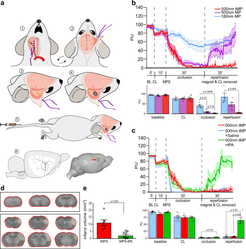
Experimental stroke models in rodents are essential for mechanistic studies and therapeutic development. However, these models have several limitations negatively impacting their translational relevance. Here we aimed to develop a minimally invasive thrombotic stroke model through magnetic particle delivery that does not require craniotomy, is amenable to reperfusion therapy, can be combined with in vivo imaging modalities, and can be performed in awake mice. We found that the model results in reproducible cortical infarcts within the middle cerebral artery (MCA) territory with cytologic and immune changes similar to that observed with more invasive distal MCA occlusion models. Importantly, the injury produced by the model was ameliorated by tissue plasminogen activator (tPA) administration. We also show that MCA occlusion in awake animals results in bigger ischemic lesions independent of day/night cycle. Magnetic particle delivery had no overt effects on physiologic parameters and systemic immune biomarkers. In conclusion, we developed a novel stroke model in mice that fulfills many requirements for modeling human stroke.


Conflict of interest statement

Competing interests: C.I. serves on the scientific advisory board of Broadview Ventures. The other authors declare no competing interests.

Figures

Fig. 1
Fig. 1. Surgical scheme and assessment of magnetic particle stroke.
a Surgical strategy for MCA stroke using magnetic particles: (1) Right common carotid artery surgically isolated and held by nylon thread; (2) Animal turned supine and scalp excised to remove right temporal muscle, exposing temporal bone; (3) Thinning of temporal bone to ~70 µm over MCA using burr drill; (4) Placement of magnet over thinned area; (5) Tail vein injection of 250 µl tMP in saline; (6) Accumulation of particles in distal branches of the MCA. b Laser Doppler flowmetry measuring relative cerebral blood flow (CBF) changes as perfusion units (PU) in the MCA territory before surgery, following carotid ligation (CL), tMP injection (MPS), 30 min occlusion, and reperfusion after the removal of both magnet and CL. Effect of particle size and composition on LDF (180 nm n = 4, 500 nm MP n = 9, 500 nm tMP n = 9; bar graphs show binned time points. Occlusion and reperfusion groups showing binned data points from final 10 min of recordings). One hundred eighty nanometer particles failed to provide sufficient decrease in CBF. Five-hundred-nanometer particles provided sufficient drop in CBF with immediate reperfusion upon magnet removal. Five-hundred-nanometer thrombin particles (tMP) provide desired sustained drop in CBF following magnet removal. One-way ANOVA with Tukey’s post-hoc test or Kruskal–Wallis test with Dunn’s post-hoc test as appropriate. Data are presented as mean values ± SEM. c LDF trace of 500 nm tMP with and without administration of bolus 10 mg/kg tPA modeling therapeutic reperfusion (500 nm tMPS alone n = 9; 500 nm tMP with tPA or saline, n = 5). Occlusion and reperfusion groups showing binned data points from final 10 min of recordings. One-way ANOVA with Tukey’s post-hoc test or Kruskal–Wallis test with Dunn’s post-hoc test as appropriate. Data are presented as mean values ± SEM. d, Cresyl Violet staining of 30 µm thick coronal brain sections collected at 300 µm intervals. Infarct outlined in yellow. Top panel is sham (magnet and carotid ligation surgery without particles). e Infarct volume at 48 h with and without 10 mg/kg tissue plasminogen activator (tPA). Unpaired two-sided t-test (n = 7/group). Data are presented as mean values ± SEM. Source data are provided as a Source Data file.
Fig. 2
Fig. 2. Longitudinal perfusion imaging shows endogenous reperfusion after tMP stroke.
a Longitudinal Laser Speckle Imaging (LSI) during surgery and 5 days following stroke. Cerebral Blood Flow (CBF) as perfusion units (PU) of ipsilateral and contralateral middle cerebral artery/affected cortex: Baseline (BL) 10 min, CCA ligation (CL) 10 min, tail vein administration of 500 nm thrombin magnetic particles (tMP) 30 min occlusion. Magnet and carotid ligation removed (Reperfusion) 10 min. Values were normalized and binned for last 5 min for all stages, with exception of final 10 min for tMP injection. tMPS shows a gradual increase of CBF to be restored to ipsilateral cortex by day 5; n = 4/group, unpaired two-sided t-test. Values are mean ± SEM. b Sham surgery and recovery (tMP but no magnet) shows CBF (PU) restored after CL is removed; n = 5/group, unpaired two-sided t-test. Values are mean ± SEM. Left of top trace shows zoomed-in representative images with right middle cerebral artery (RMCA) outlined by dashed line. R rostral C caudal, LMCA left middle cerebral artery. Source data are provided as a Source Data file.
Fig. 3
Fig. 3. Motor-sensory impairment following tMP stroke.
a Hanging wire test shows motor, grip strength, and balance impairments in MP stroke compared to sham mice at 48 h (n, sham = 8, tMPS = 9, tMPS + tPA = 7; Kruskal–Wallis with Dunn’s post-hoc test). b Corner tests show sensorimotor perception impairments in MP stroke compared to sham at 48 h (sham n = 8, tMPS n = 7, tMPS + tPA n = 7; Kruskal–Wallis with Dunn’s post-hoc test). c Time-to-contact and time-to-removal adhesive tape test between sham (receiving particles but no magnet) and stroke mice (particles and magnet). Notice significant increased time to remove adhesive at 3 days following stroke (n = 6/group; two-way ANOVA with Tukey’s post-hoc test). All values are mean ± SEM. Source data are provided as a Source Data file.
Fig. 4
Fig. 4. Longitudinal profile of immune cell infiltration after tMP stroke.
Flow cytometry shows infiltration of peripheral immune cells after tMP stroke. a Cell counts for ipsi-and contralateral cortex at 1, 2, and 7 days following tMP stroke (1 d ipsi n = 5, 1 d contra n = 5, 2 d ipsi n = 6, 2 d contra n = 6, 7 d ipsi n = 6, 7 d contra n = 5; two-way ANOVA and unpaired two-sided t-test for hemisphere effects). Data are presented as mean values ± SEM. b Gating strategy flow scheme is shown for cortices at 2 days. Plots gated on DAPI and CD45hi compare both hemispheres. Subsequent plots below show ipsilateral cortex only. EC endothelial cells, N neutrophils, T T cells, B B cells, NK natural killer cells. Source data are provided as a Source Data file.
Fig. 5
Fig. 5. Optimization for awake stroke: tMP stroke using permanent unilateral coil and magnet.
Animals underwent a short surgery (~25 min) under isoflurane (1.5%) for permanent placement of carotid coil and magnet (as outlined in methods), followed by injection of thrombin particles. a Instead of temporary ligation with nylon suture, animals received unilateral permanent placement of 0.18 mm Ø coil to the common carotid artery that is ipsilateral to cortex of intended injury and magnet placement (magnet left in place). di digastric muscle, scm sternocleidomastoid muscle, aa aortic arch, cc common carotid artery, t trachea. b 48-h infarct volume of tMPS compared to tMPS using coil both under anesthesia (tMPS n = 7, tMPS coil n = 15; unpaired two-sided t-test, p value). Forty-eight-hour corner and hanging wire behavior test between tMPS and tMPS using coil was not significant (corner test: tMPS n = 9, tMPS coil n = 13; hanging wire test: tMPS n = 11, tMPS coil n = 13; Mann–Whitney test). Data are presented as mean values ± SEM. c Animals were anesthetized (1.5% isoflurane) for surgery and LSI measurement. Animals were monitored for CBF as perfusion units (PU) following a baseline reading (BL), the unilateral coil placement on the carotid ipsilateral of permanent magnet placement (CC), following injection of particles and consequent tMP stroke (tMPS), 4 h following stroke, and up to 5 days. Sham animals followed identical surgical procedure and particle injection except for magnet placement (tMP). n = 4–5/group, unpaired two-sided t-test or Mann–Whitney test as appropriate. Data are presented as mean values ± SEM. Source data are provided as a Source Data file.
Fig. 6
Fig. 6. Effects of anesthesia and circadian cycle on tMP stroke.
a Coil and magnet placement surgeries were performed mid-morning and injected as one group (in order of surgeries) at zeitgeber times (ZT) 6-7 or at (ZT) 13-14 (anesthesia (ZT6-7) n = 10, awake (ZT6-7) n = 10, anesthesia (ZT13-14) n = 8, awake (ZT13-14) n = 10; 2-way ANOVA). b Representative cresyl violet staining shown of 48 h post awake daytime coil tMPS, (top) outlined in blue and 48 h post isoflurane anesthetized daytime coil tMPS (bottom) outlined in red. Infarct territory outlined in yellow. c Paired time-to-particle injection following coil and magnet surgery. Animals were injected in the order of surgery, completed within 1–2 h of full recovery (n = 6/group). d, Infarct volumes of mice shown as time from end of surgery to particle injection (day n = 7, night = 6; Kruskal–Wallis test shows no effect of time between surgery and stroke induction). Individual values and mean ± SEM are shown. Source data are provided as a Source Data file.

Update of

References

    1. Virani, S. S. et al. Heart disease and stroke statistics—2020 update: a report from the American Heart Association. Circulation141, e139–e596 (2020). - PubMed
    1. Campbell, B. C. V. et al. Ischaemic stroke. Nat. Rev. Dis. Prim.5, 70 (2019). - PubMed
    1. Virani, S. S. et al. Heart disease and stroke statistics—2021 update. Circulation143, e254–e743 (2021). - PubMed
    1. Bhaskar, S., Stanwell, P., Cordato, D., Attia, J. & Levi, C. Reperfusion therapy in acute ischemic stroke: dawn of a new era? BMC Neurol.18, 8 (2018). - PMC - PubMed
    1. Hussein, H. M. et al. Occurrence and predictors of futile recanalization following endovascular treatment among patients with acute ischemic stroke: a multicenter study. AJNR Am. J. Neuroradiol.31, 454–458 (2010). - PMC - PubMed

MeSH terms

Substances

LinkOut - more resources