. 2025 May 10;15(1):16359.
doi: 10.1038/s41598-025-98765-8.

Multi-parametric quantitative evaluation of murine cervical remodeling during pregnancy and postpartum

Affiliations

Multi-parametric quantitative evaluation of murine cervical remodeling during pregnancy and postpartum

Yan Yan et al. Sci Rep. .

Abstract

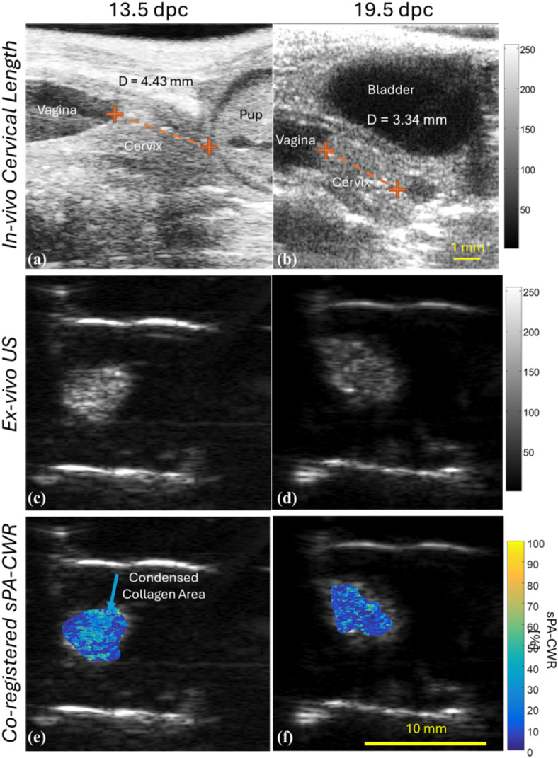
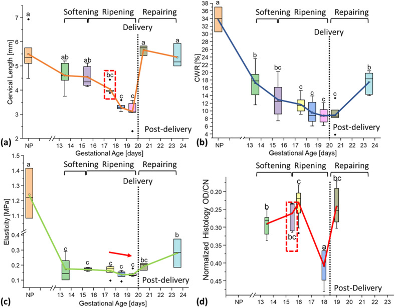
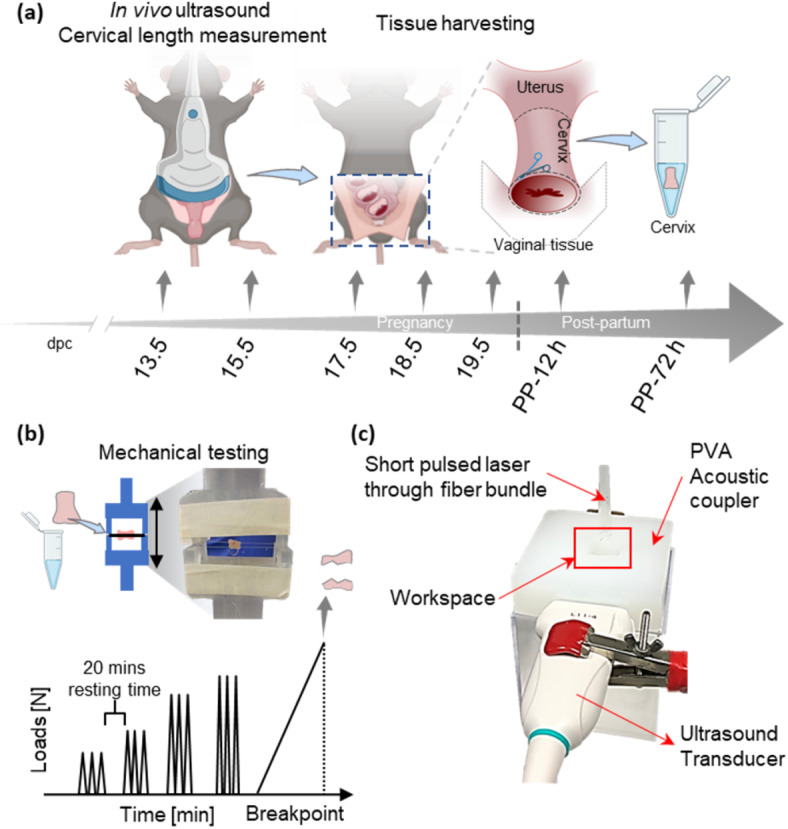
Cervical remodeling during pregnancy is a critical process that, if untimely, can lead to complications such as preterm birth (PTB). This study introduces a novel multi-parametric approach combining non-invasive imaging modalities to quantify cervical tissue changes during pregnancy and postpartum in a murine model. By integrating ultrasound-based measurements of cervical length, photoacoustic imaging of the collagen-to-water ratio, and elastography for tissue elasticity alongside histological assessments, this method provides a comprehensive evaluation of cervical remodeling. The findings reveal that combining these parameters significantly improves the accuracy of gestational age prediction compared to individual measurements, with a tri-parametric model achieving 85.3% prediction accuracy compared to 65.4% accuracy with histological analysis alone. This approach not only enhances the understanding of cervical remodeling but also holds potential as a minimally invasive, point-of-care diagnostic tool for early detection of cervical ripening and PTB risk. Ultimately, these advancements could inform clinical strategies for pregnancy management and labor induction, improving maternal and neonatal outcomes.

Keywords: Biomarkers; Cervical remodeling; Elasticity; Gestational age; Non-invasive; Photoacoustic; Ultrasound.


Conflict of interest statement

Competing interests: The authors declare no competing interests.

Figures

Fig. 1
Fig. 1
In vivo cervical length measurements (US) and ex vivo tissue composition imaging (PA). Panels (a) and (b) show representative sonographic measurements of cervical length at 13.5 and 19.5 dpc, respectively. Panels (c) and (d) present US images of excised cervical tissue at 13.5 and 19.5 dpc, respectively. Subsequent panels (e) and (f) showcase co-registered US and sPA-CWR images for tissue composition evaluations.
Fig. 2
Fig. 2
Multi-parametric quantification of murine cervical samples. (a) Cervical length changes during remodeling. The cervical ripening began at 17.5 days post coitum, marked by a notably shortened cervix and substantial alterations in cervical length over the next two days. Post-delivery, cervical lengths quickly reverted to those seen in non-pregnant samples, with minimal fluctuations over the subsequent three days. This suggests the existence of cervical changes that are not detectable through morphological imaging. (b) Spectroscopic Photoacoustic (sPA) estimation of cervical tissue composition as the Collagen-to-Water Ratio (CWR) percentage. The results indicated a trend similar to cervical length—however, the first significantly lower CWR aligned with biological expectations. Moreover, the gradual change in the collagen-to-water ratio after delivery until three days postpartum suggests that the cervical tissue undergoes a tissue-repairing process. (c) Elasticity changes in cervical samples in terms of their breaking modulus. This illustrated the same trend as other modalities but could not identify significant changes during gestation. After delivery, there was a significant increase in cervical elasticity, confirming the cervical repair process, similar to that indicated by sPA quantifications. (d) Histology quantifications. Optical density (OD; mean + standard error of the mean [SEM]; n = 3–12/group) of polarized light birefringence of Picrosirius red-stained sections as normalized to cell nuclei (CN) density/mouse in the same region. It confirmed that the collagen network in the cervical tissue had significantly gone through remodeling. It was associated with a significantly higher normalized OD at 15.5 dpc, followed by a reversed OD, indicating the cervixes were ripened. After delivery, the OD reduced back to a similar level as middle gestation, revealing the repairing process of the cervix.
Fig. 3
Fig. 3
Experimental procedure. (a) Pregnant mice underwent in vivo assessment of cervical length using ultrasound at 13.5, 15.5, 17.5, 18.5-, and 19.5-days post coitum (dpc, n = 8 each dpc), as well as within 12 h of delivery, and at 72–96 h postpartum (pp, n = 6 each pp). Non-pregnant mice were also included (n = 6). (b) Schematic of mechanical testing for the cervical elastic modulus. (c) The spectroscopic photoacoustic (sPA) imaging process for cervical tissue composition as the collagen-to-water ratio (sPA-CWR).

Similar articles

References

    1. Liu, L. et al. Global, regional, and national causes of under-5 mortality in 2000-15: an updated systematic analysis with implications for the Sustainable Development Goals. Lancet388(10063), 3027–3035. 10.1016/S0140-6736(16)31593-8 (2016). - PMC - PubMed
    1. Chawanpaiboon, S. et al. Global, regional, and National estimates of levels of preterm birth in 2014: a systematic review and modelling analysis. Lancet Glob. Health7(1), e37–e46. 10.1016/S2214-109X(18)30451-0 (2019). - PMC - PubMed
    1. Lamont, R. F. et al. Commentary on a combined approach to the problem of developing biomarkers for the prediction of spontaneous preterm labor that leads to preterm birth, (in English). Placenta98, 13–23. 10.1016/j.placenta.2020.05.007 (2020). - PubMed
    1. Elovitz, M. A. et al. Cervicovaginal microbiota and local immune response modulate the risk of spontaneous preterm delivery. Nat. Commun.10(1), 1305. 10.1038/s41467-019-09285-9 (2019). - PMC - PubMed
    1. Ngo, T. T. M. et al. Noninvasive blood tests for fetal development predict gestational age and preterm delivery. Science360(6393), 1133–1136. 10.1126/science.aar3819 (2018). - PMC - PubMed