. 2025 May 10;8(1):727.
doi: 10.1038/s42003-025-08103-x.

Molecular basis of TRPV3 channel blockade by intracellular polyamines

Affiliations

Molecular basis of TRPV3 channel blockade by intracellular polyamines

Jingying Zhang et al. Commun Biol. .

Abstract

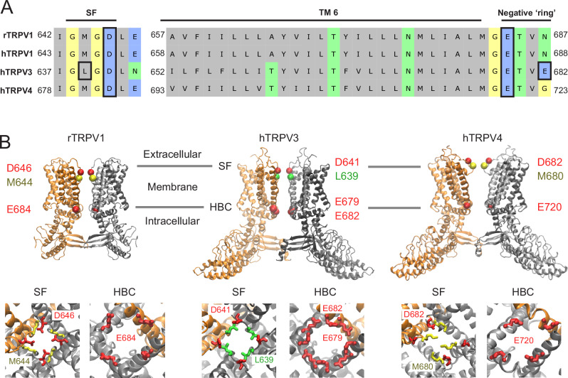
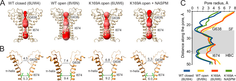
ThermoTRPV1-4 channels are involved in the regulation of multiple physiological processes, including thermo- and pain perception, thermoregulation, itch, and nociception and therefore tight control of their activity is a critical requirement for correct perception of noxious stimuli and pain. We previously reported a voltage-dependent inhibition of TRPV1-4 channels by intracellular polyamines that could be explained by high affinity spermine binding in, and passage through, the permeation path. Here, using electrophysiology and cryo-electron microscopy, we elucidate molecular details of TRPV3 blockade by endogenous spermine and its analog NASPM. We identify a high-affinity polyamine interaction site at the intracellular side of the pore, formed by residues E679 and E682, with no significant contribution of residues at the channel selectivity filter. A cryo-EM structure of TRPV3 in the presence of NASPM reveals conformational changes coupled to polyamine blockade. Paradoxically, although the TRPV3 'gating switch' is in the 'activated' configuration, the pore is closed at both gates. A modified blocking model, in which spermine interacts with the cytoplasmic entrance to the channel, from which spermine may permeate, or cause closure of the channel, provides a unifying explanation for electrophysiological and structural data and furnishes the essential background for further exploitation of this regulatory process.


Conflict of interest statement

Competing interests: The authors declare no competing interests.

Figures

Fig. 1
Fig. 1. Conductive pores of TRPV channels.
(A) Amino acid sequences in the pore and inner cavity regions of TRPV channels. The mostly hydrophobic inner cavity-lining S6 is flanked by a charged SF extracellular loop and C-terminal regions. Two unconserved residues in TRPV3 – L639 (in SF) and E682 as well as highly conserved negatively charged residues in SF and in C-terminal negatively charged ‘ring’ are highlighted with black boxes. (B) Potential polyamine interaction sites. Top row: open structures of rat TRPV1 (PDB: 7L2M), human TRPV3 (PDB: 8V6N), and human TRPV4 (PDB: 8T1D). Only two opposing monomers are shown for clarity. Two hypothesized interaction site at the SF (labeled red for acid residues and yellow for methionine in TRPV1 and TRPV4 or green for leucine in TRPV3) and the C-terminal negatively charged ‘ring’ at the HBC (labeled red for glutamic acid) are highlighted. Bottom row: Close up views of these regions are shown for each channel viewed from the cytoplasm through the membrane plane.
Fig. 2
Fig. 2. Spermine block of WT and mutant hTRPV3 channels.
A–F Representative traces of the 2-APB-induced hTRPV3 currents (WT and indicated mutants), measured using a voltage-step protocol (A, top) in absence (black traces, controls) and presence (color-coded traces) of 100 μM cytoplasmic spermine. G Averaged (n = 6 for WT, n = 7 for L639M, n = 5 for D641N, ± SE) Grel-V relationships for WT hTRPV3 (solid red line) and selectivity filter L639M (dashed violet line) and D641N (dashed dark yellow line) mutants in the presence of 100 μM cytoplasmic spermine. H Averaged (n = 6 for WT, n = 5 for all mutants, ± SE) Grel-V relationships for WT hTRPV3 (red line), single E679Q mutant (blue line), single E682Q mutant (green line), double E679Q/E682Q mutant (orange line), and triple E679Q/E682Q/D641N mutant (dashed dark yellow line) in presence of 100 μM cytoplasmic spermine. Datasets were fitted with the sum of two Boltzmann distributions except E679Q/E682Q/D641N mutant, which was fitted with a linear regression. WT TRPV3 data is taken from ref. .
Fig. 3
Fig. 3. Blocking of hTRPV3-K169A by polyamines.
A Averaged (n = 6 for WT, n = 7 for K169A, n = 5 for E679Q/E682Q, n = 5 for K169A/E679Q/E682Q, ± SE) Grel-V relationships for WT TRPV3 (solid red line), TRPV3-K169A (dashed red line), TRPV3-E679Q/E682Q (solid orange line), and TRPV3-K169A/E679Q/E682Q (dashed orange line) in presence of 100 μM intracellular spermine. B Averaged (n = 5 for all, ± SE) Grel-V relationships for WT TRPV3 (solid pink line), normalized WT TRPV3 (dashed pink line), and TRPV3-K169A (solid yellow line), in the presence of 100 μM intracellular NASPM, and TRPV3-K169A in the presence of 10 μM (teal line) and 1 μM (brown line) intracellular NASPM. Similarity of K169A and normalized WT Grel-V relationships suggests voltage-independent inactivation mode of WT TRPV3 in the presence of NASPM, which is absent for K169A mutant. Datasets were fitted with Boltzmann distributions (Function 1). Unscaled WT TRPV3 data is taken from ref. . C Averaged (n = 5 for all, ± SE) Grel-V relationships for TRPV3-K169A (solid red line), TRPV3-K169A-E679Q (solid blue line), TRPV3-K169A-E682Q, and TRPV3-K169A (solid orange line), in the presence of 100 μM intracellular NASPM, as well as TRPV3-K169A in presence of 10 μM intracellular NASPM (teal line). Datasets were fitted with Boltzmann distributions (Function 1), fitting results are presented in Table S2. (D) Molecular structures of spermine and NASPM, respectively.
Fig. 4
Fig. 4. Cryo-EM structure of TRPV3-K169A in nanodiscs with NASPM.
A Cryo-EM density map. Subunits are uniquely colored and lipid-like densities are colored in yellow. B Side view of the modeled TRPV3-K169A structure. Electron density of the distal C-terminal domain and finger 5 of ARD are labeled in red. C Bottom view of (B). C-terminal ‘gating switch’ (arrow) adopts clearly an α-helical conformation, as in open conformation of the channel. Superposition of the overall structure (D) and the pore region (E) of TRPV3-K169A + NASPM (green) with TRPV3-K169A open (PDB: 6UW6, red) and WT TRPV3 closed (PDB: 6UW4, blue) structures. RMSD values for TRPV4-K169A + NASPM vs open (6UW6) and closed (6UW4) structures are presented in (D). Constrictions at SF (G638) and HBC (I674), as well as the π-helical bulge in S6 helix are labeled in (E). Only two opposing subunits are shown for clarity in (D, E).
Fig. 5
Fig. 5. Structure of the conductive pore of TRPV3-K169A in the presence of NASPM.
A The pores of human TRPV3 in the absence of agonists (PDB: 6UW4) and in the presence of 2-APB (PDB: 8V6N), TRPV3-K169A in the absence of agonists (PDB: 6UW6), and TRPV3-K169A in the presence of 100 µM of NASPM (this work), calculated by the MOLEonline server, shown as a colored surface representing electric potential (ChimeraX). Only two opposing subunits are shown for clarity. B Distances between constriction-forming diagonal sidechains at SF residue G638 and HBC residue I674 (Å). Only two opposing subunits are shown for clarity. C Water-accessible dimensions of K169A conduction pore in the presence of NASPM (green), compared to the WT closed (6UW4, blue), WT open (8V6N, yellow), and K169A open (6UW6, red), structures of TRPV3. Profiles are calculated with the program HOLE. Gray area represents an approximate location of the channel inner cavity. Major SF and HBC constrictions at G638 and I674 respectively, are labeled.
Fig. 6
Fig. 6. Proposed molecular mechanism of PA block of TRPV channels.
A Modified kinetic model of TRPV3 inhibition by intracellular PAs. The model assumes that spermine interaction blocks the open channel. TRPV3-bound PA may either return to the cytoplasm, or permeate the channel and exit to the extracellular side, or force the channel to close. All transitions are reversible, however extracellular PA concentration is assumed to be zero. Relative conductance (Grel, Function 2) is a function of intracellular PA and eight parameters (constants ka1, ka-1, ka2, KC, and apparent charges associated with them), where KC = kb1 / kb-1 and represents an equilibrium binding constant of PA bound to the closed TRPV channel. B Schematic representation of the hypothesized molecular mechanism of TRPV channel block by spermine. Key acidic residues (D641 above the SF and E679/E682 below the HBC) are labeled in red, spermine is presented as blue rods, and potassium ions are presented as blue beads. Open TRPV3 in the absence of spermine with both SF and HBC wide open, is permeable to potassium ions. Intracellular spermine (or NASPM) interacts with E679/E682 and blocks permeation. Spermine unblock can occur by return to the cytoplasm or by permeation to the outside. While interacting, spermine promotes channel closure. Model prediction of experimental data for WT and mutant TRPV3 in the presence of 100 µM intracellular spermine (C, n = 6 for WT, n = 5 for all mutants, ± SE) or NASPM (D, n = 5 for all, ± SE), data points from Figs. 2 and 3. Parameters were constrained as noted in Table 3. Data points labeled as empty circles (K169A/E679Q/E682Q) were excluded from analysis.

Similar articles

  • Blockade of TRPV channels by intracellular spermine.
    Maksaev G, Yuan P, Nichols CG. Maksaev G, et al. J Gen Physiol. 2023 May 1;155(5):e202213273. doi: 10.1085/jgp.202213273. Epub 2023 Mar 13. J Gen Physiol. 2023. PMID: 36912700 Free PMC article.
  • Gating of human TRPV3 in a lipid bilayer.
    Deng Z, Maksaev G, Rau M, Xie Z, Hu H, Fitzpatrick JAJ, Yuan P. Deng Z, et al. Nat Struct Mol Biol. 2020 Jul;27(7):635-644. doi: 10.1038/s41594-020-0428-2. Epub 2020 Jun 22. Nat Struct Mol Biol. 2020. PMID: 32572252 Free PMC article.
  • Conformational dynamics in TRPV1 channels reported by an encoded coumarin amino acid.
    Steinberg X, Kasimova MA, Cabezas-Bratesco D, Galpin JD, Ladron-de-Guevara E, Villa F, Carnevale V, Islas L, Ahern CA, Brauchi SE. Steinberg X, et al. Elife. 2017 Dec 5;6:e28626. doi: 10.7554/eLife.28626. Elife. 2017. PMID: 29206105 Free PMC article.
  • Interactions of polyamines with ion channels.
    Williams K. Williams K. Biochem J. 1997 Jul 15;325 ( Pt 2)(Pt 2):289-97. doi: 10.1042/bj3250289. Biochem J. 1997. PMID: 9230104 Free PMC article. Review.
  • TRPV3.
    Yang P, Zhu MX. Yang P, et al. Handb Exp Pharmacol. 2014;222:273-91. doi: 10.1007/978-3-642-54215-2_11. Handb Exp Pharmacol. 2014. PMID: 24756710 Review.

References

    1. Igarashi, K. & Kashiwagi, K. The functional role of polyamines in eukaryotic cells. Int J. Biochem. Cell Biol.107, 104–115 (2019). - PubMed
    1. Bekebrede, A. F. et al. The molecular and physiological effects of protein-derived polyamines in the intestine. Nutrients12. (2020). - PMC - PubMed
    1. Dever, T. E. & Ivanov, I. P. Roles of polyamines in translation. J. Biol. Chem.293, 18719–18729 (2018). - PMC - PubMed
    1. Malpica-Nieves, C. J. et al. The involvement of polyamine uptake and synthesis pathways in the proliferation of neonatal astrocytes. Amino Acids52, 1169–1180 (2020). - PMC - PubMed
    1. Nakamura, A. et al. Symbiotic polyamine metabolism regulates epithelial proliferation and macrophage differentiation in the colon. Nat. Commun.12, 2105 (2021). - PMC - PubMed

LinkOut - more resources