Review
. 2025 May 10;25(1):175.
doi: 10.1186/s12935-025-03793-z.

Bone marrow microenvironment in myelodysplastic neoplasms: insights into pathogenesis, biomarkers, and therapeutic targets

Affiliations
Review

Bone marrow microenvironment in myelodysplastic neoplasms: insights into pathogenesis, biomarkers, and therapeutic targets

Forouzan Bahmani et al. Cancer Cell Int. .

Abstract

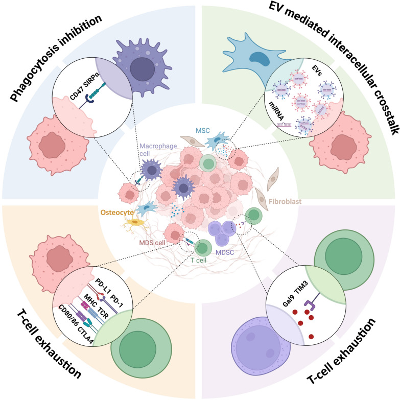
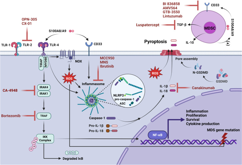
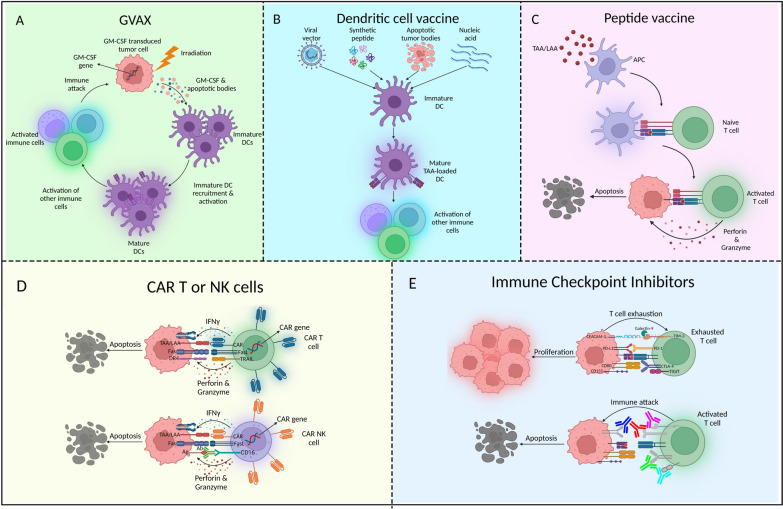
Myelodysplastic neoplasms (MDS) represent a heterogeneous group of malignant hematopoietic stem and progenitor cell (HSPC) disorders characterized by cytopenia, ineffective hematopoiesis, as well as the potential to progress to acute myeloid leukemia (AML). The pathogenesis of MDS is influenced by intrinsic factors, such as genetic insults, and extrinsic factors, including altered bone marrow microenvironment (BMM) composition and architecture. BMM is reprogrammed in MDS, initially to prevent the development of the disease but eventually to provide a survival advantage to dysplastic cells. Recently, inflammation or age-related inflammation in the bone marrow has been identified as a key pathogenic mechanism for MDS. Inflammatory signals trigger stress hematopoiesis, causing HSPCs to emerge from quiescence and resulting in MDS development. A better understanding of the role of the BMM in the pathogenesis of MDS has opened up new avenues for improving diagnosis, prognosis, and treatment of the disease. This article provides a comprehensive review of the current knowledge regarding the significance of the BMM to MDS pathophysiology and highlights recent advances in developing innovative therapies.

Keywords: Bone marrow microenvironment; Hematopoietic stem and progenitor cells; Ineffective hematopoiesis; Inflammation; Innovative therapies; Myelodysplastic neoplasms.


Conflict of interest statement

Declarations. Ethics approval and consent to participate: Not applicable. Consent for publication: Not applicable. Competing interests: The authors declare no competing interests.

Figures

Fig. 1
Fig. 1
Overview of the dynamic interactions among cellular components in the MDS microenvironment. T cell exhaustion results from: Excessive expression of PD-1 and CTLA-4 on T cells, which have the potential to interact with PD-L1 and CD80/86 on MDS cells, as well as elevated Gal9 levels secreted by MDSCs, which bind to TIM3 on T cells. MDS cells inhibit phagocytosis by upregulating the “don’t eat me” ligand CD47, which interacts with its receptor on the surface of macrophages, SIRPα. MDS and MSCs produce EVs containing miRNAs, which play important roles in accelerating the progression of MDS. MSC: mesenchymal stromal cell; MDSC: myeloid-derived suppressor cells; EVs: extracellular vesicles; miRNAs: microRNAs; PD-1: programmed death 1; PD-L1: programmed death-ligand 1; CTLA-4: cytotoxic T-lymphocyte associated protein 4; TIM-3: T-cell immunoglobulin and mucin domain 3; GAL9: galectin 9; SIRPα: signal-regulatory protein alpha
Fig. 2
Fig. 2
Inflammatory signaling pathways involved in MDS and associated therapeutic targets. S100A8/A9 bind to TLR4 and CD33 and initiate the assembly of the NLRP3 inflammasome. The binding of S100A8/A9 to TLR4 also activates NF-kB through IRAK1/TRAF6/NF-κB signaling pathway, which results in the production of proinflammatory cytokines (pro-IL-1β and pro-IL-18). S100A8/A9 promote NOX activation, leading to excessive production of ROS and the subsequent activation of NLPR3 and inflammasome assembly. Inflammasomes recruit ASCs to form complexes that facilitate the conversion of pro-caspase-1 to caspase-1. Mature and activated caspase-1 cleaves pro-IL-1β and pro-IL-18 into their bioactive forms, which induce pyroptosis. TLR: toll-like receptor; TIRAP: toll-interleukin-1 receptor domain-containing adaptor protein; IRAK: interleukin receptor-associated kinase; TRAF: tumor necrosis factor receptor-associated factor; IκK: inhibitor of κB kinase; NF-κB: nuclear factor kappa B; NLRP3: nucleotide-binding domain and leucine-rich repeat pattern recognition receptor; ASC: apoptosis-associated speck-like protein containing a caspase-recruitment domain; GSDMD: gasdermin D; NOX: nicotinamide-adenine dinucleotide phosphate oxidase; ROS: reactive oxygen species; Ub: ubiquitin; TGF-β: transforming factor-β; IL: interleukin
Fig. 3
Fig. 3
Novel immunotherapy approaches in MDS. A Genetically modified tumor cells are used in GVAX vaccine to release GM-CSF, which enhances the immune response against tumor cells. GVAX promotes DC recruitment and activation to increase the ability of the immune system to recognize and destroy tumor cells. Apoptotic bodies produced by irradiated tumor cells are taken up by DCs, contributing to DC maturation. B Dendritic cell vaccines are generated by loading patient-derived DCs with TAA/LAA to trigger a targeted immune response against tumor cells. These vaccines can be prepared by expressing TAA/LAA on MHC-I/II in DCs loaded with peptides, nucleic acids, viral vectors, or apoptotic tumor bodies. C Peptide vaccines are based on the identification of epitopes that cause antitumor immune responses specific to TAA/LAA. D CAR-T or -NK cells are genetically engineered cells designed to express CARs targeting TAA/LAA on tumor cells. CAR immune cells induce apoptosis in tumor cells by releasing cytotoxic molecules such as IFNγ, perforin, and granzymes and activating death receptor pathways (Fas-FasL and TRAIL-DR4). E ICIs inhibit ICPs such as PD-L1, TIGIT, TIM-3, and CTLA-4, which tumor cells use to exhaust T cells and avoid detection. By inhibiting these immune checkpoints, T cells can recognize and attack tumor cells. GM-CSF, granulocyte‒macrophage colony‒stimulating factor; GVAX: GM-CSF-transduced tumor cell vaccines; DC: dendritic cell; TAA: tumor-associated antigen; LAA: leukemia-associated antigen; MHC: major histocompatibility complex; CAR: chimeric antigen receptor; FasL: Fas ligand; TRAIL: Tumor necrosis factor (TNF)-related apoptosis-inducing ligand; DR: death receptor; ICIs: Immune checkpoint inhibitors; ICPs: immune checkpoint proteins; PD-1: programmed death-1; PD-L1: programmed death-ligand 1; TIM-3: T cell Ig- and mucin-domain-containing molecule-3; TIGIT: T cell immunoreceptor with immunoglobulin and ITIM domain; CTLA-4: Cytotoxic T-lymphocyte-associated protein 4

Similar articles

References

    1. Tefferi A, Vardiman JW. Myelodysplastic syndromes. N Engl J Med. 2009;361(19):1872–85. - PubMed
    1. Sekeres MA. The epidemiology of myelodysplastic syndromes. Hematol Oncol Clin. 2010;24(2):287–94. - PubMed
    1. Khoury JD, Solary E, Abla O, Akkari Y, Alaggio R, Apperley JF, et al. The 5th edition of the World Health Organization classification of haematolymphoid tumours: myeloid and histiocytic/dendritic neoplasms. Leukemia. 2022;36(7):1703–19. - PMC - PubMed
    1. Lin L, Du L, Cao K, Huang Y, Yu P, Zhang L, et al. Tumour cell-derived exosomes endow mesenchymal stromal cells with tumour-promotion capabilities. Oncogene. 2016;35(46):6038–42. - PMC - PubMed
    1. Soto CA, Lo Celso C, Purton LE, Frisch BJ. From the niche to malignant hematopoiesis and back: reciprocal interactions between leukemia and the bone marrow microenvironment. J Bone Miner Res Plus. 2021;5(10): e10516. - PMC - PubMed

LinkOut - more resources