. 2025 Jun;12(23):e2417605.
doi: 10.1002/advs.202417605. Epub 2025 May 11.

SCORT-Cas13d Nanotherapy Precisely Targets the 'Undruggable' Transcription Factor HoxB13 in Metastatic Prostate Cancer In Vivo

Affiliations

SCORT-Cas13d Nanotherapy Precisely Targets the 'Undruggable' Transcription Factor HoxB13 in Metastatic Prostate Cancer In Vivo

Zhifen Cui et al. Adv Sci (Weinh). 2025 Jun.

Abstract

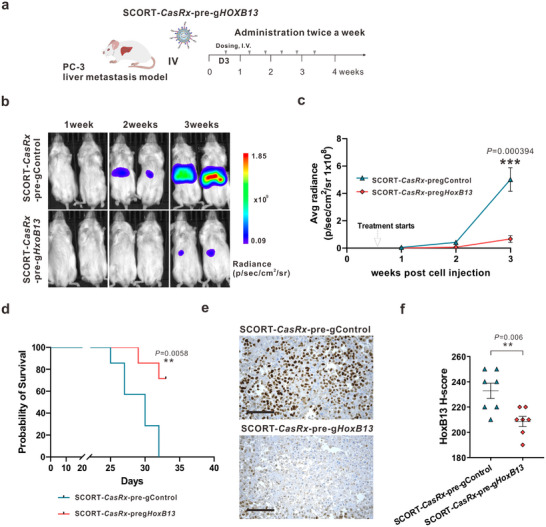
Metastatic cancer, the primary cause of cancer mortality, frequently exhibits heightened dependence on certain transcription factors (TFs), which serve as master regulators of oncogenic signaling yet are often untargetable by small molecules. Selective Cell in ORgan Targeting (SCORT) nanoparticles are developed for precise CRISPR/Cas13d mRNA and gRNA delivery to metastatic cancer cells in vivo, aiming to knock down the undruggable oncogenic TF HoxB13. In prostate cancer liver metastasis models driven by HoxB13, repeated systemic SCORT-Cas13d-gHoxB13 treatment significantly decreases HoxB13 expression, reduces metastasis, and extends mouse survival. Prolonged treatment shows no significant impact on major organ function, histology or immune markers. Mechanistically, SCORT-Cas13d-gHoxB13 treatment suppresses metastatic tumor proliferation and angiogenesis while promoting apoptosis by regulating multiple gene pathways. Unexpectedly, it inhibits the non-canonical, EMT-independent oncogenic function of Snail. These findings suggest that SCORT-Cas13d-gHoxB13 can effectively and safely target the undruggable HoxB13 in metastatic prostate cancer, positioning CRISPR/Cas13d as a potential treatment.

Keywords: HoxB13; SCORT nanoparticles; SCORT‐Cas13d nanotherapy; mechanistic insights into therapeutic action; metastatic prostate cancer; undruggable oncogenic transcription factors.


Conflict of interest statement

Q.W., Z.C., and Y.D. are inventors on a patent filed by Duke University that relates to the research reported in this paper. J.H. is a consultant for or owns shares in the following companies: Kingmed, MoreHealth, OptraScan, Genetron, Omnitura, Vetonco, York Biotechnology, Genecode, VIVA Biotech, and Sisu Pharma, and received grants from Zenith Epigenetics, BioXcel Therapeutics, Inc., and Fortis Therapeutics.

Figures

Figure 1
Figure 1
CasRx‐mediated specific HoxB13 knockdown inhibits proliferation and invasion of both AR+ and AR‐ CRPC cells. a) Representative HoxB13 immunoreactivity in normal human prostate, ADPC, and mCRPC tissues. Scale bar: 200 µm (upper panels);100 µm (lower panels). b) H‐score of HoxB13 nuclear staining in human tissues. The bar shows the median of each group, while each dot represents an individual sample. P‐values were calculated by One‐way ANOVA. **** p < 0.0001. c) HoxB13 mRNA level in 293FT cells after transfection with CasRx plasmid and different pre‐gRNAs designed to target HoxB13. HoxB13 transcript levels are normalized by 18s‐rRNA. Data are presented as mean ± SEM with n = 6 biologically independent replicates. d) HoxB13 mRNA level in 293FT cells after transfection with CasRx or luciferase mRNA and pre‐gControl or pre‐gHoxB13. Data are presented as mean ± SEM with n = 6 biologically independent replicates. P‐values were calculated by a two‐tailed Student's t‐test. **** p < 0.0001. e) Volcano plot of gene expression changes between CasRx‐pre‐gHoxB13 and CasRx‐pre‐gControl transfected 293FT cells for 24 h. RNA sequencing was performed in biological triplicates. Significantly differentially expressed genes (Fold change>2, q‐value<0.01) are shown in red. f‐h) LNCaP95 (upper), and PC‐3 cells (lower) were incubated with mock, empty LNPs (eLNPs), LNPs encapsulating CasRx mRNA and pre‐gControl or pre‐gHoxB13, respectively, and subjected for analysis. HoxB13 transcript levels. Data are presented as mean ± SEM with n = 5 biologically independent replicates (f). Cell proliferation. Data are presented as mean ± SD with n = 5 biologically independent replicates (g). Cell invasion. The number of invaded cells was quantified (left), and representative images are shown on the right (100x magnification). Data are presented as mean ± SEM of three representative fields from one of three biologically independent experiments (h). P‐values were calculated by One‐way ANOVA. ** p < 0.01, **** p < 0.0001.
Figure 2
Figure 2
Construction of SCORT LNPs for specific targeting of CRPC cells in vitro and in vivo. a) Schematic of SCORT LNPs generation through microfluidic mixing and E3 aptamer conjugation. b) Composition of LNP candidates. c) Bioluminescence signal in LNCaP95, PC‐3, and AML12 cells after 24 h incubation with luciferase mRNA‐encapsulated LNP candidates modified with no aptamer (No Apt), a control aptamer (+Apt‐Control), or E3 aptamer (+Apt‐E3). The experiment was conducted with n = 5 biologically independent replicates. Data are presented as mean ± SEM. P‐value was calculated by One‐way ANOVA. **** p < 0.0001, ns, not significant. D) Schematic of the establishment of LNCaP95 liver metastasis model and distribution of mCherry mRNA‐encapsulated LNPs in major organs. e) Representative images of cell classification mapping (left panel) and mCherry‐positive cells (right panel) by IMC analysis. f) Percentage of mCherry‐positive cells in different cell types. Each red point represents the distribution in an individual mouse (n = 4, one batch of SCORT‐mCherry per mouse). Data are presented as mean ± SEM. P value was calculated by One‐way ANOVA. **** p < 0.0001.
Figure 3
Figure 3
SCORT‐CasRx‐pre‐gHoxB13 treatment suppresses liver metastasis and improves survival in AR+ CRPC liver metastasis mouse model. a) Schematic illustration of the experimental design. The CRPC liver metastasis mouse model was established by hemi‐spleen injection of LNCaP95 cells stably expressing luciferase (LNCaP95‐Luc). One week after cell engraftment, mice received I.V. administration of DPBS, SCORT LNPs, SCORT‐CasRx‐pre‐gControl, and SCORT‐CasRx‐pre‐gHoxB13, receptively, twice a week for 6.5 weeks (n = 7). b) Representative bioluminescence imaging of the whole animal on week 3, 5, 7 after cancer cell engraftment. c) Cumulative luciferase counts during the time course. Data are presented as mean ± SEM. P‐values were calculated by One‐way ANOVA, ** p < 0.01. d) Kaplan‐Meier survival curves. P values were determined by the log‐rank (Mantel‐Cox) test. ** p < 0.01. e–g) Three days after the last treatments, liver metastatic tumors were subjected to analysis. HoxB13 transcript levels. Data are presented as mean ± SEM. P values were calculated by One‐way ANOVA, **** p < 0.0001 (e). Representative images of HoxB13 protein immunostaining and the corresponding H&E staining of the same area. Scale bar: 60 µm. (f). The percentage (%) of tumor cells with HoxB3 knockdown in the SCORT‐CasRx‐pre‐gHoxB13 group. Data is presented as mean ± SEM.(g).
Figure 4
Figure 4
SCORT‐CasRx‐pre‐gHoxB13 treatment inhibits liver metastasis and improves survival in AR‐ liver metastasis mouse model. a) Schematic illustration of the experimental design. The PC‐3 liver metastasis mouse model was established by hemi‐spleen injection of PC‐3 Red‐FLuc cells. Three days after cancer cell engraftment, mice received I.V. administration of SCORT‐CasRx‐pre‐gControl, or SCORT‐CasRx‐pre‐gHoxB13, respectively, twice a week for 3 weeks (n = 7). b) Representative bioluminescence imaging of the whole animal on 1, 2, and 3 weeks following cancer cell injection. c) Cumulative luciferase counts during the time course. Data are presented as mean ± SEM. P values were calculated by a two‐tailed Student's t‐test, *** p < 0.001. d) Kaplan‐ Meier survival curves. P values were determined by the log‐rank (Mantel‐Cox) test. ** p < 0.01. e) Representative images of HoxB13 protein immunostaining. Three days after the final treatment, the liver with metastatic PC‐3 tumors was subjected to IHC analysis. Scale bar: 50 µm. f) H‐score for HoxB13 protein. Data are presented as mean ± SEM. P values were calculated by two‐tailed Student's t‐test, ** p < 0.01.
Figure 5
Figure 5
Repeated treatment with SCORT‐CasRx‐pre‐gHoxB13 is well tolerated in immunocompetent mice. CD‐1 mice were given I.V. administrations of DPBS, SCORT LNPs, SCORT‐CasRx‐pre‐gControl, and SCORT‐CasRx‐pre‐gHoxB13, respectively, twice a week for 6.5 weeks (n = 8). a) Change in body weight. Body weight at the start of treatment was set at 100%. b) Hepatic and renal functions were not impaired by SCORT‐CasRx‐pre‐gHoxB13 treatment. ALT, alanine transaminase; AST, aspartate aminotransferase; BUN, blood urea nitrogen; CREAT, creatinine. Data are presented as box and whisker plots, where the box represents the median (center line) with bounds of the 25th to 75th percentiles. The whiskers extend to 1.5 times the interquartile range. P values were calculated by One‐way ANOVA. ns, not significant. c) No substantial histopathological changes were observed in the indicated organ tissues following SCORT‐CasRx‐pre‐gHoxB13 treatment. Scale bar: 100 µm.
Figure 6
Figure 6
Direct cellular responses and transcriptional changes following SCORT‐CasRx‐pre‐gHoxB13 treatment in AR+ CRPC liver metastasis mouse model. a) Schematic illustration of the experimental design. Five weeks after LNCaP95 cell engraftment, mice received two doses of either SCORT‐CasRx‐pre‐gControl or SCORT‐CasRx‐pre‐gHoxB13 with a 3‐day interval. Tumors were isolated three days after the second dose and subjected to analysis (n = 6). b) HoxB13 transcript level. Data are presented as mean ± SEM. P values were calculated by One‐way ANOVA, *** p<0.001. c) Representative immunostaining images. Scale bar 50 µm. d) Volcano plot of gene expression changes between SCORT‐CasRx‐pre‐gControl and SCORT‐CasRx‐pre‐gHoxB13 treated groups. A total of 1,660 upregulated genes and 1,879 downregulated genes were identified and are highlighted in red. e) Validation of HoxB13, SNAI1 (encoding Snail) and CDH1 (encoding E‐cadherin) gene expression changes in metastatic tumor samples by qRT‐PCR. f) Identified top KEGG pathways by Cistrome GO from DEGs. Pathways associated with positive scores are enriched from the upregulated genes and vice versa. The size of the point represents the number of genes, whereas the color represents the ‐log10 (FDR) value.
Figure 7
Figure 7
Schematic overview of the SCORT–Cas13d–a pre‐gHoxB13 approach for targeting metastatic prostate cancer in the liver. Intravenously administered SCORT nanoparticles carry Cas13d mRNA and pre‐guide RNAs (pre‐gHoxB13), which preferentially accumulate in HoxB13‐dependent prostate cancer metastases rather than in normal liver cells. Within metastatic tumor cells, the Cas13d mRNA is translated, and pre‐gHoxB13 is processed to generate mature gHoxB13 guides. The resulting Cas13d–gHoxB13 complexes knock down HoxB13 transcripts, leading to reduced HoxB13 protein expression. Notably, the systemic treatment showed no significant adverse effects on major organs (e.g., heart, liver, spleen, and lung) and did not raise immune markers, underscoring the approach's safety. Immunohistochemistry (IHC) confirmed efficient HoxB13 knockdown in vivo, and survival analyses demonstrated improved outcomes following repeated SCORT–Cas13d–pre‐gHoxB13 administration. Transcriptomic profiling further revealed that SCORT–Cas13d–pre‐gHoxB13 modulates multiple gene pathways (Figure 6), reducing tumor proliferation, angiogenesis, and metabolic activity while promoting apoptosis. Overall, these findings suggest that SCORT–Cas13d–pre‐gHoxB13 is a precise, safe, and effective method for silencing the “undruggable” transcription factor HoxB13 in metastatic prostate cancer.

References

    1. a) Chaffer C. L., Weinberg R. A., Science 2011, 331, 1559; - PubMed
    2. b) Steeg P. S., Nat. Rev. Cancer 2016, 16, 201. - PMC - PubMed
    1. Sung H., Ferlay J., Siegel R. L., Laversanne M., Soerjomataram I., Jemal A., Bray F., CA Cancer J Clin 2021, 71, 209. - PubMed
    1. Ganesh K., Massague J., Nat. Med. 2021, 27, 34. - PMC - PubMed
    1. Darnell J. E. Jr., Nat. Rev. Cancer 2002, 2, 740. - PubMed
    1. Bradner J. E., Hnisz D., Young R. A., Cell 2017, 168, 629. - PMC - PubMed

LinkOut - more resources