

C-shaped root canal systems in the bilateral mandibular first molars: a case report and literature review
- PMID: 40350452
- PMCID: PMC12067870
- DOI: 10.1186/s12903-025-06060-9
C-shaped root canal systems in the bilateral mandibular first molars: a case report and literature review
Abstract
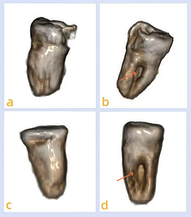
Background: Mandibular first molars typically exhibit complex root and canal anatomy. A thorough understanding of their morphological variations is crucial for endodontists to achieve successful root canal treatment. A C-shaped root canal system is a unique anatomical variation characterized by a C-shaped or semi-circular cross-sectional morphology, observed predominantly in mandibular second molars. The prevalence rate of the C-shaped root canal system is approximately 2.7-48.7% in the mandibular second molar, while the condition is rarely seen in mandibular first molars, particularly in bilateral cases.
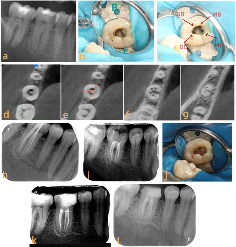
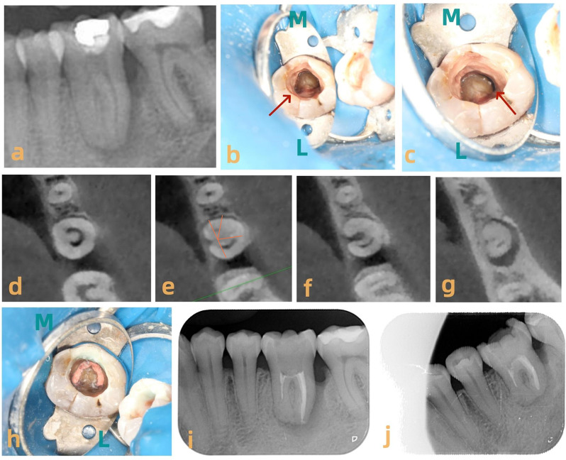
Case presentation: This report details an uncommon case of a C-shaped root canal system in bilateral mandibular first molars: the right molar exhibited a fused root with two separate mesial and two distal canals, while the left molar displayed a single oval mesial canal and a semicolon-shaped distal canal. To our knowledge, this is the first report of the presence of four separate canals within a fused C-shaped root in the mandibular first molar.
Conclusions: While C-shaped root variations are detected in mandibular first molars, a thorough knowledge of normal root canal anatomy and associated variations presents a significant challenge for clinicians in terms of successful endodontic treatment.
Keywords: C-shaped root canal system; Mandibular first molar; Root canal treatment, CBCT.
© 2025. The Author(s).
Conflict of interest statement
Declarations. Ethics approval and consent to participate: This study did not involve human or animal subjects’ experiment, and thus ethical review and approval were waived. Informed consent was obtained from the patient for the treatment and publication of this case, with permission to use clinical and radiographic images. Consent for publication: Written informed consent for publication from patient has been taken. Competing interests: The authors declare no competing interests.
Figures

References
-
- Vertucci FJ. Root Canal anatomy of the human permanent teeth. Oral Surg Oral Med Oral Path. 1984;58:589–99. - PubMed
-
- Kirici DO, Koc S. Middle distal Canal of mandibular first molar: A case report and literature review. Niger J Clin Pract. 2019;22(2):285–8. - PubMed
-
- Tredoux S, Warren N, Buchanan GD. Root and Canal configurations of mandibular first molars in a South African subpopulation. J Oral Sci. 2021;63(3):252–6. - PubMed
-
- Marceliano-Alves MF, Lima CO, Bastos LGDPMN, Bruno AMV, Vidaurre F, Coutinho TM, Fidel SR, Lopes RT. Mandibular mesial root Canal morphology using micro-computed tomography in a Brazilian population. Aust Endod J. 2019;45(1):51–6. - PubMed
Publication types
MeSH terms
Grants and funding
- NO.202208010746/the Science and Technology Development Project of Shandong Province Health and Medicine
- NO.202208010746/the Science and Technology Development Project of Shandong Province Health and Medicine
- NO.202208010746/the Science and Technology Development Project of Shandong Province Health and Medicine
LinkOut - more resources
Full Text Sources
