. 2025 May 11;16(1):4369.
doi: 10.1038/s41467-025-59623-3.

Basal-shift transformation leads to EGFR therapy-resistance in human lung adenocarcinoma

Affiliations

Basal-shift transformation leads to EGFR therapy-resistance in human lung adenocarcinoma

Taro Shinozaki et al. Nat Commun. .

Abstract

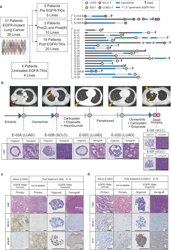
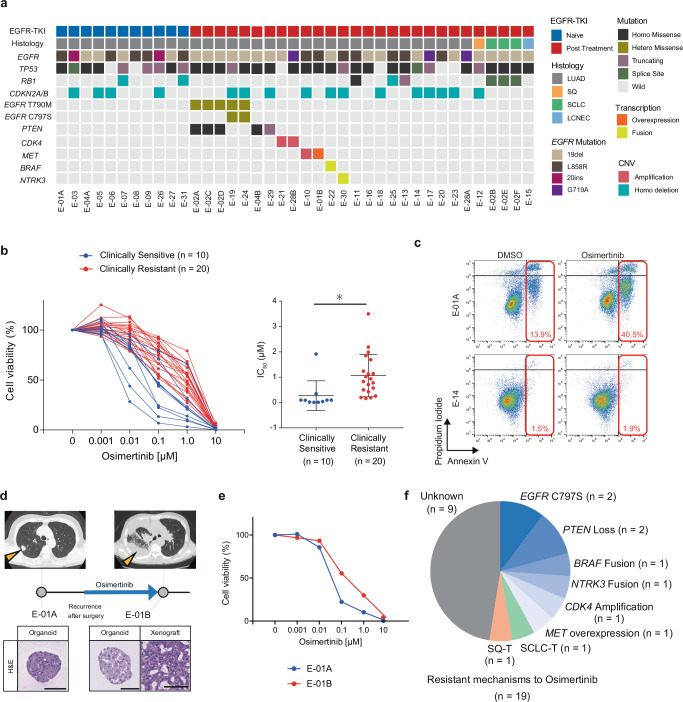
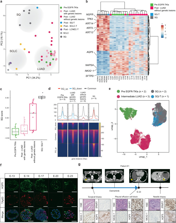
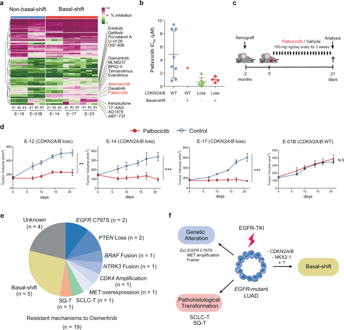
Although EGFR tyrosine kinase inhibitors (EGFR-TKIs) are effective for EGFR-mutant lung adenocarcinoma (LUAD), resistance inevitably develops through diverse mechanisms, including secondary genetic mutations, amplifications and as-yet undefined processes. To comprehensively unravel the mechanisms of EGFR-TKI resistance, we establish a biobank of patient-derived EGFR-mutant lung cancer organoids, encompassing cases previously treated with EGFR-TKIs. Through comprehensive molecular profiling including single-cell analysis, here we identify a subgroup of EGFR-TKI-resistant LUAD organoids that lacks known resistance-related genetic lesions and instead exhibits a basal-shift phenotype characterized by the hybrid expression of LUAD- and squamous cell carcinoma-related genes. Prospective gene engineering demonstrates that NKX2-1 knockout induces the basal-shift transformation along with EGFR-target therapy resistance. Basal-shift LUADs frequently harbor CDKN2A/B loss and are sensitive to CDK4/6 inhibitors. Our EGFR-mutant lung cancer organoid library not only offers a valuable resource for lung cancer research but also provides insights into molecular underpinnings of EGFR-TKI resistance, facilitating the development of therapeutic strategies.


Conflict of interest statement

Competing interests: T.S. is an inventor on several patents related to organoid culture. We declare that none of the authors have competing financial or non-financial interests as defined by Nature Portfolio.

Figures

Fig. 1
Fig. 1. Establishment of an EGFR-mutant Lung Cancer Organoid Library (ELCOL).
a Summary of ELCOL. Treatment course for patients with clinical resistance to EGFR-TKI is shown on the right. Circle indicates the timing of tissue sampling and organoid derivation and is colored according to the histology. Thick line shows the treatment period. LUAD; lung adenocarcinoma, SQ-T; squamous transformation, SCLC-T; small cell lung cancer transformation, LCNEC-T; large cell neuroendocrine carcinoma transformation. 1st/2nd generation EGFR-TKIs include Gefitinib, Erlotinib, Afatinib and Dacomitinib. b Example of longitudinal organoid establishment from a patient with LUAD and SCLC-T (E-02). The computed tomography image at each sampling timepoint is shown on the top, and yellow arrowheads indicate lung cancer lesions. Representative images of hematoxylin and eosin (H&E) staining of the organoids and xenografted tumors (bottom). Scale bar: 100 μm. c Representative H&E staining and NKX2-1 and ΔNp63 immunostaining of the primary tumors, organoids and xenografted tumors of the E-12 line with SQ-T. Scale bar: 100 μm. d Representative H&E staining and NKX2-1 and SYP immunostaining of the primary tumors, organoids and xenografted tumors of the E-15 line with LCNEC-T. Scale bar: 100 μm.
Fig. 2
Fig. 2. Genetic determinants of EGFR-TKI resistance in lung cancer organoids.
a Summary of genetic alterations identified in ELCOL (n = 39 organoid lines). Genes known to be associated with sensitivity to EGFR-TKI are selected. EGFR T790M and C797S mutations that confer resistance to 1st/2nd generation EGFR-TKI and Osimertinib, respectively, are shown independently. b Sensitivity of lung cancer organoids to Osimertinib. The organoids were grouped according to the response of the original tumor to Osimertinib. Cell viability is shown as the ratio of ATP abundance between treated and untreated samples in quadruplicate. Clinically sensitive or resistant lines derive from a tumor judged to be PR/CR or PD, respectively, in RECIST. The IC50 value of each organoid line is shown on the right. Each dot shows one line. *p = 0.0123, t-test (two-sided). Data are shown as mean ± S.D. Source data are provided as a Source Data file. c Apoptosis analysis of E-01A (Osimertinib sensitive) and E-14 organoids (Osimertinib resistant) treated with DMSO or Osimertinib (1 μM, 72 hours) using flow cytometry. The number (%) indicates the proportion of Annexin V-positive cells. d Organoid derivation from the E-01 patient before and after the development of Osimertinib resistance. Yellow arrowheads show the sampled tumors. Scale bar: 100 μm. e Osimertinib sensitivity in pre- (E-01A) and post- (E-01B) Osimertinib lines. 0.1 μM ****p = 1.85 × 10⁻⁶, 1 μM ****p = 3.12 × 10⁻⁵, t-test (two-sided). Data are shown as mean ±S.D. The experiment was performed with four technical replicates. f The proportion of organoid lines with genetically defined mechanisms of Osimertinib resistance and histological transformation in Osimertinib-treated lines (n = 19). Source data are provided as a Source Data file.
Fig. 3
Fig. 3. Basal-shift transformation in EGFR-TKI resistant LUAD organoids.
a Principal component analysis of EGFR-mutant lung cancer organoid transcriptomes. The transcriptomes of primary SQ (n = 5) and SCLC lines (n = 5) were included for reference. b Genes differentially expressed between primary SQ (n = 5), SQ-T (n = 1), pre-EGFR-TKIs LUAD (n = 7) and post-EGFR-TKIs LUAD without genetic lesions (n = 11) (FDR < 5.0 ×10⁻⁵ in DESeq2). Expressions in post-EGFR-TKIs LUAD lines without known genetic lesions are also shown. SQ and LUAD genes are co-expressed in LUAD without genetic lesions. c SQ-related gene scores for EGFR-mutant organoid lines. Pre-EGFR-TKIs (n = 7 lines), LUAD with genetic lesions (n = 12 lines), LUAD without genetic lesions (n = 11 lines), SQ-T (n = 1 line), SQ (n = 5 lines). Each dot shows SQ-related gene scores for one organoid line. Box plots represent the median (center line), upper and lower quartiles (box limits) and 1.5× interquartile range (whiskers). Source data are provided as a Source Data file. d Coverage of SQ specific ATAC peaks in EGFR-mutant organoid lines. The mean coverage of each peak is shown per subtype. SQ_up indicates upregulated genes in SQ (red). SQ_down indicates downregulated genes in SQ (blue). e scRNA-seq analysis and UMAP embedding of treatment-naïve LUAD, intermediate LUAD and SQ(-T) organoids. The samples were integrated using Harmony. The cell numbers for the UMAP plot are 1797 for E-01A (pre EGFR-TKIs), 4574 for E-05 (pre EGFR-TKIs), 3740 for E-14 (intermediate LUAD), 6924 for E-17 (intermediate LUAD), 1338 for E-20 (intermediate LUAD), 3280 for KOR386 (SQ), 4710 for KOR484 (SQ) and 6777 for E-12 (SQ-T). f Immunofluorescence TAp63 and AQP5 staining in E-13, E-14, E-17, E-20 and E-23 organoid lines with EGFR-TKI resistance and without known genetic lesions. Scale bar: 50 μm. g Representative H&E staining and NKX2-1, ΔNp63 and AQP5 immunostaining of E-23 patient tumors. Scale bar: 100 μm.
Fig. 4
Fig. 4. NKX2-1 loss mediates EGFR-TKI resistance.
a Bar graphs showing the proportion of CDKN2A/B loss in basal-shift (n = 5) and non-basal-shift (n = 17) EGFR-mutant organoid lines. Source data are provided as a Source Data file. b Representative pictures of immunohistochemistry for CDKN2A protein in the E-12 and E-23 primary tissues before EGFR-TKI treatment. Scale bar: 100 μm. c Enriched and de-enriched motifs in the basal-shift LUAD lines (n = 5) compared to the pre-treatment LUAD lines (n = 6). The motifs were ranked based on the adjusted p values in motif enrichment analysis. d NKX2-1 staining in E-13, E-14, E-17, E-20 and E-23 organoid lines. LUAD and SQ-T lines were used as positive and negative controls, respectively. Scale bar: 100 μm. e Additional knockout of NKX2-1(N) in TP53 (T) and CDKN2A (C) double knockout human alveolar organoids. Confirmation of NKX2-1 knockout in TCN organoids by sanger sequencing (bottom). f Representative NKX2-1 and TAp63 staining in normal alveolar, alveolar_TC and alveolar_TCN lines. Scale bar: 100 μm. At least five independent organoids were evaluated with similar results. g Immunofluorescence staining of TAp63 and AQP5 in an alveolar_TCN organoid. Scale bar: 50 μm. At least five independent organoids were evaluated with similar results. h PCA analysis of lung cancer organoid transcriptomes including genetically engineered alveolar organoids (alveolar_TC and alveolar_TCN). i The growth of the indicated organoid lines in the complete medium containing EGF (E), IGF-1 (I) and FGF-2 (F), and the condition without EIF. The figure indicates the organoid area relative to the control (complete). The experiments were performed in quadruplicate for each condition, technical replicates. Scale bar: 1 mm. Source data are provided as a Source Data file.
Fig. 5
Fig. 5. Therapeutic Effect of CDK4/6 Inhibitors on EGFR-mutant Lung Cancer with CDKN2A/B Loss.
a A heatmap showing the therapeutic landscape of 54 compounds in basal-shift (E-14, E-17 and E-23) and non-basal-shift organoid (E-01B and E-16) lines. Percent growth inhibition for each compound versus DMSO is shown in triplicate. Source data are provided as a Source Data file. b IC50 values of Palbociclib for the four distinct groups: (1) non-basal-shift organoids with CDKN2A/B WT, (2) basal-shift organoids with CDKN2A/B WT, (3) non-basal-shift organoids with CDKN2A/B loss, (4) basal-shift organoids with CDKN2A/B loss. Each dot shows one line. Data are shown as mean ± S.D. Each dot represents the IC50 value for each line obtained from four technical replicates. Source data are provided as a Source Data file. c Overview of Palbociclib treatment in mouse xenografts. d The effect of Palbociclib on xenografts of CDKN2A/B loss (E-12, E-14 and E-17) or CDKN2A/B WT (E-01B) organoids. n = 5 tumors for each condition. Data are shown as mean ± SD. E-12 **p = 0.0021, E-14 ***p = 0.0008, E-17 ***p = 0.0004, t-test (two-sided). N.S. not significant. Source data are provided as a Source Data file. e Mechanisms of Osimertinib resistance in ELCOL incorporating basal-shift. Source data are provided as a Source Data file. f The role of basal-shift and other mechanisms in the acquisition of EGFR-TKI resistance.

References

    1. Paez, J. G. et al. EGFR mutations in lung cancer: correlation with clinical response to gefitinib therapy. Science304, 1497–1500 (2004). - PubMed
    1. Lynch, T. J. et al. Activating mutations in the epidermal growth factor receptor underlying responsiveness of non-small-cell lung cancer to gefitinib. N. Engl. J. Med.350, 2129–2139 (2004). - PubMed
    1. Mok, T. S. et al. Gefitinib or carboplatin-paclitaxel in pulmonary adenocarcinoma. N. Engl. J. Med.361, 947–957 (2009). - PubMed
    1. Rosell, R. et al. Erlotinib versus standard chemotherapy as first-line treatment for European patients with advanced EGFR mutation-positive non-small-cell lung cancer (EURTAC): a multicentre, open-label, randomised phase 3 trial. Lancet Oncol.13, 239–246 (2012). - PubMed
    1. Sequist, L. V. et al. Phase III study of afatinib or cisplatin plus pemetrexed in patients with metastatic lung adenocarcinoma with EGFR mutations. J. Clin. Oncol.31, 3327–3334 (2013). - PubMed

MeSH terms

LinkOut - more resources