. 2025 May 11;16(1):373.
doi: 10.1038/s41419-025-07703-z.

Digoxin promotes anoikis of circulating cancer cells by targeting Na+/K+-ATPase α3-isoform

Affiliations

Digoxin promotes anoikis of circulating cancer cells by targeting Na+/K+-ATPase α3-isoform

Yoshihisa Numata et al. Cell Death Dis. .

Abstract

Circulating cancer cells (CCCs) are closely related to the process of distant metastasis. In early step of the metastasis cascade, CCCs must evade the detachment-induced cell death (anoikis) for their survival. Here, we examined whether Na+/K+-ATPase α3-isoform (α3NaK) in CCCs contributes to avoidance of anoikis. In CCCs isolated from gastric cancer patients, α3NaK was predominantly localized in the plasma membrane (PM), but it moved to the cytoplasm when the CCCs were attached to culture dishes. The CCCs showed significant expression of integrin α5 but not fibronectin, one of components of the extracellular matrix (ECM). In human gastric cancer MKN45 cells, digoxin (20 and 50 nM), a cardiac glycoside, significantly inhibited the enzyme activity and translocation (from cytoplasm to PM) of α3NaK, while they had no significant effect on ubiquitous Na+/K+-ATPase α1-isoform (α1NaK) in the PM. The translocation of α3NaK required the loss of ECM components from the cells. Additionally, digoxin significantly enhanced caspase 3/7 activity, as well as the expression of cleaved caspase 3, while reducing the viability of detached (floating) cells. In the MKN45 xenograft mouse model, intraperitoneal administration of digoxin (2 mg/kg/day) significantly decreased the number of CCCs and suppressed their liver metastasis. Our results suggest that α3NaK plays an essential role in the survival of CCCs in gastric cancer, and that digoxin enhances anoikis in detached (metastatic) gastric cancer cells by inhibiting the α3NaK translocation from cytoplasm to PM, thereby reducing CCCs. Targeting α3NaK may be a promising therapeutic strategy against CCC survival.


Conflict of interest statement

Competing interests: The authors declare no competing interests.

Figures
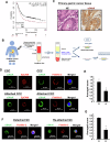
Fig. 1
Fig. 1. Expression of α3NaK in CCCs of patients with gastric cancer.
A Kaplan-Meier Plotter analysis of α3NaK in gastric cancer. B A schematic overview of the experimental procedure for isolation of CCCs from patients with gastric cancer. CCCs were isolated and enriched using the RosetteSep™ Human CD45 Depletion Cocktail and Lymphoprep, which depletes white and red blood cells by density centrifugation with tetrameric antibody complexes recognizing CD45. The CD45(-) cells, including CCCs, were fixed with 4% paraformaldehyde on the glass-based dish. When indicated, the cells were attached to a poly-L-lysine-coated dish before fixation. C Hematoxylin and eosin (HE) staining and immunohistochemistry using anti-α3NaK antibody were performed in primary gastric cancer tissues. Scale bar, 10 μm. D Immunocytochemistry was performed using antibodies for α3NaK (green), EpCAM (a marker for CCCs; red), and flotillin-2 (a marker for PM; red) in CCCs and attached CCCs of gastric patients. Scale bars, 10 μm. E The distribution of α3NaK in detached (D) and attached (A) CCCs were scored as 0 (predominantly at cytoplasm), 1 (both at cytoplasm and PM), and 2 (predominantly at PM) in (D). Averaged scores in detached (n = 18) and attached (n = 29) CCCs were shown. *p < 0.05. F Immunocytochemistry was performed using antibodies for α3NaK (green), EpCAM (red), and flotillin-2 (red) in detached and re-attached gastric cancer cells (GC) obtained by enzyme digestion of primary cancer tissue. Scale bars, 10 μm. G The distribution of α3NaK in detached (D) and re-attached (R) gastric cancer cells obtained by enzyme digestion of primary cancer tissue were scored as 0 (predominantly at cytoplasm), 1 (both at cytoplasm and PM), and 2 (predominantly at PM) in (F). Averaged scores in detached (n = 64) and re-attached (n = 90) cells were shown. *p < 0.05.
Fig. 2
Fig. 2. Digoxin inhibits the translocation of α3NaK from cytoplasm to the PM, and promotes anoikis in MKN45 cells.
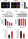
A Immunocytochemistry was performed using antibodies for α3NaK (green) and flotillin-2 (red) in detached and re-attached MKN45 cells. Cells were detached by the treatment with the solution containing 0.25% trypsin and 10 mM EDTA. Scale bars, 10 μm. B The distribution of α3NaK in detached (D) and re-attached (R) MKN45 cells were scored as 0 (predominantly at cytoplasm), 1 (both at cytoplasm and PM), and 2 (predominantly at PM) in (A). CF Cell surface biotinylation was performed in detached MKN45 cells. Effects of digoxin (2, 20, and 50 nM) on the surface expression level of α3NaK in detached MKN45 cells. (-) indicates cells not treated with digoxin. Typical images of Western blots using antibodies for α3NaK (110 kDa; C) and α1NaK (100 kDa; E) in the total lysates (input) and biotinylation samples (surface) were shown. Quantification of the surface expression level of α3NaK in (C) and (E) (D, F). n = 3–4. *p < 0.05; **p < 0.01. G The effect of digoxin (20 nM) on caspase 3/7 activity of detached (floating) MKN45 cells. The activity in the digoxin-treated cells was compared with untreated cells (control) (n = 6). **p < 0.01. H Cell viability was assessed by measuring mitochondrial activity. The effect of digoxin (20 nM) on cell viability was examined in detached (floating) MKN45 cells. The activity in the digoxin-treated cells was compared with untreated cells (control) (n = 6). **p < 0.01.
Fig. 3
Fig. 3. Inhibition of α3NaK activity by digoxin in MKN45 cells and gastric cancer tissues.
A Na+/K+-ATPase activities in the samples were measured at two different Na+ concentrations; α1NaK predominantly contributes to total Na+/K+-ATPase activity at 20 mM of Na+, and both α1NaK and α3NaK contribute to total activity at 120 mM of Na+. Digoxin (20 and 50 nM)-sensitive Na+/K+-ATPase activities of the membrane fractions of MKN45 cells (B) and human gastric cancer tissues (C) were measured in the 20 mM-Na+ and 120 mM-Na+ solutions. (-) indicates cells not treated with digoxin. n = 3-4. *p < 0.05; **p < 0.01.
Fig. 4
Fig. 4. Expression of ECM-related components in CCCs.
A Surface expression levels of α3NaK and α1NaK in detached MKN45 cells under the presence (+) and absence (−) of ECM components (fibronectin, collagen, and laminin; 15 μg/ml). Representative Western blot images using antibodies against α3NaK (110 kDa) and α1NaK (100 kDa) in the total lysates (input) and biotinylation samples (surface) were shown in the upper panels. In the lower panels, surface expression level of α3NaK and α1NaK was quantified. n = 4. **p < 0.01. B Western blotting was performed using anti-integrin α5 (140 kDa) or fibronectin (260 kDa) antibody in gastric cancer tissues. C Immunocytochemistry was conducted with antibodies against integrin α5 and fibronectin in gastric cancer tissues. Scale bars, 10 μm. Immunocytochemistry was performed using antibodies against EpCAM (red) and integrin α5 (D; green) or fibronectin (E; green) in CCCs from gastric patients. Scale bars, 10 μm.
Fig. 5
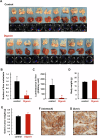
Fig. 5. Digoxin inhibits liver metastasis in the MKN45 orthotopic xenograft model.
A The series of images showed the growth of transplanted gastric cancers in the stomach (top) and their metastasis to the liver (middle) in each orthotopic xenograft mouse treated with or without digoxin (2 mg/kg/day). Ex vivo bioluminescence imaging of the liver (bottom) further identifies liver metastases. Arrowheads indicate metastatic gastric cancers in the liver. B The numbers of liver metastasis were compared between control (n = 9) and digoxin-treated (n = 10) mice. *p < 0.05. C Bioluminescence intensities were quantified ex vivo in the liver of control (n = 9) and digoxin-treated (n = 10) mice. *p < 0.05. D Body weights were compared between control (n = 9) and digoxin-treated (n = 10) mice. E Tumor weights isolated from stomachs were compared between control (n = 9) and digoxin-treated (n = 10) mice. Immunohistochemistry using anti-α3NaK antibody was performed in primary gastric cancer tissues in the stomach (F) and metastatic cancer tissues in the liver (G). Scale bars, 10 μm.
Fig. 6
Fig. 6. Digoxin decreases the number of CCCs in the MKN45 orthotopic xenograft model.
A A schematic overview of the experimental procedure for isolation of CCCs from mice. Immunocytochemistry was performed using antibodies for α3NaK (green), EpCAM (red; B), and flotillin-2 (red; C) in CCCs. Scale bars, 10 μm. D The number of CCCs was compared between control (n = 9) and digoxin-treated (n = 10) mice. **p < 0.01.
Fig. 7
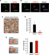
Fig. 7. Digoxin decreases the number of CCCs in the MKN45 subcutaneous xenograft model.
Immunocytochemistry was performed using antibodies for α3NaK (green), EpCAM (red; A), and flotillin-2 (red; B) in CCCs. C Immunohistochemistry using anti-α3NaK antibody was performed in subcutaneous cancer tissue derived from plantation of MKN45 cells. Scale bars, 10 μm. D The number of CCCs was compared between control (n = 7) and digoxin-treated (n = 6) mice. **p < 0.01. E Typical images of subcutaneous cancer tissues derived from plantation of MKN45 cells were shown (left). Tumor weights of the subcutaneous cancer tissues were compared between control (n = 7) and digoxin-treated (n = 6) mice (right).
Fig. 8
Fig. 8. Scheme of the effect of digoxin on the anoikis resistance of CCCs.
Digoxin inhibits the translocation of α3NaK from cytoplasm to the PM upon cancer cell detachment, and promote anoikis in the CCCs, suppressing liver metastasis of gastric cancer.

References

    1. Kinami S, Saito H, Takamura H. Significance of lymph node metastasis in the treatment of gastric cancer and current challenges in determining the extent of metastasis. Front Oncol. 2021;11:806162. - PMC - PubMed
    1. Riihimäki M, Hemminki A, Sundquist K, Sundquist J, Hemminki K. Metastatic spread in patients with gastric cancer. Oncotarget. 2016;7:52307–16. - PMC - PubMed
    1. Xu J, Shen L, Shui Y, Yu W, Guo Q, Yu R, et al. Patterns of recurrence after curative D2 resection for gastric cancer: Implications for postoperative radiotherapy. Cancer Med. 2020;9:4724–35. - PMC - PubMed
    1. Montagnani F, Crivelli F, Aprile G, Vivaldi C, Pecora I, De Vivo R, et al. Long-term survival after liver metastasectomy in gastric cancer: systematic review and meta-analysis of prognostic factors. Cancer Treat Rev. 2018;69:11–20. - PubMed
    1. Zhang Y, Lin Y, Duan J, Xu K, Mao M, Wang X. A population-based analysis of distant metastasis in stage IV gastric cancer. Med Sci Monit. 2020;26:e923867. - PMC - PubMed

MeSH terms