. 2025 Jun;24(6):e70080.
doi: 10.1111/acel.70080. Epub 2025 May 12.

Reducing HuD Levels Alleviates Alzheimer's Disease Pathology in 5xFAD Mice

Affiliations

Reducing HuD Levels Alleviates Alzheimer's Disease Pathology in 5xFAD Mice

Eunbyul Ji et al. Aging Cell. 2025 Jun.

Abstract

Alzheimer's disease (AD) is the most common neurodegenerative pathology in older persons. The accumulation of amyloid β (Aβ) plaques is a major contributor to AD development. The RNA-binding protein HuD/ELAVL4 has been implicated in the formation of Aβ plaques, but its role in AD is unclear. Here, we report that ablation of HuD from CAMK2A+ neurons (HuDcKO) in the 5xFAD mouse model of AD results in a significant reduction of Aβ plaques and the alleviation of some AD-associated behaviors. Given the lack of effective therapies for AD, we propose that reducing HuD levels or function can contribute to diminishing Aβ plaque formation and AD-associated pathology.

Keywords: Elavl4; 5xFAD; Alzheimer's disease; HuD; aβ plaques; homecage activity.


Conflict of interest statement

The authors declare no conflicts of interest.

Figures
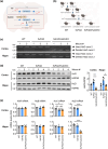
FIGURE 1
FIGURE 1
Generation and characterization of 5xFAD/HuDcKO mice. (a) Strategy to ablate the HuD/Elavl4 gene by the loxP‐Cre system. Exon 2 of the HuD gene, flanked by two loxP sites, was designed to be excised by Cre recombinase under the control of the Camk2a promoter. Red arrows indicate the primers amplifying the floxed HuD exon 2 for genotyping. (b) Breeding scheme to generate HuDcKO mice in the 5xFAD background. (c) Recombination at the HuD locus was confirmed by PCR amplification of genomic DNA isolated from cerebral cortex (Cortex) and hippocampus (Hippo) of 9‐month‐old (9 m.o.) female mice using the primers indicated in panel a (red). (d) The levels of HuD protein from 9 m.o. female mice were assessed by western blot analysis using HSP90 as a loading control. Signal intensities were quantified using ImageJ. The numbers above the lanes in (c, d) indicate individual mice in each genotype group (biological replicates). (e) RNA was isolated from 9 m.o. female mice, and the levels of mRNAs encoding proteins in the Hu family and Gapdh mRNA (used for normalization) were measured by RT‐qPCR analysis. Data in (d, e) represent the means ± SEM. Statistical significance (* p < 0.05; ***p < 0.001; ****p < 0.0001) was assessed with Student's t‐test.
FIGURE 2
FIGURE 2
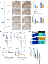
HuDcKO inhibits AD traits in 5xFAD. (a) The presence of plaques containing Aβ was analyzed by IHC with antibodies detecting Aβ peptides ranging in length between 37 and 42 kDa (Aβs, top) or specifically detecting Aβ42 (bottom) in 7 m.o. and 9 m.o. female mice. Aβ plaque signals were quantified as % area using ImageJ (right). Scale bars, 200 μm. (b) The concentration of Aβ40 and Aβ42 was measured in brain lysate from 7 m.o. female WT (n = 2), 5xFAD (n = 3), and 5xFAD/HuDcKO (n = 3) using BioPlex. (c–e) Homecage activity test was performed with 9 m.o. female mice that were WT (n = 9), 5xFAD (n = 10), or 5xFAD/HuDcKO (n = 7). (c) Daily homecage activity, shown in heatmap actograms, represents the distance traveled (m) per minute; the indents in days 0 and 5 reflect the fact that the recordings began and ended in mid‐morning, respectively. (d) Distance traveled in homecage was recorded during 12‐h light or dark phases across five nights. (e) Average distance traveled per night in the last three nights of testing, after mice had acclimated to the new surroundings. Data in (a,b,d,e) represent the means ± SEM. Statistical significance (*p < 0.05; **p < 0.01; ****p < 0.0001) was assessed using Student's t‐test or Tukey's test. Greek letters denote significant Tukey's pairwise comparisons between genotypes: α, WT versus 5xFAD; β, WT versus 5xFAD/HuDcKO; γ, 5xFAD versus 5xFAD/HuDcKO.

References

    1. Akamatsu, W. , Fujihara H., Mitsuhashi T., et al. 2005. “The RNA‐Binding Protein HuD Regulates Neuronal Cell Identity and Maturation.” Proceedings of the National Academy of Sciences of the United States of America 102, no. 12: 4625–4630. 10.1073/pnas.0407523102. - DOI - PMC - PubMed
    1. Amadio, M. , Pascale A., Wang J., et al. 2009. “nELAV Proteins Alteration in Alzheimer's Disease Brain: A Novel Putative Target for Amyloid‐Beta Reverberating on AbetaPP Processing.” Journal of Alzheimer's Disease 16, no. 2: 409–419. 10.3233/jad-2009-0967. - DOI - PMC - PubMed
    1. Ambrée, O. , Touma C., Görtz N., et al. 2006. “Activity Changes and Marked Stereotypic Behavior Precede Abeta Pathology in TgCRND8 Alzheimer Mice.” Neurobiology of Aging 27, no. 7: 955–964. 10.1016/j.neurobiolaging.2005.05.009. - DOI - PubMed
    1. Blinkouskaya, Y. , and Weickenmeier J.. 2021. “Brain Shape Changes Associated With Cerebral Atrophy in Healthy Aging and Alzheimer's Disease.” Frontiers of Mechanical Engineering 7: 705653. 10.3389/fmech.2021.705653. - DOI - PMC - PubMed
    1. Bolognani, F. , and Perrone‐Bizzozero N. I.. 2008. “RNA‐Protein Interactions and Control of mRNA Stability in Neurons.” Journal of Neuroscience Research 86: 481–489. 10.1002/jnr.21473C. - DOI - PubMed

LinkOut - more resources