Case Reports
. 2025 Jun;65(6):1132-1144.
doi: 10.1111/trf.18270. Epub 2025 May 12.

Vesiculation as potential novel pathogenic mechanism in autoimmune hemolytic anemia

Affiliations
Case Reports

Vesiculation as potential novel pathogenic mechanism in autoimmune hemolytic anemia

Esther C W de Boer et al. Transfusion. 2025 Jun.

Abstract

Background: Autoimmune hemolytic anemia (AIHA) is typically mediated by immunoglobulin G (IgG) or immunoglobulin M (IgM) antibodies, and more rarely by immunoglobulin A (IgA). The mechanism of red blood cell (RBC) destruction in IgA-mediated AIHA is not well understood. We report a case of severe AIHA with intravascular hemolysis, positive for IgA. Hemolysis did not subside despite multiple transfusions and treatment lines, leading to a fatal outcome. Here, we set out to investigate underlying pathophysiological mechanisms.

Study design and methods: To investigate the underlying pathophysiological methods, standard hematological methods were used, as well as erythrophagocytosis assays and flow cytometry using patient RBCs and plasma, to investigate anti-RBC antibodies, complement activation, and vesiculation.

Results: Blood smear analysis revealed significant heterogeneity in RBC size and volume, and the presence of ghost cells, indicating RBC damage. While the patient's RBCs were found opsonized with IgA and IgG autoantibodies, phagocytosis by neutrophils was not induced in vitro, nor did sensitized donor RBCs with patient plasma. Using flow cytometry, we detected vesicles in the patient's plasma and observed patient plasma-induced vesiculation of healthy donor RBC. Patient plasma showed marked complement activation, and the vesicles in the patient plasma were also complement-opsonized, as well as bound by IgA, IgG, and IgM.

Conclusions: Based on these findings, we suggest vesiculation of RBCs as evidenced by the presence of vesicles and ghost cells in the patient, and subsequent complement activation induced by the vesicles. This could have driven the aggravation of the disease in this patient, resulting in a fatal outcome.

Keywords: AIHA/drug‐induced IHA; hematology—red cells; immunology (other than RBC serology).


Conflict of interest statement

Masja de Haas delivers paid consultancy for JnJ and educational services for BioRad. Josephine M. I. Vos has received the following as institutional honoraria: research support from Beigene and AbbVie/Genmab; advisory board/consultancy fees from Sanofi and Janssen; and speaker fees from BMS, Sanofi, Beigene, Novartis, and Amgen.

Figures

FIGURE 1
FIGURE 1
(A) Lithium heparin blood collection tube with gel separator, centrifuged, showing hemolytic plasma indicative of intravascular hemolysis. (B) Monospecific direct antiglobulin test (DAT) showing strong positivity for IgA (3+) and positivity for IgG, IgM, C3c, and C3d as well as control (1+). Visually, the IgM column is slightly more positive than control column. (C) Blood smear at onset of AIHA, showing spherocytes and ghost cells (blue arrows). (D) Course of hemoglobin (Hb, g/dL, solid line) and lactate dehydrogenase (LDH, IU/L, dotted line) during hospitalization. Transfused red blood cell (RBC) units (orange triangles) and start of therapies are marked at the administered time point. t = 1, t = 2, and t = 3 represent the timing of blood samples used in experimental assays. IvIg = intravenous immunoglobulins. [Color figure can be viewed at wileyonlinelibrary.com]
FIGURE 2
FIGURE 2
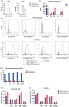
AI‐based IFC erythrophagocytosis by neutrophils. (A) Flow cytometric detection of C3 deposition, IgA and IgG binding in healthy and AIHA patient RBCs (t = 2). Histogram (left panel) and gMFI (right panel) are shown. (B and D) AI‐based IFC phagocytosis assay by primary neutrophils. AI analysis was performed on 5000 single‐image events collected per sample. The graphs show the percentage of events in different classes. Anti‐GPA opsonized RBCs were used as a positive control for phagocytosis. (B) Phagocytosis performed with healthy donor and patient RBCs (n = 1). (D) Phagocytosis performed with non‐sensitized and sensitized healthy donor RBCs with either healthy donor plasma or patient plasma (n = 1). (C and E) Cytospins representative of each phagocytosis condition for healthy and patient RBCs (C) and sensitized healthy donor RBCs with healthy donor or patient plasma (E). Phagocytic events are indicated by the red arrows. [Color figure can be viewed at wileyonlinelibrary.com]
FIGURE 3
FIGURE 3
RBC‐derived vesiculation. (A) Flow cytometry plots of healthy and AIHA whole blood samples at t = 2 showing the presence of particles with low SSC‐A and FCS‐A in the AIHA patient. (B) Gating on the particles with low FSC‐A and SSC‐A, patient plasma and eluate reveal the presence of small particles which are partially CD235a‐positive, n = 1. (C) CD235a+ events from patient plasma t = 1 and t = 2 are smaller on FSC‐A and SSC‐a than healthy donor RBCs. (D) Healthy donor RBCs were incubated with patient plasma and complement‐active plasma in the Flow IAT. Flow IAT reveals that an increased number of vesicles relative to RBCs are present after 30 min incubation of healthy donor RBCs with patient plasma or RBC eluate compared to healthy controls. Representative of n = 3. (E) DiD+ particles after Flow IAT. Healthy donor RBCs were stained with DiD before Flow IAT; the presence of DiD+ particles indicates that these are donor RBC‐derived, n = 1. [Color figure can be viewed at wileyonlinelibrary.com]
FIGURE 4
FIGURE 4
Antibodies and complement deposition. (A) C3b/c and C4b/c ELISA show increased complement activation products in patient plasma compared to healthy donor plasma, especially at t = 1. (B) Vesicles stained in IgA AIHA plasma are highly positive for IgA and show a positive signal for IgG, IgM, and C3. C3b/c and C4b/c ELISA show increased complement activation products in patient plasma compared to healthy donor plasma, especially at t = 1. (C–E) Healthy donor RBCs were incubated with AIHA patient plasma and complement‐active donor plasma in the Flow IAT. Based on FSC vs. SSC, RBCs and vesicles were then separated. (C) After Flow IAT with patient RBC eluate, healthy donor RBCs (top) are only positive for IgA. On the vesicles (bottom), deposition of IgA, IgG, IgM, and C3 is found by both patient plasmas and RBC eluate. Representative of n = 3. (D, E) C3 deposition on RBCs in the Flow IAT with healthy serum only or combined with inhibitors of specific complement pathways. Classical pathway (CP) was inhibited by anti‐C1q.98, alternative pathway (AP) by anti‐FD (lampalizumab) and lectin pathway (LP) by anti‐MBL.1. (D) The ratio between vesicles and RBCs after Flow IAT did not change upon inhibition of different complement activation pathways. (E) On RBCs, low‐grade complement activation is inhibited by both CP and AP inhibition and can be reduced to background level by combining CP and AP inhibition. (F) On vesicles, complement activation is high and only inhibited by classical pathway inhibition, without an additional inhibitory effect of AP inhibition, n = 1. [Color figure can be viewed at wileyonlinelibrary.com]
FIGURE 5
FIGURE 5
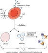
Proposed disease mechanism in the current case. Patient RBCs are bound by autoantibodies of mainly IgA, either monomeric or dimeric, and to some extent IgG and IgM class. Binding of the autoantibodies to RBCs triggers vesiculation. As a result of vesiculation, RBC ghosts and vesicles are formed. The vesicles are then strongly opsonized by complement, mainly via the classical pathway. Overall, these processes contribute to intravascular hemolysis, inflammation, and thrombotic risk. [Color figure can be viewed at wileyonlinelibrary.com]

References

    1. Berentsen S, Barcellini W. Autoimmune hemolytic anemias. N Engl J Med. 2021;385(15):1407–1419. - PubMed
    1. Loriamini M, Cserti‐Gazdewich C, Branch DR. Autoimmune hemolytic anemias: classifications, pathophysiology, diagnoses and management. IJMS. 2024;25(8):4296. - PMC - PubMed
    1. Sokol RJ, Hewitt S, Booker DJ, Bailey A. Red cell autoantibodies, multiple immunoglobulin classes, and autoimmune hemolysis. Transfusion. 1990;30(8):714–717. - PubMed
    1. Sokol RJ, Booker DJ, Stamps R, Booth JR, Hook V. IgA red cell autoantibodies and autoimmune hemolysis. Transfusion. 1997;37(2):175–181. - PubMed
    1. Bardill B, Mengis C, Tschopp M, Wuillemin WA. Severe IgA‐mediated auto‐immune haemolytic anaemia in a 48‐yr‐old woman. Eur J Haematol. 2003;70(1):60–63. - PubMed

Publication types

MeSH terms

LinkOut - more resources