

Bony Hydatidosis of Femur Head - A Rare Case Report
- PMID: 40351630
- PMCID: PMC12064242
- DOI: 10.13107/jocr.2025.v15.i05.5570
Bony Hydatidosis of Femur Head - A Rare Case Report
Abstract
Introduction: Hydatid disease occurs due to Echinococcus in humans as they are intermediate host for tapeworm. The bone involvement is rare. Insidious nature and nonspecific nature of complaints make delay in diagnosis. Surgery is the mainstay of treatment with role of chemotherapy is as an adjunct treatment modality.
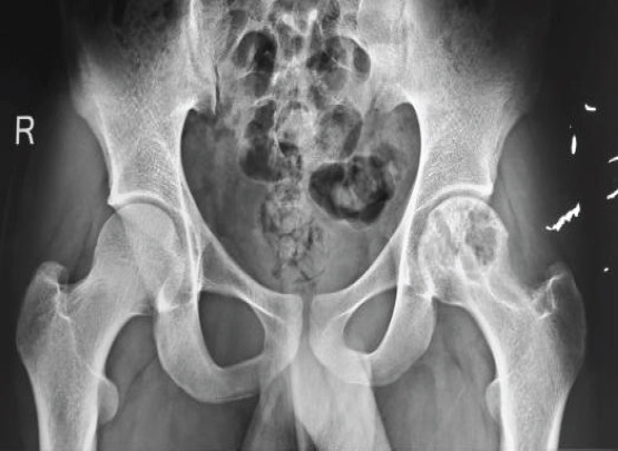
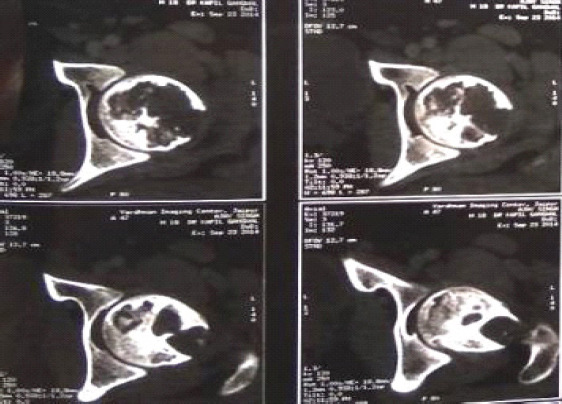
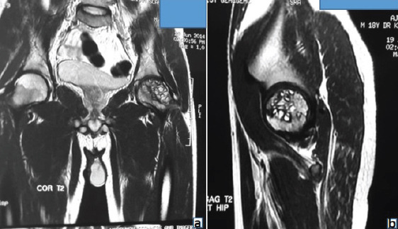
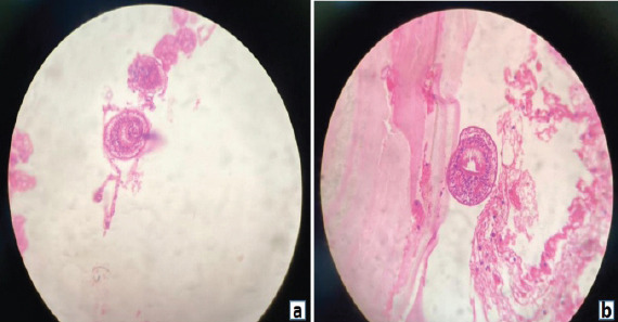
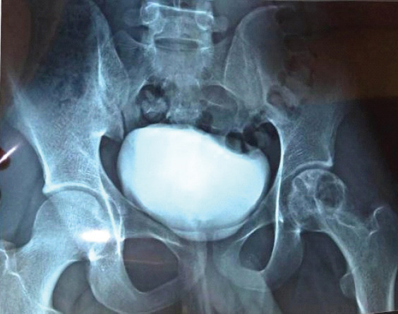
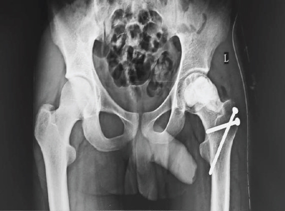
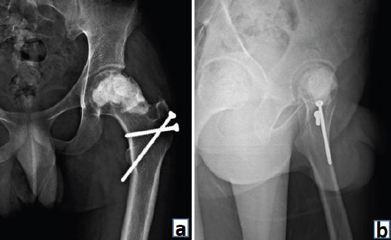
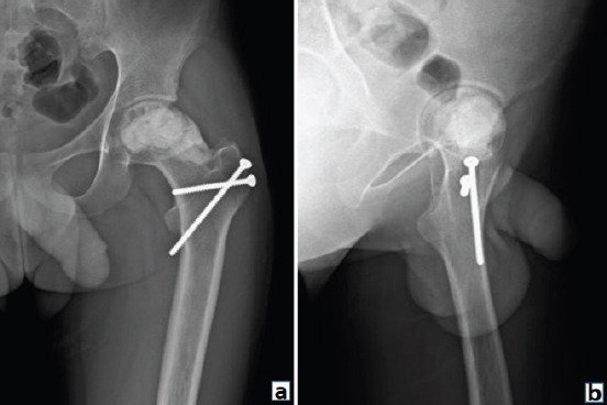
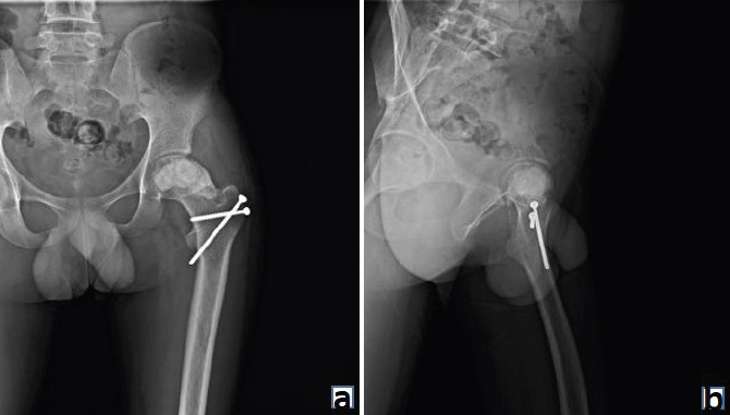
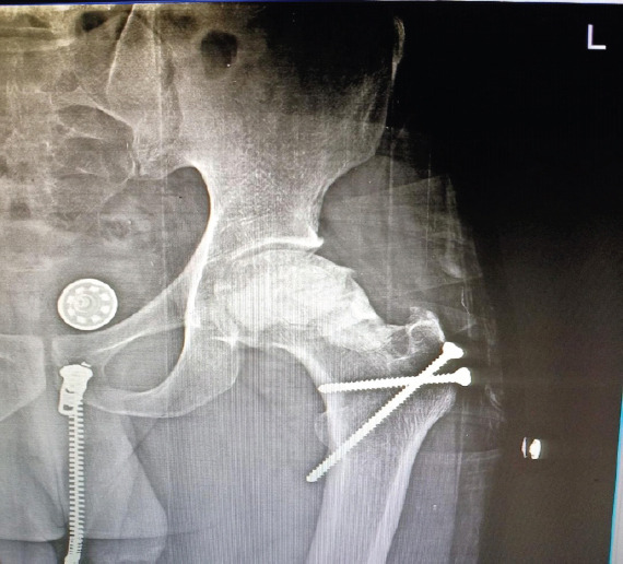
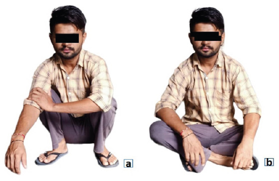
Case report: An 18-year-old male patient presented with the left groin pain and terminal restriction of hip movement for 1 year duration. The patient underwent open biopsy and histopathological examination of tissue specimen revealed hydatid cyst. The patient was started on albendazole and after 3 months, surgical curettage and removal of cysts were performed with application of bone cement. At 8 years follow-up, the patient is asymptomatic and doing well.
Conclusion: The treatment of osseous hydatid disease is challenging. High index of suspicion in endemic areas is required along with radiological and laboratory investigation to confirm the diagnosis. Timely and proper management can completely cure the patient without any residual pathology with full functional recovery.
Keywords: Echinococcus granulosus; albendazole; bony hydatidosis; femur head.
Copyright: © Indian Orthopaedic Research Group.
Conflict of interest statement
Conflict of Interest: Nil
Figures
Similar articles
-
[Hydatidosis of psoas muscle revealed by vascular axis compression in lower limb: About one case at the Ibn Sina University hospital, Rabat, Morocco].Med Trop Sante Int. 2022 Jul 18;2(3):mtsi.v2i3.2022.195. doi: 10.48327/mtsi.v2i3.2022.195. eCollection 2022 Sep 30. Med Trop Sante Int. 2022. PMID: 36284556 Free PMC article. French.
-
A rare case of recurrent spinal hydatid cyst in a 17-year-old man with neurological deficits and balance impairment.BMC Infect Dis. 2024 Dec 6;24(1):1392. doi: 10.1186/s12879-024-10286-3. BMC Infect Dis. 2024. PMID: 39639192 Free PMC article.
-
An Unusual Cause of Knee Mass: Osseous Hydatidosis.Cureus. 2021 Oct 6;13(10):e18556. doi: 10.7759/cureus.18556. eCollection 2021 Oct. Cureus. 2021. PMID: 34765341 Free PMC article.
-
Composite treatment for primary long-bone hydatidosis.Orthopedics. 2012 Dec;35(12):e1826-31. doi: 10.3928/01477447-20121120-34. Orthopedics. 2012. PMID: 23218646 Review.
-
Hydatidosis of infratemporal fossa with proptosis - an unusual presentation: a case report and review of the literature.J Med Case Rep. 2018 Oct 17;12(1):309. doi: 10.1186/s13256-018-1812-y. J Med Case Rep. 2018. PMID: 30326941 Free PMC article. Review.
References
-
- Moro P, Schantz PM. Echinococcosis:A review. Int J Infect Dis. 2009;13:125–33. - PubMed
-
- Sapkas GS, Machinis TG, Chloros GD, Fountas KN, Themistocleous GS, Vrettakos G. Spinal hydatid disease, a rare but existent pathological entity:Case report and review of the literature. South Med J. 2006;99:178–83. - PubMed
-
- Chandra SS, Kumar MM, Ramnarayanan, Vanchi PK. Hydatid disease of proximal femur:A case report. Int J Res Orthop. 2017;3:883–5.
-
- Agarwal S, Kundu ZS, Singh S, Soni N. Hydatid disease of the bone. Internet J Spine Surg. 2009;5:240–5.
Publication types
LinkOut - more resources
Full Text Sources