Simultaneous Bilateral Patellar Tendon Ruptures Treated with Primary Repair and Dermal Allograft Augmentation: A Case Report
- PMID: 40351645
- PMCID: PMC12064259
- DOI: 10.13107/jocr.2025.v15.i05.5548
Simultaneous Bilateral Patellar Tendon Ruptures Treated with Primary Repair and Dermal Allograft Augmentation: A Case Report
Abstract
Introduction: Patellar tendon ruptures are a relatively common injury encountered by orthopedic surgeons and typically only occur unilaterally. However, there are rare reports of bilateral patellar tendon ruptures occurring simultaneously, in patients with underlying systemic disorders, higher energy mechanisms, or injury or overuse in high-level athletes. When patellar tendon ruptures occur, and the extensor mechanism is disrupted, patellar tendon repair versus reconstruction is warranted to restore functionality. The use of dermal allografts for the reconstruction of chronic patellar tendon ruptures is well described; however, there is not much literature describing their use in the acute setting. This report describes the primary repair of simultaneous bilateral patellar tendon rupture with the use of the ArthroFLEX Decellularized Dermal Allograft. This is a novel use for this allograft, as it is currently indicated for use in the treatment of various tendon repairs/reconstructions as well as hallux rigidus and hip capsule reconstruction. There are no reports describing the use of the ArthroFLEX Decellularized Dermal Allograft in the acute setting as augmentation of primary patellar tendon repair in a patient with simultaneous bilateral patellar tendon ruptures in the absence of underlying systemic disease; thus, this report presents a novel use for this dermal allograft.
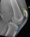
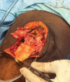
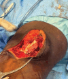
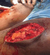
Case report: This patient is a 40-year-old African American male with no active underlying systemic diagnosis who sustained simultaneous bilateral patellar tendon ruptures from a low-energy mechanism. He subsequently underwent bilateral patellar tendon repair during which a dermal allograft augment was utilized to further strengthen this repair. In addition, a defunctioning purse string suture was used to further protect the patellar tendon repair by off loading the extensor mechanism.
Conclusion: This report adds to the body of literature surrounding the rare entity of simultaneous bilateral patellar tendon ruptures in otherwise healthy patients while also presenting a novel use for the ArthroFLEX Decellularized Dermal Allograft in the acute repair of a patellar tendon rupture. This report also supports the use of a defunctioning purse string suture to help offload the healing extensor and decrease the amount of tension across a healing tendon repair.
Keywords: ArthroFLEX decellularized dermal allograft; Simultaneous bilateral patellar tendon ruptures; dermal allograft augmentation; primary patellar tendon repair.
Copyright: © Indian Orthopaedic Research Group.
Conflict of interest statement
Conflict of Interest: Nil
Figures


References
-
- Owens B, Mountcastle S, White D. Racial differences in tendon rupture incidence. Int J Sports Med. 2007;28:617–20. - PubMed
-
- Matava MJ. Patellar tendon ruptures. J Am Acad Orthop Surg. 1996;4:287–96. - PubMed
-
- Hsu H, Siwiec RM. StatPearls. Treasure Island, FL: StatPearls Publishing; 2024. Patellar tendon rupture. - PubMed
-
- Otsubo H, Kamiya T, Suzuki T, Kuroda M, Ikeda Y, Matsumura T, et al. Repair of acute patellar tendon rupture augmented with strong sutures. J Knee Surg. 2017;30:336–40. - PubMed
Publication types
LinkOut - more resources
Full Text Sources
