

Phrenic Nerve Palsy in Anterior Cervical Discectomy and Fusion: Rare as Hen's Teeth
- PMID: 40351649
- PMCID: PMC12064271
- DOI: 10.13107/jocr.2025.v15.i05.5564
Phrenic Nerve Palsy in Anterior Cervical Discectomy and Fusion: Rare as Hen's Teeth
Abstract
Introduction: Phrenic nerve palsy is a rare but potentially serious complication. The clinical presentation can vary from being asymptomatic to severe respiratory distress requiring mechanical complication. In the Anglophone literature, there is only a single case report of bilateral phrenic nerve injury as a complication following anterior cervical discectomy and fusion (ACDF).
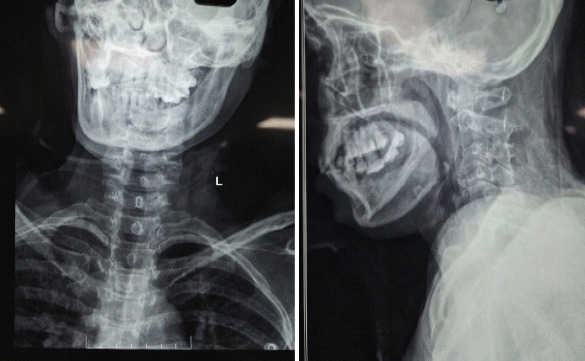
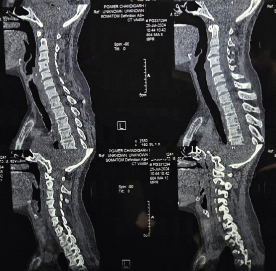
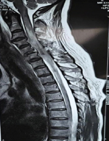
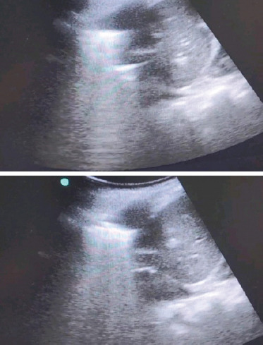
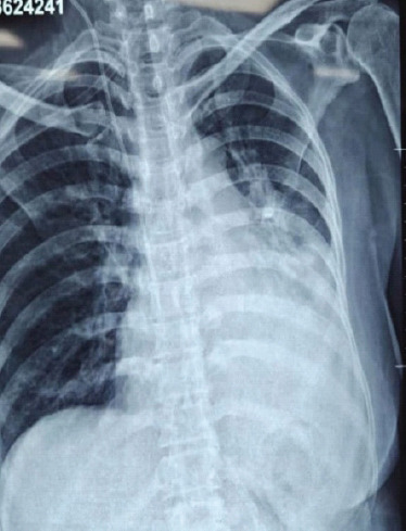
Case report: This case report describes a 52-year-old female who developed right-sided phrenic nerve palsy after undergoing ACDF for cervical spine trauma. The patient had respiratory distress immediately after surgery and Ultrasonography and X-rays revealed Rt phrenic nerve palsy. To the best of our knowledge, this is the first case of unilateral phrenic nerve palsy after ACDF at the C5-C6 level.
Conclusion: Unilateral phrenic nerve palsy probably occurred as a complication of ACDF for cervical spine trauma. Phrenic nerve palsy should be kept in mind as a serious complication of spinal surgery.
Keywords: Phrenic nerve palsy; anterior cervical discectomy and fusion; cervical spine trauma; post-operative complication; respiratory distress.
Copyright: © Indian Orthopaedic Research Group.
Conflict of interest statement
Conflict of Interest: Nil
Figures
Similar articles
-
Bilateral phrenic nerve palsy as a complication of anterior decompression and fusion for cervical ossification of the posterior longitudinal ligament.Spine (Phila Pa 1976). 2001 Jun 15;26(12):E281-6. doi: 10.1097/00007632-200106150-00029. Spine (Phila Pa 1976). 2001. PMID: 11426169
-
Phrenic Nerve Palsy After Posterior Cervical Fusion: A Case Report and Review of Literature.Clin Spine Surg. 2025 Jun 1;38(5):217-223. doi: 10.1097/BSD.0000000000001712. Epub 2024 Nov 11. Clin Spine Surg. 2025. PMID: 39527252 Review.
-
Phrenic nerve palsy after cervical laminectomy and fusion.N Am Spine Soc J. 2020 Sep 24;4:100022. doi: 10.1016/j.xnsj.2020.100022. eCollection 2020 Dec. N Am Spine Soc J. 2020. PMID: 35141599 Free PMC article.
-
Bilateral phrenic nerve palsy after posterior cervical decompression and fusion surgery: a rare event after surgery.Spinal Cord Ser Cases. 2023 Aug 12;9(1):41. doi: 10.1038/s41394-023-00595-1. Spinal Cord Ser Cases. 2023. PMID: 37573432 Free PMC article.
-
A Review of Complication Rates for Anterior Cervical Diskectomy and Fusion (ACDF).Surg Neurol Int. 2019 Jun 7;10:100. doi: 10.25259/SNI-191-2019. eCollection 2019. Surg Neurol Int. 2019. PMID: 31528438 Free PMC article. Review.
References
-
- McAfee PC, Reah C, Gilder K, Eisermann L, Cunningham B. A meta-analysis of comparative outcomes following cervical arthroplasty or anterior cervical fusion:Results from 4 prospective multicenter randomized clinical trials and up to 1226 patients. Spine (Phila Pa 1976) 2012;37:943–52. - PubMed
-
- Hitchon PW, Moritani T, Woodroffe RW, Abode-Iyamah K, Tecle NE, Noeller J, et al. C5 palsy following posterior decompression and instrumentation in cervical stenosis:Single center experience and review. Clin Neurol Neurosurg. 2018;174:29–35. - PubMed
-
- Fujibayashi S, Shikata J, Yoshitomi H, Tanaka C, Nakamura K, Nakamura T. Bilateral phrenic nerve palsy as a complication of anterior decompression and fusion for cervical ossification of the posterior longitudinal ligament. Spine (Phila Pa 1976) 2001;26:E281–6. - PubMed
Publication types
LinkOut - more resources
Full Text Sources
Miscellaneous