. 2025 Apr 1;26(2):39444.
doi: 10.31083/AP39444. eCollection 2025 Apr.

Prenatal Exposure to Valproic Acid may Alter CD200/CD200R Signaling Pathways in a Rat Model of Autism Spectrum Disorder

Affiliations

Prenatal Exposure to Valproic Acid may Alter CD200/CD200R Signaling Pathways in a Rat Model of Autism Spectrum Disorder

Xiaoou Xu et al. Alpha Psychiatry. .

Abstract

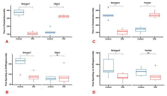
Objective: To investigate the potential toxic effects of prenatal exposure to valproic acid (VPA) on microglia-neuron communication in the brain, with a specific focus on the alterations in key molecules involved in this process, namely CX3CL1/CX3CR1 and CD200/CD200R, during the early stages of life in a rat model of autism.

Methods: Pregnant female rats were administered either sterile saline or VPA on embryonic day 12.5. The brains of the rat offspring were collected on postnatal day 30 for analysis. Immunohistochemical techniques and enzyme-linked immunosorbent assay (ELISA) were employed to assess changes in microglia-neuron crosstalk.

Results: The study revealed a significant reduction in CD200 levels within the hippocampus of rats on postnatal day 30 following prenatal exposure to VPA, indicating an impairment in CD200/CD200R signaling. Additionally, there was no observed increase in microglial numbers or any pathological alterations in the hippocampus. Additionally, no significant changes in the levels of CX3CL1 and CX3CR1 were noted in the VPA-exposed rats compared with the control group.

Conclusion: Prenatal exposure to VPA resulted in a decrease in CD200 expression within the hippocampus, potentially disrupting the communication between microglia and neurons. The findings suggest that VPA may modify the interactions between microglia and neurons, which could lead to neuroinflammation due to hyperactivated microglia. These disruptions have the potential to affect synaptic connectivity and contribute to the development of neurodevelopmental disorders, including autism. Further research is necessary to clarify the underlying mechanisms and implications for pathological conditions associated with autism spectrum disorder (ASD).

Keywords: CD200; CD200R; CX3CL1; CX3CR1; autism spectrum disorder; microglia; neuroinflammation; neuron; valproic acid.


Conflict of interest statement

The authors declare no conflict of interest.

Figures

Fig. 1.
Fig. 1.
Social deficits in the VPA group rats demonstrated across the three-chamber social test paradigm. (A) In Phase 1, VPA group rats exhibited significantly reduced interaction time with stranger 1 compared with the control group, while showing increased interaction time with the object (n = 6 for each group). (B) During Phase 1, VPA group rats spent significantly less time sniffing stranger 1 compared with the control group, while spending more time sniffing the object. (C) In phase 2, VPA group rats displayed significantly reduced interaction time with stranger 2 compared with the control group, while exhibiting increased interaction time with the familiar object. (D) During phase 2, VPA group rats spent significantly less time sniffing stranger 2 compared with the control group, while spending more time sniffing the familiar object. *p < 0.05 vs control group, **p < 0.01 vs control group. ns, no significance; VPA, valproic acid.
Fig. 2.
Fig. 2.
IBA1 expressions in the Rat Hippocampus. (A) Illustration of the quantitative immunohistochemical analysis of IBA1, representing the number of microglia in the hippocampus on postnatal day 30 (P30). No statistically significant difference was observed in the number of microglial cells between rats treated with VPA and those treated with saline (p > 0.05). (B) Depiction of the immunohistochemical photomicrographs of microglia from both the VPA group and the control group. Scale bar: 40μm. (C) Levels of IBA1 by ELISA between the VPA group and the control group (p > 0.05). There was no statistically significant difference in the expression level of IBA in the hippocampus between the two groups, indicating that exposure to VPA during pregnancy had no significant effect on the number of microglia in the hippocampus of rats offspring. ns, no significance.
Fig. 3.
Fig. 3.
CD200/CD200R expressions in the Rat Hippocampus. (A) The expression levels of CD200 by quantitative immunohistochemical analysis in rats treated with valproic acid and saline (p < 0.05). (B) Levels of CD200 by ELISA between the VPA group and the control group (p < 0.05). (C) Depiction of the corresponding images related to CD200/CD200R expression in the hippocampus from both groups. Scale bar: 40 μm. (D) The expression levels of CD200R by quantitative immunohistochemical analysis and ELISA, separatel (p > 0.05). It did not show a statistically significant difference of the level of CD200R between the two groups for VPA-treated rats and controlled rats (p > 0.05). However, the quantity of CD200 in rats treated with valproic acid was significantly lower compared with those observed in controlled rats (*p < 0.05). These results showed that exposure to VPA during pregnancy could decrease the level of CD200 of rats offspring. ns, no significance.
Fig. 4.
Fig. 4.
CX3CR1/CX3CL1 expressions in the Rat Hippocampus. (A) The expression levels of CX3CL1 by quantitative immunohistochemical analysis in rats treated with valproic acid and saline (p > 0.05). (B) Levels of CX3CL1 by ELISA between the VPA group and the control group (p > 0.05). (C) Corresponding images related to CX3CR1/CX3CL1 expression in the hippocampus from both groups. Scale bar: 40 μm. (D) The expression levels of CX3CR1 by quantitative immunohistochemical analysis and ELISA, separatel (p > 0.05). Rats administered with VPA did not exhibit a statistically significant difference in CX3CR1 expression compared with those in the control group. Similarly, the quantity of CX3CL1 in VPA-treated rats did not demonstrate a significant difference compared with the levels in the control group. It demonstrated that exposure to VPA during pregnancy had no influence on the level of CX3CR1/CX3CL1 of rats offspring. ns, no significance.
Fig. 5.
Fig. 5.
Neuronal morphological features in the hippocampus assessed using HE staining. Scale bar: 50μm. No evidence of inflammatory cell infiltration was observed in the CA1, CA2, and CA3 regions of the hippocampus in both control and VPA-treated rats. Furthermore, no pathological alterations were observed in the hippocampus of rats in either group.

References

    1. Dawson G, Rieder AD, Johnson MH. Prediction of autism in infants: progress and challenges. The Lancet. Neurology . 2023;22:244–254. doi: 10.1016/S1474-4422(22)00407-0. - DOI - PMC - PubMed
    1. Chamberlain SR, Aslan B, Quinn A, Anilkumar A, Robinson J, Grant JE, et al. Autism and gambling: A systematic review, focusing on neurocognition. Neuroscience and Biobehavioral Reviews . 2023;147:105071. doi: 10.1016/j.neubiorev.2023.105071. - DOI - PubMed
    1. Maisterrena A, Matas E, Mirfendereski H, Balbous A, Marchand S, Jaber M. The State of the Dopaminergic and Glutamatergic Systems in the Valproic Acid Mouse Model of Autism Spectrum Disorder. Biomolecules . 2022;12:1691. doi: 10.3390/biom12111691. - DOI - PMC - PubMed
    1. Usui N, Kobayashi H, Shimada S. Neuroinflammation and Oxidative Stress in the Pathogenesis of Autism Spectrum Disorder. International Journal of Molecular Sciences . 2023;24:5487. doi: 10.3390/ijms24065487. - DOI - PMC - PubMed
    1. Matta SM, Hill-Yardin EL, Crack PJ. The influence of neuroinflammation in Autism Spectrum Disorder. Brain, Behavior, and Immunity . 2019;79:75–90. doi: 10.1016/j.bbi.2019.04.037. - DOI - PubMed

LinkOut - more resources