Review
. 2025 Apr 25:16:1576133.
doi: 10.3389/fphys.2025.1576133. eCollection 2025.

From function to structure: how myofibrillogenesis influences the transverse-axial tubular system development and its peculiarities

Affiliations
Review

From function to structure: how myofibrillogenesis influences the transverse-axial tubular system development and its peculiarities

Zuzana Sevcikova Tomaskova et al. Front Physiol. .

Abstract

The transverse-axial tubular system (TATS) is the extension of sarcolemma growing to the cell interior, providing sufficient calcium signaling to induce calcium release from sarcoplasmic reticulum cisternae and stimulate the contraction of neighboring myofibrils. Interestingly, the development of TATS is delayed and matures during the post-partum period. It starts with small invaginations near the sarcolemma, proceeding to grow an irregular network that is later assembled into the notably transversally oriented tubular network. Accumulating evidence supports the idea that the development of TATS is linked to cell dimensions, calcium signaling, and increasing myofibrillar content orchestrated by electromechanical stimulation. However, the overall mechanism has not yet been described. The topic of this review is the development of TATS with an emphasis on the irregular phase of tubule growth. The traditional models of BIN1-related tubulation are also discussed. We summarized the recently described protein interactions during TATS development, mainly mediated by costameric and sarcomeric proteins, supporting the idea of the coupling sites between TATS and the myofibrils. We hypothesize that the formation and final organization of the tubular system is driven by the simultaneous development of the contractile apparatus under cycling electromechanical stimulus.

Keywords: Z-line; cardiomyocyte; costameres; myofibrillogenesis; postnatal development; sarcomere; t-tubules; transverse–axial tubular system.


Conflict of interest statement

The authors declare that the research was conducted in the absence of any commercial or financial relationships that could be construed as a potential conflict of interest.

Figures
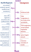
FIGURE 1
FIGURE 1
Timescale of myofibrillogenesis and tubulogenesis from newborn to adult stages of rat cardiomyocytes. The important steps of the formation of tubules and myofibrils are indicated on the time scale spanning from the day of birth (P0) to the adult stage (>P21). The development of tubules is delayed and starts on day P7, whereas the first, not oriented, myofibrils are already present during embryonic development.
FIGURE 2
FIGURE 2
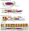
The phases of postnatal growth of the rat ventricular cardiomyocyte. (A) Hyperplastic phase (P0–P5). The cardiomyocyte has a spindle-like shape with a prominent nucleus. The immature myofibrils are localized near sarcolemma or loosely distributed in the cytoplasm. SR creates the CRUs (periphery couplings) with the sarcolemma. (B) Translational phase (P6–P14). The cardiomyocyte has a more rectangular-like shape. Two nuclei are already present in most of the cells. Short tubules are present coupled with myofibrils via costameres and longer tubules coupled with more mature myofibrils. The SR creates the CRUs with this longitudinal tubule. The scaffold of cardiomyocytes is emphasized with a Z-line connected with titin filaments surrounded by a developing network of microtubules and γ-actin. (C) Hypertrophic phase (P15–P21). The cardiomyocytes resembles the adults. The myofibrils are almost the cell volume, although they are not fully laterally aligned. TATS creates a sparse and irregular network. CRUs are present at the transversal and longitudinal elements of TATS. The transversal alignment of the tubules has started and is mediated by desmin. (D) Adult (>P21). The cardiomyocyte has a rectangular shape; the proportions of the cellular components are schematic. The aligned mature myofibrils are interwoven with the rows of mitochondria. The TATS has a prominent transversal orientation with well-developed CRUs (dyads). The scaffold of cardiomyocytes is finished by the maturation of sarcomeres and lateral alignment of all myofibrils mediated by desmin.
FIGURE 3
FIGURE 3
Scheme of a single sarcomere with proteins involved in tubulogenesis and myofibrillogenesis. The connection between tubules and sarcoplasmic reticulum, including the calcium release unit (CRU, dyad), is visualized on the left side of the sarcomere. The connection of a tubule to a Z-line, mediated by many different proteins, is depicted on both tubules. During postnatal development, the expression of these proteins varies according to the developmental stage.
FIGURE 4
FIGURE 4
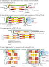
The hypothesis of tubular growth coupled with ongoing myofibrillogenesis. (A) Scalloping of sarcolemma: the first contact of premyofibrils and sarcolemma (P5–P7). The figure depicts the adhesion sites of the sarcolemma and the premyofibrils. For simplicity, one growing presarcomere of premyofibril is constructed from several Z-bodies connected with F-actin. The presarcomere is finished with myosin filaments among actin filaments. The environment of tubules is depicted differently in the left and the right tubules. The extracellular matrix (ECM) continues to both tubules. The left tubule emphasizes the talin–vinculin costamere units coupling with the Z-bodies. The right tubule shows the protein environment participating in the initial tubulogenesis as BIN1 assembled with PIP2 and CAV3 rings. The BIN1 can be pulled by microtubules. DHPR channels interacted with γ-actin filament through protein AHNAK. (B) Lateral alignment of nascent myofibrils and maturation of Z-lines (P8-P10). The tubules now grow like small transversal sarcolemmal invaginations, closely attached to the mature Z-lines. The sarcomere is now more mature (depicted by the created M-line), Z-bodies are transformed into Z-lines, and titin filaments are assembled, spanning across two Z-lines. The left tubule highlights the attachments with sarcomere Z-line proteins, strengthened by DGC costameric units (dystroglycan, spectrin, and dystrophin) pulled by γ-actin. Protein PTPN23 and nexilin improve the interaction of tubules with the Z-line. The right tubule shows the agents involved in tubulation (BIN1, dysferlin) with an emphasis on the CRU formation on tubules, provided by the assembly of cisternae of SR (jSR) with RyR2 channels facing tubular DHPRs. The tubule-jSR cleft is supported by the presence of myospryn. (C) Irregular growth of TATS (P11–P14). The image depicts the irregular growth of tubules by ongoing myofibrillogenesis. Transversal growth is induced by subsarcolemmal growth of new nascent sarcomeres (in lighter shades of original colors, in rectangles), whereas axial tubular growth is stimulated by the elongation of myofibril by the addition of sarcomere(s) in series. To simplify the image, only costameres are depicted as anchoring sites of tubules and sarcomeric Z-lines. (D) Lateral alignment of sarcomeres in cell volume (P15–P21). The left part (BEFORE) depicts the tubules and several sarcomeres before the final alignment, and the right part (AFTER) depicts the state of the final alignment of cell compartments. Desmin filaments provide the transversal scaffold for the coupled alignment of tubules and Z-lines, whereas microtubules support the axial alignment. The lateral alignment of myofibrils as the last stage of development is completed with Tcap assembly to the titin end and obscurin assembly among adjacent Z-lines and M-line-longitudinal SR interface. CRU formation is finished with the assembly of JP2 with junctin. The right tubule depicts the elongation of tubules induced by MTM1.

Similar articles

References

    1. Aherrahrou Z., Schlossarek S., Stoelting S., Klinger M., Geertz B., Weinberger F., et al. (2016). Knock-out of nexilin in mice leads to dilated cardiomyopathy and endomyocardial fibroelastosis. Basic Res. Cardiol. 111, 6. 10.1007/s00395-015-0522-5 - DOI - PubMed
    1. Ampong B. N., Imamura M., Matsumiya T., Yoshida M., Takeda S. (2005). Intracellular localization of dysferlin and its association with the dihydropyridine receptor. Acta Myol. 24, 134–144. - PubMed
    1. Anversa P., Olivetti G., Bracchi P., Loud A. V. (1981). Postnatal development of the M-band in rat cardiac myofibrils. Circulation Res. 48, 561–568. 10.1161/01.res.48.4.561 - DOI - PubMed
    1. Anversa P., Olivetti G., Loud A. V. (1980). Morphometric study of early postnatal development in the left and right ventricular myocardium of the rat. I. Hypertrophy, hyperplasia, and binucleation of myocytes. Circ. Res. 46, 495–502. 10.1161/01.res.46.4.495 - DOI - PubMed
    1. Asghari P., Schulson M., Scriven D. R., Martens G., Moore E. D. (2009). Axial tubules of rat ventricular myocytes form multiple junctions with the sarcoplasmic reticulum. Biophysical J. 96, 4651–4660. 10.1016/j.bpj.2009.02.058 - DOI - PMC - PubMed

LinkOut - more resources