Paradoxical evolution of spheno-orbital meningioma after cessation of progestin treatment
- PMID: 40353175
- PMCID: PMC12065499
- DOI: 10.25259/SNI_947_2024
Paradoxical evolution of spheno-orbital meningioma after cessation of progestin treatment
Abstract
Background: Meningioma is the most frequent primary benign intracranial tumor, with a higher incidence in women. Treatment with progesterone acetates, including cyproterone, nomegestrol, chlormadinone, promegestone, medrogestone, and medroxyprogesterone acetate, has been identified as a risk factor of meningioma, particularly in the anterior and middle cranial fossae. Discontinuation of these treatments often leads to volume stabilization or regression of the tumor.
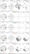
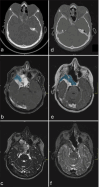
Case description: A 42-year-old woman undergoing treatment with nomegestrol acetate (NA) presented with headaches and visual loss in her right eye. She was diagnosed with a large spheno-orbital meningioma invading the sphenoid and ethmoid sinuses, associated with hyperostosis of the sphenoid wing. An initial resection was performed using an extended endonasal approach. Immunohistochemistry confirmed a chondroid meningioma, Grade II, with progestin receptor in 100% of the tumor cell nuclei and a Ki-67 proliferation index of 3-5%. NA was immediately stopped on diagnosis. Despite the cessation of the NA, the intraosseous sphenoidal part of the tumor continued to grow, leading to optic nerve compression. A second surgery was performed using a right fronto-temporo-orbito-zygomatic approach. Examination of the dura of the middle fossa showed subtle tumoral infiltration, while the Ki-67% index was estimated at 1%. Examination of the sphenoid bone demonstrated reactive hyperostosis with minimal to no tumor infiltration.
Conclusion: This case illustrates that the proliferative activity of the progestin-associated meningioma does not account for intraosseous progression within the sphenoid bone following cessation of progestin therapy. Our observations suggest an upregulation of osteogenesis in infiltrated bone, even as the dural part of the meningioma regresses.
Keywords: Histopathological correlation; Hyperostosis; Meningioma; Paradoxical evolution; Progestin; Skull base.
Copyright: © 2025 Surgical Neurology International.
Conflict of interest statement
There are no conflicts of interest.
Figures


References
-
- AbiJaoude S, Marijon P, Roblot P, Tran S, Cornu P, Kalamarides M, et al. Sustained growth of intraosseous hormone-associated meningiomas after cessation of progestin therapy. Acta Neurochir (Wien) 2021;163:1705–10. - PubMed
-
- Apra C, Roblot P, Alkhayri A, Le Guérinel C, Polivka M, Chauvet D. Female gender and exogenous progesterone exposition as risk factors for spheno-orbital meningiomas. J Neurooncol. 2020;149:95–101. - PubMed
-
- Balasch J. Sex steroids and bone: Current perspectives. Hum Reprod Update. 2003;9:207–22. - PubMed
Publication types
LinkOut - more resources
Full Text Sources
