Brain abscess mimicking a brain tumor only realized during surgery: A case report in a resource strained environment
- PMID: 40353178
- PMCID: PMC12065485
- DOI: 10.25259/SNI_67_2025
Brain abscess mimicking a brain tumor only realized during surgery: A case report in a resource strained environment
Abstract
Background: Diagnosis of brain tumors increased in sub-Saharan Africa since the advent of computed tomography (CT)-scans and magnetic resonance imaging (MRI) in these regions, enabling easy diagnosis. However, some histological types of brain tumors can be confusing, especially on CT-scan, simulating other pathologies such as inflammatory granulomas or pyogenic abscesses. MRI, in this instance, with its diffusion-weighted imaging, susceptibility weighted imaging, or perfusion imaging, is important to help with accurate diagnosis. The down side of these imaging facilities, however, is that less and less importance is accorded to proper and detailed history taking. Such a care-free attitude to history taking can be costly, especially in resource strained environments.
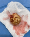
Case description: We report the case of a 06-year-old child who presented with seizures associated with headaches and vomiting. In this case, proper history taking following the surgical intervention revealed a history of head trauma after a fall with a scalp wound, which was suppurated but later progressed well. The CT scan showed a solid cystic lesion. The first component is a ring enhanced portion (hyperdense ring with the hypodense center, surrounded by edema) with central calcification located in the frontal region, and the second component is a cystic portion located in the temporal region. This lesion with dual component was more suggestive of a tumoral lesion on imaging than an abscess. The child did not benefit from further imaging due to unavailability in the region as well as the socioeconomic status of the family making them incapable of going elsewhere to do it. A decision to surgically excise the lesion was made, and during surgery, we found a well-circumscribed yellowish lesion associated with an arachnoid cyst. The capsule of the lesion was very thick, and after opening it, the content was pus combined with debris. The child did well on antibiotic therapy post-surgery. The follow-up was unremarkable.
Conclusion: Brain MRI is essential to differentiate some pyogenic brain abscesses from tumors. However, meticulous history taking is important to gather as much information as possible about any medical pathology, which would then be corroborated with the physical examination findings and imaging to increase diagnostic accuracy and minimize misdiagnosis.
Keywords: Abscess; Brain tumor; Magnetic resonance imaging.
Copyright: © 2025 Surgical Neurology International.
Conflict of interest statement
There are no conflicts of interest.
Figures


Similar articles
-
Letter to the Editor: Depression As The First Symptom Of Frontal Lobe Grade 2 Malignant Glioma.Turk Psikiyatri Derg. 2022 Summer;33(2):143-145. doi: 10.5080/u25957. Turk Psikiyatri Derg. 2022. PMID: 35730515 English, Turkish.
-
More advantages in detecting bone and soft tissue metastases from prostate cancer using 18F-PSMA PET/CT.Hell J Nucl Med. 2019 Jan-Apr;22(1):6-9. doi: 10.1967/s002449910952. Epub 2019 Mar 7. Hell J Nucl Med. 2019. PMID: 30843003
-
Brain abscess and cystic brain tumor: discrimination with dynamic susceptibility contrast perfusion-weighted MRI.J Comput Assist Tomogr. 2005 Sep-Oct;29(5):663-7. doi: 10.1097/01.rct.0000168868.50256.55. J Comput Assist Tomogr. 2005. PMID: 16163039
-
Role of diffusion-weighted imaging and proton MR spectroscopy in distinguishing between pyogenic brain abscess and necrotic brain tumor.Acta Neurol Taiwan. 2004 Sep;13(3):107-13. Acta Neurol Taiwan. 2004. PMID: 15508936 Review.
-
Pituitary abscess following endoscopic endonasal drainage of a suprasellar arachnoid cyst: Case report and review of the literature.J Clin Neurosci. 2019 Oct;68:322-328. doi: 10.1016/j.jocn.2019.07.081. Epub 2019 Aug 8. J Clin Neurosci. 2019. PMID: 31402262 Review.
References
-
- Bodilsen J, Dalager-Pedersen M, Van De Beek D, Brouwer MC, Nielsen H. Risk factors for brain abscess: A nationwide, population-based, nested case-control study. Clin Infect Dis. 2020;71:1040–6. - PubMed
-
- Brouwer MC, Coutinho JM, Van De Beek D. Clinical characteristics and outcome of brain abscess: Systematic review and meta-analysis. Neurology. 2014;82:806–13. - PubMed
-
- Brouwer MC, Tunkel AR, McKhann GM, 2nd, Van De Beek D. Brain abscess. N Engl J Med. 2014;371:447–56. - PubMed
-
- Chuang JM, Lin WC, Fang FM, Huang YJ, Ho JT, Lu CH. Bacterial brain abscess formation in post-irradiated patients: What is the possible pathogenesis? Clin Neurol Neurosurg. 2015;136:132–8. - PubMed
-
- Dahal T, Bhardwaj S, Sharma P. Pyogenic-cerebral (brain) abscess. J Neurol Neurosci. 2022;13:408.
Publication types
LinkOut - more resources
Full Text Sources
