Review
. 2025 Dec;16(1):2503423.
doi: 10.1080/21505594.2025.2503423. Epub 2025 May 15.

Pathogenicity and virulence of Chlamydia trachomatis: Insights into host interactions, immune evasion, and intracellular survival

Affiliations
Review

Pathogenicity and virulence of Chlamydia trachomatis: Insights into host interactions, immune evasion, and intracellular survival

Alix McCullough et al. Virulence. 2025 Dec.

Abstract

Chlamydia trachomatis is an obligate intracellular pathogen and the leading cause of bacterial sexually transmitted infections and infectious blindness worldwide. All Chlamydia species share a unique biphasic developmental cycle, alternating between infectious elementary bodies (EBs) and replicative reticulate bodies (RBs). The pathogenesis of C. trachomatis is driven by a sophisticated arsenal of adhesins, conventional type III secretion system effector proteins, and inclusion membrane proteins that subvert host cellular processes to establish infection and promote survival. In this review, we highlight the molecular mechanisms underlying C. trachomatis infection, focusing on key stages of its developmental cycle, including adhesion, invasion, replication, and egress. We delve into its interactions with host cytoskeletal structures, immune signaling pathways, and intracellular trafficking systems, as well as its strategies for immune evasion and persistence. Understanding these mechanisms offers critical insights into C. trachomatis pathogenesis and identifies promising avenues for therapeutic and vaccine development.

Keywords: Chlamydia; Inc; T3SS; adhesion; effector; host pathogen interactions.


Conflict of interest statement

No potential conflict of interest was reported by the author(s).

Figures
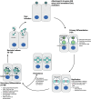
Figure 1.
Figure 1.
C. trachomatis developmental cycle. Infectious elementary bodies (EBs) secrete type III secretion system (T3SS) effector proteins to facilitate host cell invasion. Once inside the host cell, EBs undergo primary differentiation to convert into replicative reticulate bodies (RBs). After multiple rounds of replication, RBs asynchronously convert back into EBs (secondary differentiation) and are released from the host cell via lysis or extrusion. The newly infectious EBs then infect additional cells, perpetuating the infection cycle. In response to stress during replication, RBs can convert to persistent aberrant bodies (ABs), which exhibit reduced immune recognition and increased resilience. ABs are capable of generating infectious progeny through budding after removal of the stressor.
Figure 2.
Figure 2.
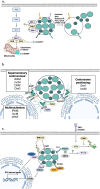
Role of C. trachomatis effectors and adhesins in manipulating host cell processes. (a) Key adhesins and host receptors facilitating C. trachomatis attachment. MOMP and OmcB mediate interaction with host heparan sulfate (HSPG), while LPS interacts with CFTR and Ctad1 interacts with integrin β1, stimulating ERK signaling. (b) Early secreted effectors TarP and TmeA orchestrate cytoskeletal rearrangements to promote bacterial uptake and manipulate host cellular dynamics. TarP activates the Arp2/3 complex via Rac1 signaling and Wave2, while TmeA directly activates Arp2/3 through N-WASP binding and promotes pedestal formation via TOCA-1. Dyn2 is recruited by TarP and polymerized by TmeA. TmeB antagonizes Arp2/3, likely reversing cytoskeletal changes post-invasion. TepP recruits crk, CrkL, PI3K, and Eps8 to the inclusion, with Eps8 contributing to the breakdown of epithelial tight junctions. (c) During mid-cycle, C. trachomatis establishes ER-inclusion contact sites via IncD, IncV, and IncS. It co-opts small GTPases such as arfs, Rabs, and RhoA through Inc proteins like CpoS and InaC, which contributes to inclusion growth, stability, and nutrient acquisition. IncE manipulates Snx5/6 and STX7/12 to recruit hybrid vesicles, while IncA facilitates homotypic inclusion fusion. Centrosome repositioning near the inclusion is mediated by IncM, IPAM, and Dre1. (d) Late in the infection cycle, bacterial release occurs via extrusion or lysis. Extrusion is driven by post-translational modifications of Inc proteins CT228, InaC, and IPAM by Cdu1. CT228 and MrcA regulate extrusion by interacting with MYPT1 and ITPR3, respectively. C. trachomatis effectors CteG, Pgp4, and CPAF contribute to host cell egress via lysis.
Figure 3.
Figure 3.
C. trachomatis secreted proteins manipulate host cell viability, centrosome dynamics and the immune response to promote infection. (a) The anti-apoptotic protein mcl-1 is upregulated via MAPK signaling, stabilized by PI3K/AKT pathways, and de-ubiquitinated by Cdu1. Ectopically expressed MOMP localizes to the mitochondria and inhibits apoptosis. C. trachomatis inclusion membrane stability is maintained by inc proteins, including CT383, IncC, CpoS, and IncS, which prevent premature lysis and promote host cell survival. (b) The inc protein IPAM induces cytokinesis defects and promotes supernumerary centrosome formation, while Dre1 interacts with dynactin to position centrosomes around the inclusion. IncM contributes to multinucleation and centrosome localization by interacting with CCDC146. CPAF and CteG also induce centrosome amplification, with CteG targeting host centrin-2, a regulator of centriole duplication, to promote supernumerary centrosome formation and intracellular survival. (c) TepP recruits scaffolding proteins and PI3K to the inclusion, modulating host chemokine expression to diminish neutrophil recruitment. The effector Cdu1 inhibits NF-κB signaling by stabilizing IκBα, while Tri1 displaces TRAF7-associated kinases to potentially suppress NF-κB signaling. Other effectors, such as CpoS and GarD, suppress STING and RNF213 pathways to evade interferon responses. CT226 recruits LRRF1 to the inclusion membrane, potentially regulating innate immune responses.

References

    1. Luu LDW, Kasimov V, Phillips S, et al. Genome organization and genomics in Chlamydia: whole genome sequencing increases understanding of chlamydial virulence, evolution, and phylogeny. Front Cell Infect Microbiol. 2023;13:1178736. doi: 10.3389/fcimb.2023.1178736 - DOI - PMC - PubMed
    1. Burton MJ, Mabey DC, Brooker S.. The global burden of trachoma: a review. PLOS Neglected Trop Dis. 2009;3(10):e460. doi: 10.1371/journal.pntd.0000460 - DOI - PMC - PubMed
    1. Huai P, Li F, Chu T, et al. Prevalence of genital chlamydia trachomatis infection in the general population: a meta-analysis. BMC Infect Dis. 2020;20(1):589. doi: 10.1186/s12879-020-05307-w - DOI - PMC - PubMed
    1. Huai P, Li F, Li Z, et al. Prevalence, risk factors, and medical costs of chlamydia trachomatis infections in Shandong Province, China: a population-based, cross-sectional study. BMC Infect Dis. 2018;18(1):534. doi: 10.1186/s12879-018-3432-y - DOI - PMC - PubMed
    1. Gupta K, Harrison SA, Davis NA, et al. Prevalence of chlamydia trachomatis infection in young women and associated predictors. Sex Transm Dis. 2021;48(8):529–29. doi: 10.1097/OLQ.0000000000001372 - DOI - PMC - PubMed

Publication types

Substances

LinkOut - more resources