. 2025 Dec;14(1):2505649.
doi: 10.1080/22221751.2025.2505649. Epub 2025 May 30.

Clade 2.3.4.4b highly pathogenic H5N1 influenza viruses from birds in China replicate effectively in bovine cells and pose potential public health risk

Affiliations

Clade 2.3.4.4b highly pathogenic H5N1 influenza viruses from birds in China replicate effectively in bovine cells and pose potential public health risk

Junlong Xiong et al. Emerg Microbes Infect. 2025 Dec.

Abstract

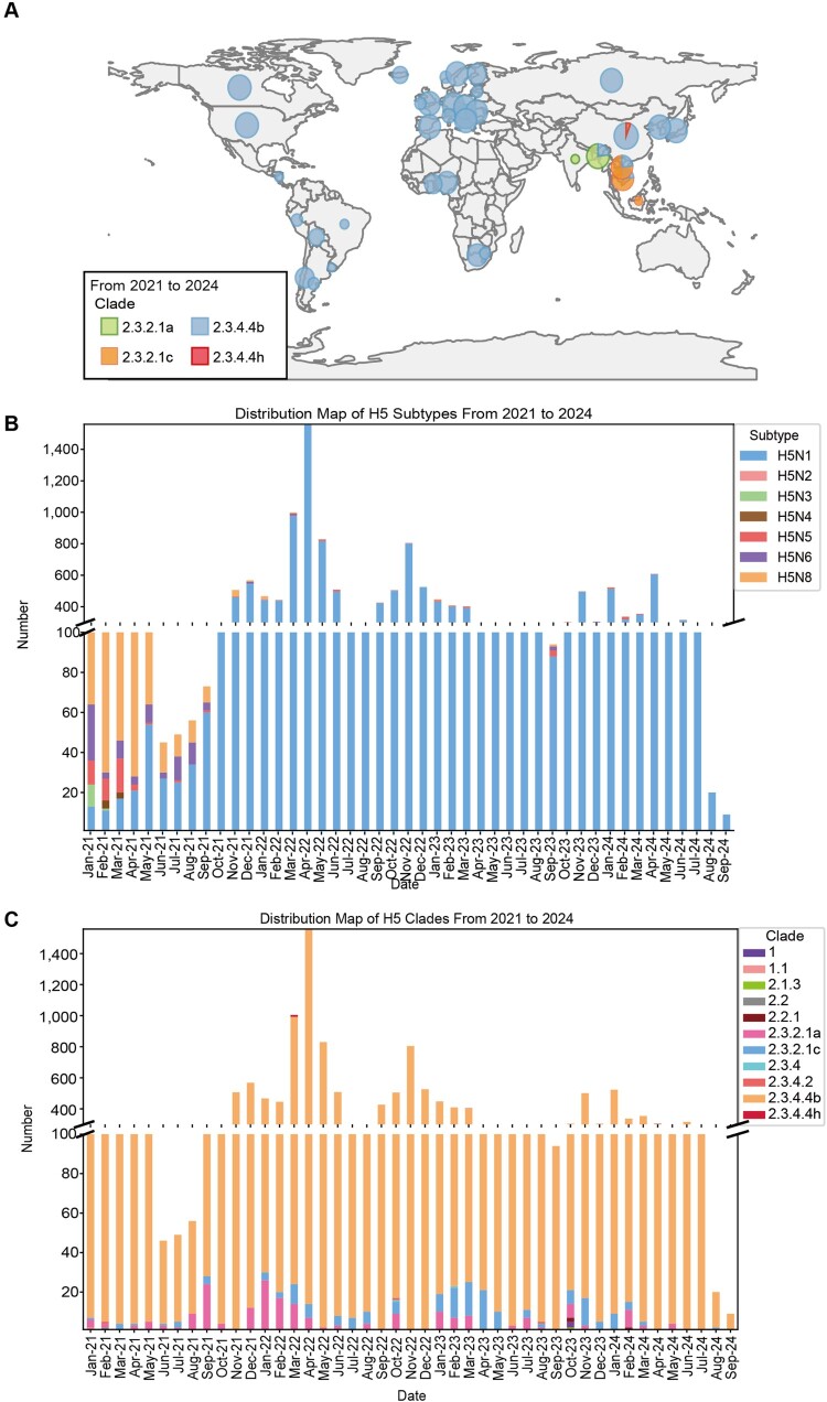
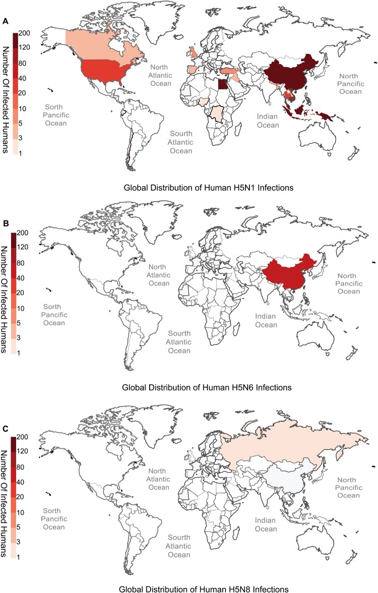
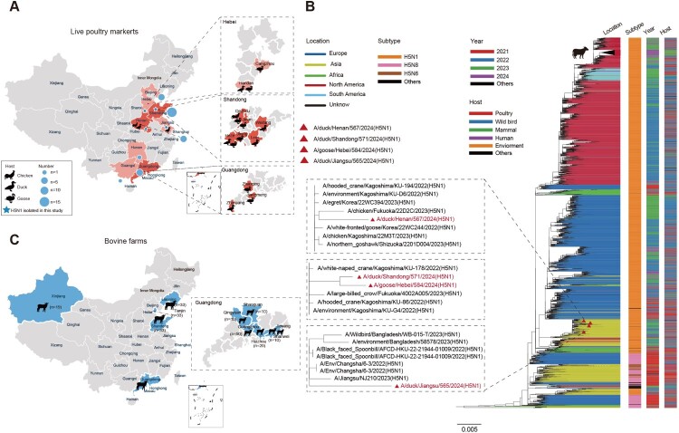
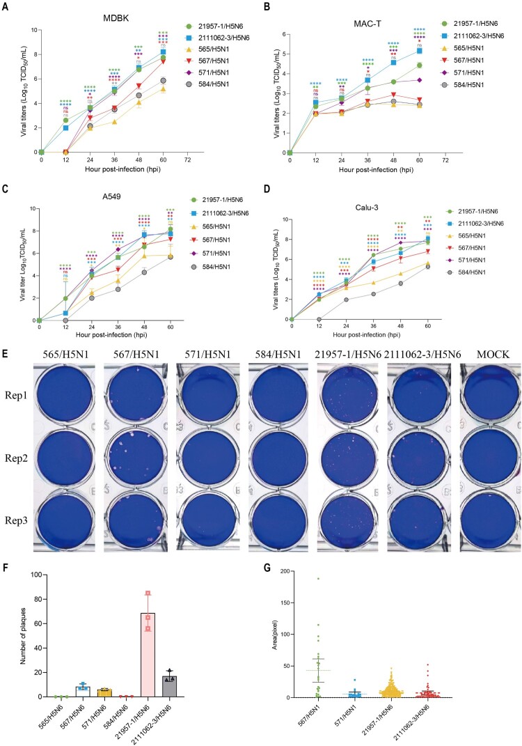
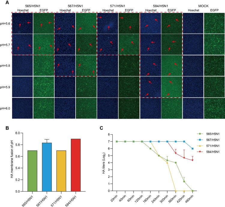
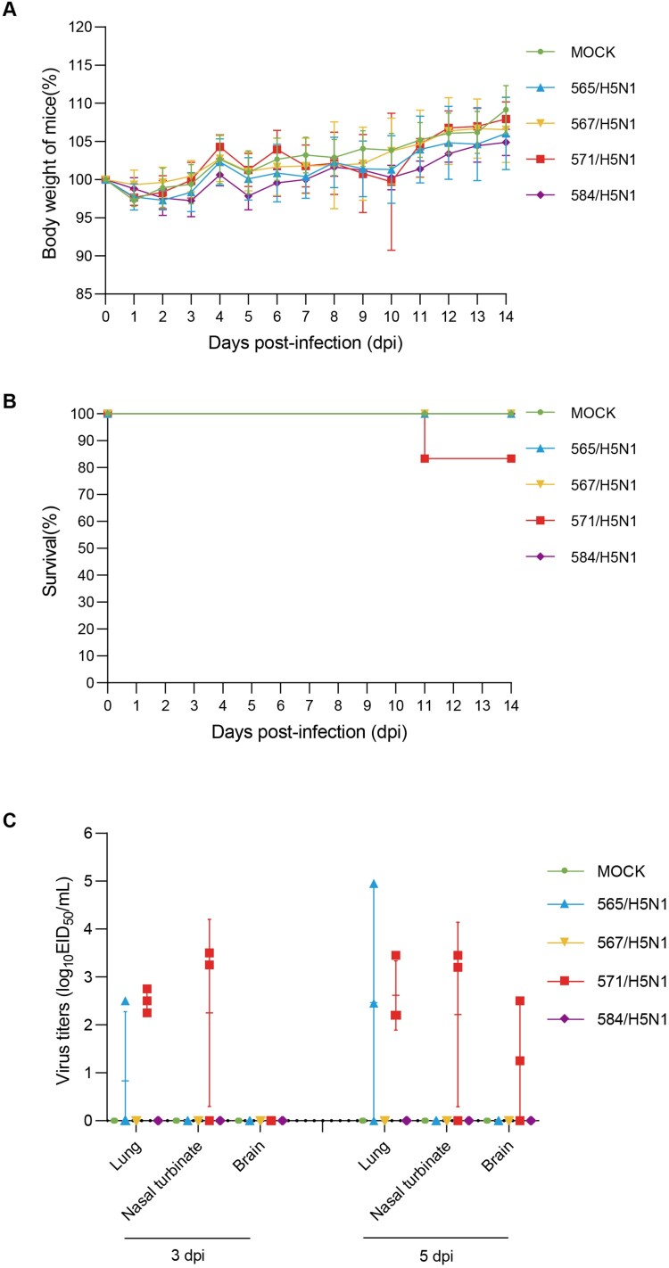
In February 2024, H5N1 highly pathogenic avian influenza viruses (HPAIVs) of clade 2.3.4.4b were first reported in dairy cows in the USA. Subsequent multiple outbreaks on dairy farms and sporadic human infections have raised substantial public health concerns. In the same year, four H5N1 HPAIVs of clade 2.3.4.4b were isolated from ducks and geese in live poultry markets (LPMs) spanning seven provinces in China. Evolutionary analysis demonstrated that these viruses had undergone two genetic reassortments with H5 influenza viruses from wild birds in different countries. Except for 565/H5N1, the other three viruses exhibited over 99% genetic homology with avian-origin H5N1 HPAIVs from South Korea and Japan. Notably, 571/H5N1 demonstrated high replication efficiency in bovine-derived cells, particularly in bovine mammary epithelial (MAC-T) cells, and caused 16.7% (1/6) mortality in mice at a dose of 105 EID50/50 μL, indicating its zoonotic potential. Given the potential cross-species transmission risk of H5N1 HPAIVs to cattle herds, we collected 228 serum samples from 12 cattle farms across five provinces and conducted serological testing to investigate seroprevalence of H5N1 HPAIVs in Chinese cattle herds. All tested samples were negative, indicating no widespread infection in the sampled cattle populations. However, infections in cattle from other regions cannot be ruled out. Nevertheless, due to the high mutability of H5N1 HPAIVs, enhanced surveillance of avian influenza viruses is critical to ensure timely responses to potential outbreaks.

Keywords: H5N1; cattle; cross-species transmission; evolution; influenza virus; zoonotic potential.


Conflict of interest statement

No potential conflict of interest was reported by the author(s).

Figures

Figure 1.
Figure 1.
Global prevalence of H5 influenza viruses during 2021–2024. (A) Global distribution of H5 influenza viruses of different subclades in different continents. The map was drawn by R software. Green represents branch 2.3.2.1a, blue represents branch 2.3.4.4b, yellow represents branch 2.3.2.1c, and red represents branch 2.3.4.4h. (B) The number of NA subtypes of global H5 influenza viruses during 2021–2024. Blue represents H5N1, pink represents H5N2, green represents H5N3, light yellow represents H5N4, red represents H5N5, purple represents H5N6, and dark yellow represents H5N8. (C) The number of subclades of global H5 influenza viruses during 2021–2024. The colour-coded classification system assigns specific hues to each subclade: Purple represents Subclade 1, while Pink indicates Subclade 1.1. Subclade 2.1.3 is marked in Green, and another Grey shade designates Subclade 2.2. Brown corresponds to Subclade 2.2.1, followed by Light Pink for Subclade 2.3.2.1a. Subclade 2.3.2.1c is highlighted in Dark Blue, while Blue denotes Subclade 2.3.4. Additionally, Red signifies Subclade 2.3.4.2, Yellow represents Subclade 2.3.4.4b, and Dark Red is used for Subclade 2.3.4.4h.
Figure 2.
Figure 2.
Global distribution of human infections caused by H5 avian influenza virus subtypes. Maps show the cumulative number of laboratory-confirmed human infections with H5N1, H5N6, and H5N8 subtypes, based on GISAID records. (A) The top panel illustrates the global distribution of H5N1 infections, with high numbers reported in China and several Southeast Asian and African countries. (B) The middle panel displays H5N6 infections, predominantly reported in China. (C) The bottom panel shows H5N8 infections, mainly reported in Russia. Colour intensity reflects the total number of infected individuals in each country, with darker shades indicating more cases. Data were retrieved from the GISAID EpiFlu™ database by filtering for human-origin H5 subtype entries.
Figure 3.
Figure 3.
Sampling of H5 influenza viruses in birds and cattle of China during 2024 and phylogenetic analysis of H5 influenza viruses. (A) Sampling sites of H5 influenza viruses isolated from chickens, ducks, and geese in LPMs of China in 2024. (B) Phylogenetic tree of HA genes of H5 influenza viruses from 2021 to 2024. (C) Sampling sites of serum samples collected from 12 bovine farms of China in 2024. All branch lengths were scaled according to the numbers of substitutions per site. Maximum likelihood (ML) phylogenies for the codon alignment of HA gene segments were estimated using the GTR + G nucleotide substitution model in the IQ-TREE. Node support was determined by nonparametric bootstrapping with 1,000 replicates. The phylogenetic tree was graphically illustrated in the FigTree (version 1.4.3) programme (http://tree.bio.ed.ac.uk/software/figtree/). In the phylogenetic tree, four H5N1 isolated in the study were marked with red delta. It highlights the location of the virus in which the dairy cows were infected. Various line colours indicate the location of virus isolation; coloured rectangular bars on the right side of the tree indicate the subtype, the year and the host corresponding to the virus at each position in the tree, respectively.
Figure 4.
Figure 4.
Viral replication and virulence of different H5 avian influenza viruses. Growth curves after inoculation of each H5 avian influenza viruses (21957-1/H5N6, 2111062-3/H5N6, 565/H5N1, 567/H5N1, 571/H5N1, 584/H5N1) into MDBK cells (A), MAC-T cell (B), A549 cells (C) and Calu-3 cells (D). The supernatants of infected cells were collected at the indicated time points. Data are represented as means ± SEM. The experimental data were analysed using an independent samples t-test. Prior to data analysis, normality was confirmed by the Shapiro-Wilk test (p > 0.05), and homogeneity of variances between groups was verified by Levene’s test (p = 0.12), meeting the assumptions for the t-test. Independent samples t-tests were performed to determine whether significant differences existed between other virus strains and the 565/H5N1 virus (A-B) or the 584/H5N1 virus (C-D). Significance levels are denoted as *p < 0.05, **p < 0.01, and ***p < 0.001. (E) Plaque imaging of the six H5 avian influenza viruses in MDBK cells was conducted. Plaque counting (F) and size measurements (G) were performed using Image J software, and graphs were created using GraphPad Prism 9.
Figure 5.
Figure 5.
HA acid stability and thermal stability. The acid stability of hyaluronic acid (HA) was assessed using the syncytia assay. Syncytia formation was observed in Vero-GFP cells infected with influenza viruses at different pH values. (A) shows the syncytia formation for viruses 565/H5N1, 567/H5N1, 571/H5N1, and 584/H5N1 at pH values ranging from 5.6–6.0, with red arrows indicating syncytia formation. The images were captured using a fluorescence microscope (Nikon) with a scale bar of 200 μm. A bar graph (B) depicts the pH at which syncytia formation occurred for each virus based on three replicate samples. The changes in hemagglutination titer for viruses 565/H5N1, 567/H5N1, 571/H5N1, and 584/H5N1 after incubation at 50°C are shown in (C), with each sample performed in triplicate.
Figure 6.
Figure 6.
Pathogenicity of four H5N1 influenza viruses in mice. Groups of twelve mice were intranasally inoculated with four H5N1 viruses (dose: 105 EID50/50 µL). At 3 and 5 dpi, three mice per group were randomly euthanized for organ collection (lungs, brains, and nasal turbinates). (a) Body weight changes of infected mice. (b) Survival rates of infected mice. (c) Viral titers in the lungs, brains, and nasal turbinates at 3 and 5 dpi.

Similar articles

References

    1. Meade PS, Bandawane P, Bushfield K, et al. . Detection of clade 2.3.4.4b highly pathogenic H5N1 influenza virus in New York city. J Virol. 2024 Jun 13;98(6):e0062624. - PMC - PubMed
    1. Restori KH, Septer KM, Field CJ, et al. . Risk assessment of a highly pathogenic H5N1 influenza virus from mink. Nat Commun. 2024 May 15;15(1):4112. - PMC - PubMed
    1. Xie R, Edwards KM, Wille M, et al. . The episodic resurgence of highly pathogenic avian influenza H5 virus. Nature. 2023 Oct;622(7984):810–817. - PubMed
    1. Maemura T, Guan L, Gu C, et al. . Characterization of highly pathogenic clade 2.3.4.4b H5N1 mink influenza viruses. EBioMedicine. 2023 Nov;97:104827. - PMC - PubMed
    1. Pulit-Penaloza JA, Brock N, Belser JA, et al. . Highly pathogenic avian influenza A(H5N1) virus of clade 2.3.4.4b isolated from a human case in Chile causes fatal disease and transmits between co-housed ferrets. Emerg Microbes Infect. 2024 Dec;13(1):2332667. - PMC - PubMed

MeSH terms