. 2025 Jun 17;99(6):e0037425.
doi: 10.1128/jvi.00374-25. Epub 2025 May 12.

IFNβ absence compensates for LAT functions in latency reactivation and T cell exhaustion

Affiliations

IFNβ absence compensates for LAT functions in latency reactivation and T cell exhaustion

Shaohui Wang et al. J Virol. .

Abstract

Type I interferons (IFNs) generate strong antiviral immunity following viral infection in mice and humans. These type I IFNs are encoded by at least 14 IFNα genes and a single IFNβ gene. We showed that the absence of one of the IFNα genes (IFNα2A-/-) affected latency levels in herpes simplex virus type 1 (HSV-1) ocularly infected mice but not in infected control mice, and the absence of IFNα2A did not affect viral reactivation in ocularly infected mice. Since the role of IFNβ in HSV-1 latency reactivation and the potential effect of latency-associated transcript (LAT) on IFNβ activity is not known, we ocularly infected IFNβ-/- mice with different doses of LAT-plus [LAT(+)] and LAT-minus [LAT(-)] viruses. Wild-type (WT) control mice and IFNβ-/- mice were infected similarly. Virus titers in the eye, viral and cellular transcripts in the eye and trigeminal ganglia (TG) of infected mice on days 3 and 5 post-infection, eye disease, survival, latency-reactivation levels, and T cell exhaustion were measured in latently infected mice. Virus replication and viral and cellular transcripts in the eye of infected IFNβ-/- mice were similar to those in WT mice, while eye disease and survival in IFNβ-/- mice differed significantly from WT mice. WT mice infected with LAT(-) virus showed reduced latency, slower reactivation, and less T cell exhaustion than mice infected with LAT(+) virus. However, using different doses of each virus, latency levels, time of reactivation, and T cell exhaustion were similar between LAT(+) and LAT(-) viruses. These results suggest that the absence of IFNβ expression compensates for the function of LAT with regard to levels of latency, T cell exhaustion, and reactivation but does not affect viral and cellular transcripts during primary infection.IMPORTANCEInterferon β (IFNβ) is a type I interferon that plays an important role in controlling primary herpes simplex virus type 1 (HSV-1) infection. To evaluate the importance of IFNβ on HSV-1 latency reactivation and its relationship to LAT, we infected IFNβ-/- mice with LAT(+) and LAT(-) viruses. In the absence of IFNβ, latency levels in mice infected with LAT(-) virus were similar to those of mice infected with LAT(+) virus. The absence of IFNβ also reduced the time of reactivation in mice infected with LAT(-) virus to that of LAT(+) virus. Our results show a strong correlation between the functions of LAT and IFNβ during latent but not primary stages of HSV-1 infection.

Keywords: CD8; IFNβ; PD-1; exhaustion; eye disease; knockout; latency; ocular infection; reactivation; type 1 interferon; virus replication.


Conflict of interest statement

The authors declare no conflict of interest.

Figures

Fig 1
Fig 1
Virus titers in the eyes of infected mice. IFNβ-/- and WT control mice were ocularly infected with 2 × 10⁵ PFU/eye of LAT(+) and LAT(-) viruses. Tear films were collected on days 1 to 5 PI, and virus titers were determined by standard plaque assay. Each point represents mean titers of 16 eyes for WT and 14 eyes for IFNα β-/- mice from two separate experiments.
Fig 2
Fig 2
Viral gene expression in corneas and TG during primary mouse infection. IFNβ-/- and WT mice were ocularly infected with 2 × 10⁵ PFU/eye of LAT(+) and LAT(-) viruses as in Fig. 1. Corneas (panels A, B, and C) and TG (panels D, E, and F) were harvested on days 3 and 5 PI. Total RNA was extracted, and ICP0 (panels A and D), gB (panels B and E), and gK (panels C and F) copy numbers were measured by qRT-PCR and normalized with GAPDH. Each point represents the mean ± SEM of 10 corneas and 10 TG for each day.
Fig 3
Fig 3
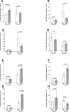
Effect of IFNβ absence on expression of immune genes during primary infection. RNA isolated on days 3 and 5 PI from corneas (panels A–D) and TG (panels E–H) of IFNβ-/- (gray bars) and WT (open bars) infected mice as described in Fig. 2 above were used to measure the expression of CD4 (panels A and E), CD8α (panels B and F), IFNγ (panels C and G), and IFNα2A (panels D and H). qRT-PCR was performed as described above, and GAPDH was used as the endogenous control. Corneas and TG from uninfected IFNβ-/- and WT mice were used for normalization. Each bar represents the mean fold change in expression of 10 corneas and 10 TG on each day.
Fig 4
Fig 4
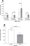
gB DNA levels in TG of latently infected mice. (A) Latency in IFNβ-/- infected mice. IFNβ-/- mice were ocularly infected with 1 × 10³, 1 × 10⁴, 1 × 10⁵, or 2 × 10⁵ PFU/eye of LAT(+) and LAT(-) viruses. TG from surviving mice was harvested on day 28 PI, and total TG DNA was isolated as described in Materials and Methods. gB DNA copy number was measured by qPCR using a standard curve generated with pAc-gB1 DNA. GAPDH expression was used to normalize relative levels of gB DNA expression. Each bar represents the mean ± SEM from 18, 12, 12, and 10 TG infected with 1 × 10³, 1 × 10⁴, 1 × 10⁵, and 2 × 10⁵ PFU/eye of LAT(+), respectively. Latency in mice infected with LAT(-) virus is based on 16 TG for 1 × 10³ PFU/eye, 14 TG for 1 × 10⁴ PFU/eye, 12 TG for 1 × 10⁵ PFU/eye, and 12 TG for 2 × 10⁵ PFU/eye; and (B) latency in WT infected mice. WT mice were ocularly infected with 2 × 10⁵ PFU/eye of LAT(+) and LAT(-) viruses. TG were isolated on day 28, DNA was extracted, and HSV-1 gB DNA levels were determined as above. Each bar represents the mean ± SEM from 46 TG for LAT(+) virus and 23 TG for LAT(-) virus from three independent experiments.
Fig 5
Fig 5
Effects of IFNβ absence on CS and explant reactivation. (A) CS in infected mice. Severity of CS on surviving mice infected with 2 × 10⁵ PFU/eye of LAT(+) and LAT(-) viruses was determined on day 28 PI as described in Materials and Methods. Each bar represents the mean ± SEM from 36 eyes [IFNβ-/-, LAT(+)], 26 eyes [IFNβ-/-, LAT(-)], 30 eyes [WT, LAT(+)], and 26 eyes [WT, LAT(-)]. (B) Virus reactivation in infected mice. IFNβ-/- and WT control mice were ocularly infected with 1 × 10⁵ PFU/eye of LAT(+) and LAT(-) viruses as above. TG from infected mice were harvested on day 28 PI, and an explant reactivation assay was performed. Data points indicate the day of virus reactivation. Each point is the mean ± SEM from 20 TG for IFNβ-/- mice infected with LAT(+) virus, 26 TG for IFNβ-/- mice infected with LAT(-) virus, and 12 TG for WT groups infected with LAT(+) and LAT(-) viruses. Data for IFNβ-/- infected mice are based on two independent experiments, while data from WT infected mice are based on one experiment.
Fig 6
Fig 6
Effects of IFNβ absence on T cell exhaustion in TG of latently infected mice. IFNβ-/- and WT mice were ocularly infected with 2 × 10⁵ PFU/eye of LAT(+) and LAT(-) viruses as above. TG from latently infected mice were individually isolated on day 28 PI, and qRT-PCR was performed using total RNA as described in Materials and Methods. CD8α (panel A) and PD-1 (panel B) expressions in naive mice were used as a baseline control to estimate relative expression of each transcript in TG of latently infected mice. GAPDH expression was used to normalize relative expression of each transcript. Each bar represents the mean ± SEM from 23 TG for WT mice infected with LAT(+), 12 TG for WT mice infected with LAT(-), 10 TG for IFNβ-/- mice infected with LAT(+), and 14 TG for IFNβ-/- mice infected with LAT(+) viruses.

References

    1. Matzinger P. 1994. Tolerance, danger, and the extended family. Annu Rev Immunol 12:991–1045. doi: 10.1146/annurev.iy.12.040194.005015 - DOI - PubMed
    1. Biswas PS, Rouse BT. 2005. Early events in HSV keratitis—setting the stage for a blinding disease. Microbes Infect 7:799–810. doi: 10.1016/j.micinf.2005.03.003 - DOI - PubMed
    1. Jamali A, Hu K, Sendra VG, Blanco T, Lopez MJ, Ortiz G, Qazi Y, Zheng L, Turhan A, Harris DL, Hamrah P. 2020. Characterization of resident corneal plasmacytoid dendritic cells and their pivotal role in herpes simplex keratitis. Cell Rep 32:108099. doi: 10.1016/j.celrep.2020.108099 - DOI - PMC - PubMed
    1. Stark GR, Kerr IM, Williams BR, Silverman RH, Schreiber RD. 1998. How cells respond to interferons. Annu Rev Biochem 67:227–264. doi: 10.1146/annurev.biochem.67.1.227 - DOI - PubMed
    1. Yin Y, Favoreel HW. 2021. Herpesviruses and the type III interferon system. Virol Sin 36:577–587. doi: 10.1007/s12250-020-00330-2 - DOI - PMC - PubMed

MeSH terms

Substances