. 2025 Jun 17;99(6):e0021925.
doi: 10.1128/jvi.00219-25. Epub 2025 May 12.

Murine norovirus allosteric escape mutants mimic gut activation

Affiliations

Murine norovirus allosteric escape mutants mimic gut activation

Michael B Sherman et al. J Virol. .

Abstract

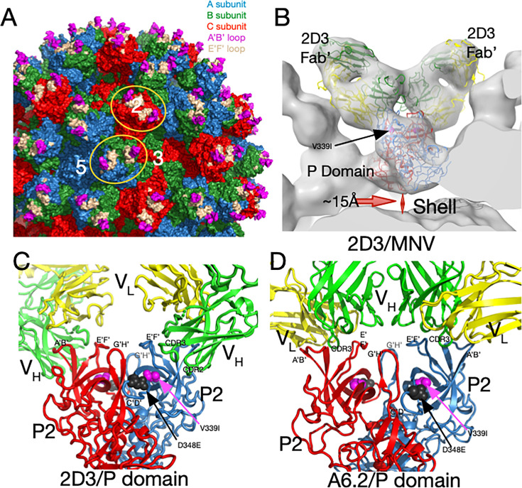
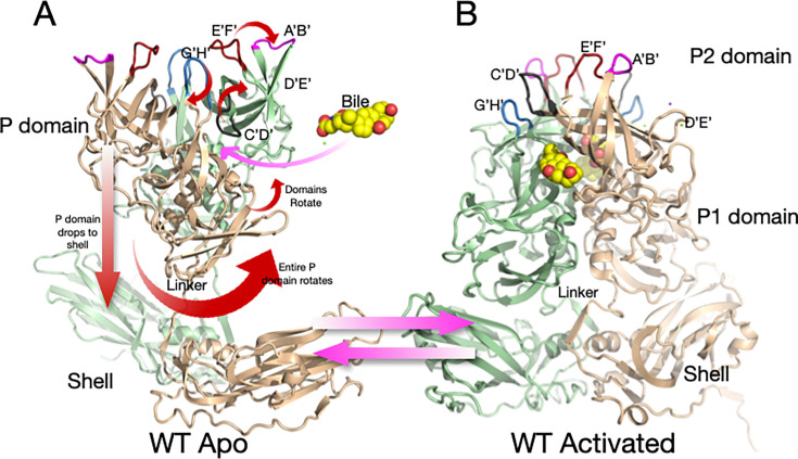
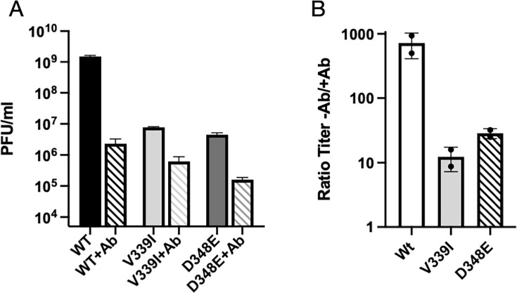
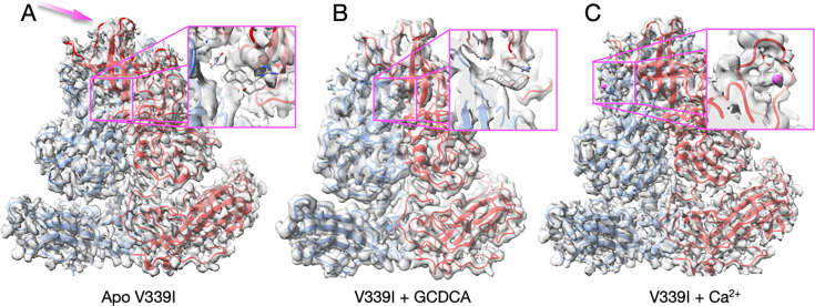
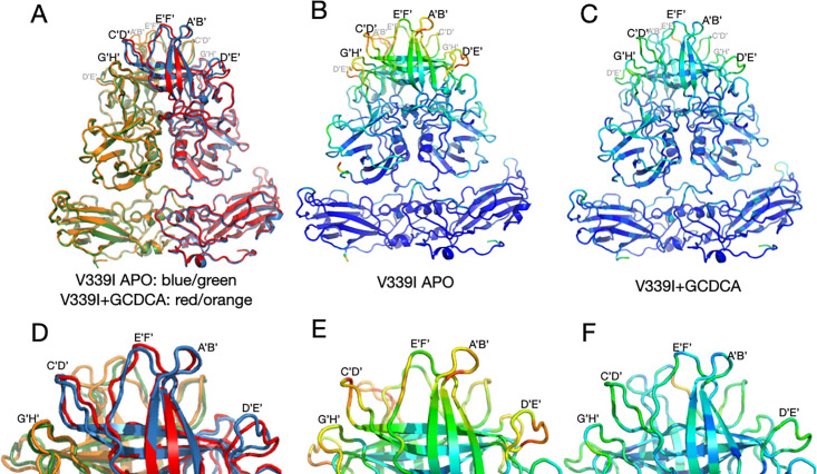
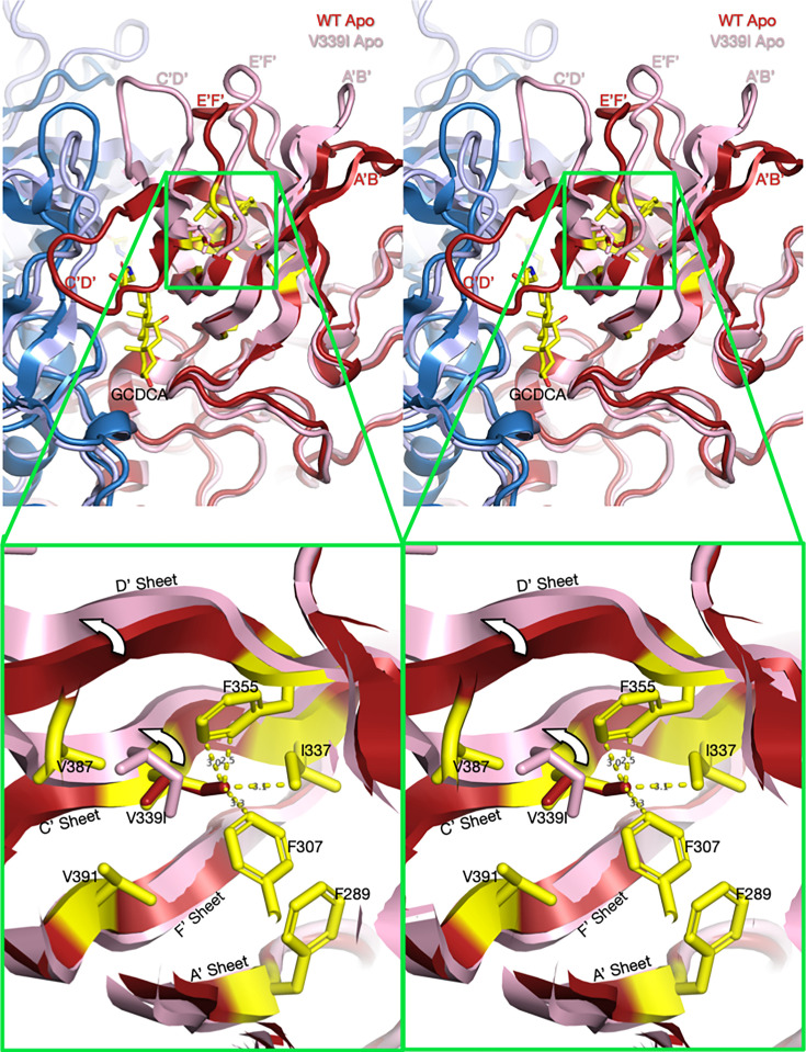
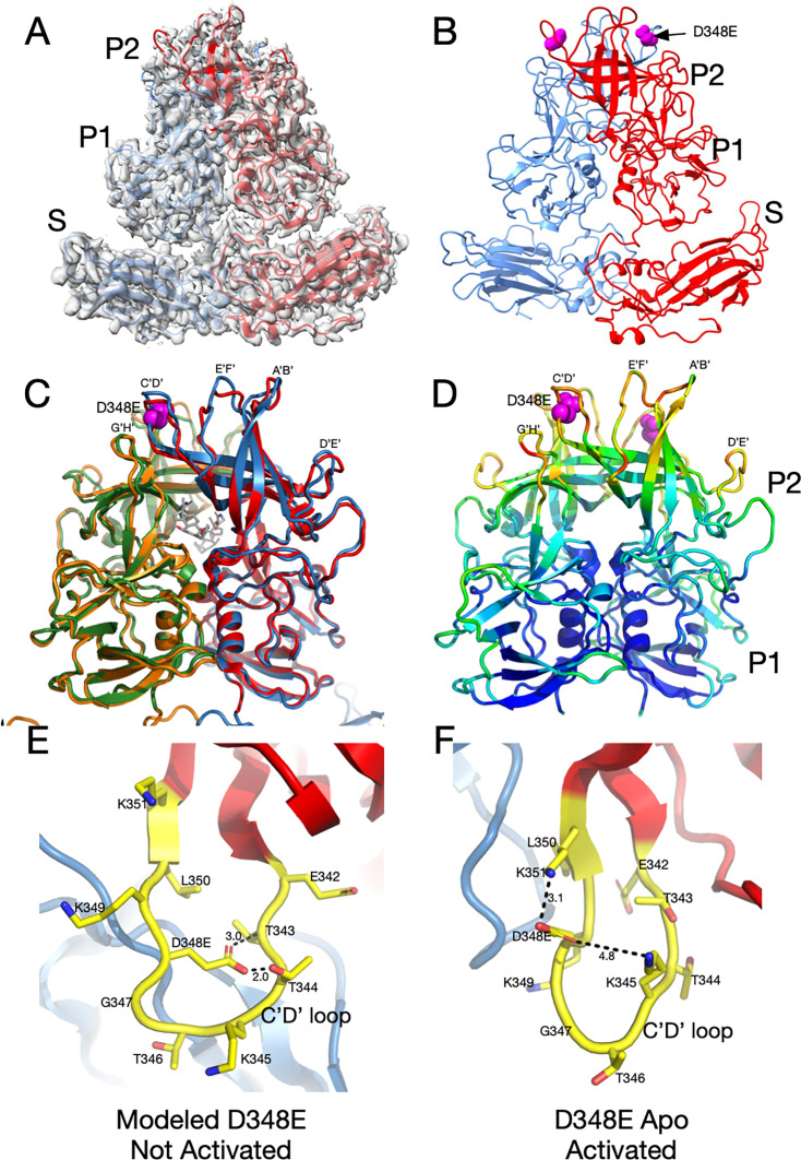
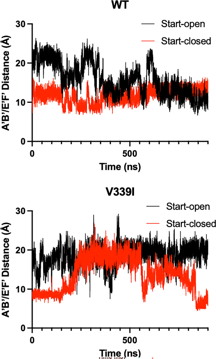
Murine norovirus (MNV) undergoes large conformational changes in response to the environment. The T=3 icosahedral capsid is composed of 180 copies of ~58 kDa VP1 that has N-terminal (N), shell (S), and C-terminal protruding (P) domains. In phosphate-buffered saline, the P domains are loosely tethered to the shell and float ~15 Å above the surface. At conditions found in the gut (i.e., low pH with high metal ion and bile salt concentrations), the P domain rotates and drops onto the shell with intra P domain changes that enhance receptor interactions while blocking antibody binding. Two of our monoclonal antibodies (2D3 and 4F9) have broad strain recognition, and the only escape mutants, V339I and D348E, are located on the C'D' loop and ~20 Å from the epitope. Here, we determined the cryo-EM structures of V339I and D348E at neutral pH +/-metal ions and bile salts. These allosteric escape mutants have the activated conformation in the absence of gut triggers. Since this conformation is not recognized by antibodies, it explains how these mutants evade antibody recognition. Dynamic simulations of the P domain further suggest that movement of the C'D' loop may be the rate-limiting step in the conformational change and that V339I increases the motion of the A'B'/E'F' loops compared to the wild-type (WT), facilitating the transition to the activated state. These findings have important implications for norovirus vaccine design since they uncover a form of the viral capsid that should lend superior immune protection against subsequent challenge by wild-type virus.IMPORTANCEImmune protection from norovirus infection is notoriously transient in both humans and mice. Our results strongly suggest that this is likely because the "activated" form of the virus found in gut conditions is not recognized by antibodies created in the circulation. By reversibly presenting one structure in the gut and a completely different antigenic structure in circulation, the gut tissue can be infected in subsequent challenges, while extraintestinal organs are protected. We find here that allosteric escape mutants to the most broadly neutralizing antibodies thwart recognition by transitioning to the activated state without the need for gut triggers (i.e., bile, low pH, or metal ions). These findings are significant because it is now feasible to present the activated form of the virus to the immune system (for example, as a vaccine) to better protect the gut tissue for longer periods of time.

Keywords: antibodies; cryo-EM; escape; norovirus.


Conflict of interest statement

The authors declare no conflict of interest.

Figures

Fig 1
Fig 1
MNV capsid structure and antibody complexes. (A) A surface-rendered section of the MNV capsid showing VP1–3 colored blue, green, and red, respectively. A/B subunits form dimers around the fivefold axes, while the adjacent C/C subunits form dimers at the icosahedral twofold axes. Also noted is the location of a fivefold, threefold, and twofold axis. At the tip of the P domain, the A’B’ and E’F’ antigenic loops are colored mauve and tan, respectively. Example A/B and C/C P domain dimers are highlighted by orange ellipses. (B) Previous cryo-EM structure (13) of the 2D3 Fab’/MNV complex and the associated pseudo-atomic model (C). 2D3 binds to the outermost tip of the P domain and makes nearly identical contacts as the antibody A6.2 (panel D) (13). Residue V339I is highlighted in mauve and D348E in black. In panels C and D, the two P domains are colored red and blue, and the antibody heavy and light chains are colored green and yellow, respectively. The model from this ~8Å map only approximates the antibody/MNV complex but is sufficient to show that the 2D3 epitope lies between the A’B/E’F’ loops. (D) Shown here is the 3Å cryo-EM structure of the A6.2 Fab/MNV P domain complex. Note that the Fabs in both antibody complexes have nearly the same P domain contacts, even though the escape mutations to 2D3 are distal to the bound antibodies and A6.2 escape mutants.
Fig 2
Fig 2
Shown here is the structure of wt MNV-1 in PBS at pH 7.4 (A) and the structural changes that occur during activation (B). In PBS, the P domain floats above the shell and is highly flexible. The addition of metal ions (i.e., Mg2+ or Ca2+), bile salts, or acidic buffers causes motions noted by the red arrows, resulting in the structure shown in B.
Fig 3
Fig 3
A6.2 neutralization of wt and the V339I and D348E escape mutants. (A) The titer of the three viruses in the presence and absence of 50 µg/mL monoclonal antibody A6.2. (B) Since the viruses all had different initial titers, the ordinate axis in this graph shows the titer in the absence of the antibody divided by the titer in the presence of the antibody. As shown here, wt was neutralized by more than three logs, while both mutants were only neutralized by one log.
Fig 4
Fig 4
Cryo-EM structures of the V339I mutant virus apo (A), complexed with GCDCA (B), or calcium (C). (A) In contrast to the apo wt virus (Fig. 2A), the P domain is contracted onto the shell even in the absence of bile salts or metal ions. The one notable difference is that the outermost loops in apo V339I (panel A, mauve arrow) are less ordered than in the presence of either GCDCA or Ca2+. The inset figure shows where GCDCA would bind if it were present. The lack of density demonstrates this was indeed an apo sample. (B) The V339I+GCDCA structure is also in the activated state, and the inset shows clear density for the bound bile salt. The outermost loops are better ordered than in the apo form. (C) The density and structure of V339I+Ca2+. As with the GCDCA complex, the outer loops are better ordered. The inset shows calcium bound to the G’H’ loop and was not observed in either the bile-bound or apo forms of V339I.
Fig 5
Fig 5
Comparisons of the various V339I structures. (A) Overlay of the apo V339I structure (blue/green) onto the V339I+GCDCA structure (red/orange). While the shell and the P1 domains are essentially identical, the structures of the loops at the tip of the P2 domain diverge, with the density of the loops in the apo structure being weaker than in the bile-bound structure. (B and C) Shown here is the per-residue CC for the bile-bound (B) and apo (C) mapped onto the structure as B-factors using the formula 100(1 CC). While shell and P1 domains for both structures match their respective densities very well, the outer loops of the apo structure are not well-defined (red and yellow colors). Panels D, E, and F are enlarged images of the loops in A, B, and C, respectively. Note that bile stabilizes the conformations of the outer loops as per the shift from red/yellow to blue/green.
Fig 6
Fig 6
Comparisons of the per-residue correlation coefficients among the three structures. (A) Shown here are the per-residue CC for the C subunits in the presence and absence of bile or calcium ions. The locations of the outermost loops are noted at the top of the graph, and the P1 domain is noted by the orange bars. Although the apo form is in the activated state, the loops are significantly less ordered than when bile or calcium is present. (B) Shown here are the per-residue CC of the A versus C subunits in the apo V330I structure. While both structures have highly mobile loops, the A subunit appears less ordered in several loops and from residue 400 onward.
Fig 7
Fig 7
Stereo diagrams of the area surrounding the V339I mutation and possible mechanism for activation (Videos S7 and S8). The top stereo pair shows a wt apo P domain dimer (4, 24) in deep red and blue. Overlaid on that structure is the apo V339I structure in pink and pale blue. To help define the location and orientation of the figure, the bound GCDCA from the V339I/GCDCA structure has been added. Note that V339I lies at the N-terminal side of the C’D’ loop that moves up drastically in the wt virus at low pH or when metal ions or bile salts are added. In the magnified view, V339 was replaced with an Ile (red) in the apo wt structure. As noted in this figure, the extra methyl group of the isoleucine would be too close to F335, I337, and F307 in this tightly packed hydrophobic pocket. However, in the actual apo V339I structure, the C’ and D’ β-strands move up, away from the core, and make room for the larger isoleucine side chain.
Fig 8
Fig 8
Structure of the D348E allosteric escape mutant and possible mechanism of activation (Videos S9 and S10). (A) The 3Å EM density of apo D348E and the corresponding model. The two subunits in the dimer are colored red and blue. Note that, as with V339I, the P domain of apo D348E is resting on the shell in the activated conformation. (B) Shown here is the apo D348E structure alone with the location of the mutation site denoted by the mauve spheres. (C) An overlay of the apo D348E structure (blue/green) with the cryo-EM structure of wild-type with GCDCA bound (red/orange). The location of the D348E mutation is noted by the mauve spheres, and the bound GCDCA (wild-type structure) is represented by the gray model. (D) This is the P domain portion of the apo D348E structure colored according to the CC, ranging from blue (1.0) to red (0.0). (E) Shown here is a modeled structure of D348E in the unactivated conformation (Video S9). As with V339I, since the D348E mutation forces the virus into the activated conformation, the mutation needed to be modeled into the wt apo conformation. D348 was mutated in COOT and assigned the most likely rotamer position. The distances in this hypothetical model are only shown for reference. The larger sidechain collides with the T343 and T344, and therefore the mutant C’D’ loop cannot adopt this conformation. (F) Shown here is the actual structure of Apo D348E that is in the activated conformation where the C’D’ loop is lifted away from the shell (Video S10). This places D348E out of the plane of the loop, and it extends upward where there is sufficient space and places D348E into a favorable electrostatic environment adjacent to K351 and K345.
Fig 9
Fig 9
Movement in the A’B’/E’F’ loops during molecular dynamic simulations. The simulations were started with the apo crystal structure of the P domain where the A subunit (black lines) is in the “open” position and the B subunit (red lines) is in the “closed” position with an average distance of ~20Å and ~10Å, respectively. With the wt structure, the two subunits converged to approximately the closed conformation (Video S5). In contrast with the V339I structure (wt with V339 replaced with an isoleucine), the V339I A subunit (open) does not converge to the closed conformation but does briefly sample an approximate conformation during the calculation (Video S6). V339I appears to destabilize the conformation of the A’B’/E’F’ loops.
Fig 10
Fig 10
Possible function of the reversible activation process in MNV. We propose that this reversible activation may have evolved to leverage the extreme conditions in the gut to avoid immune recognition. The gut has a low pH with high concentrations of metal ions and bile salts. Each of these conditions causes the collapse of the P domain onto the shell (red virion) and buries epitopes at the tip while enhancing receptor binding (5, 29, 30). In the epithelium, passive transport of bile salts and metals allows for enhanced infection, while simultaneously bile salts dampen the immune response (43). As the virus drains into the lymphatic system, those metabolites dissipate, and the virus adopts the apo structure (blue virion). The immune system therefore only sees this apo structure and therefore does not recognize the activated form in the gut. In this way, the virus can reinfect the gut in subsequent challenges without requiring escape mutations to avoid the antibody response. However, since the immune system recognizes the apo form, extraintestinal sites are protected in subsequent challenges.

Update of

Similar articles

References

    1. Prasad BVV, Hardy ME, Dokland T, Bella J, Rossmann MG, Estes MK. 1999. X-ray crystallographic structure of the Norwalk virus capsid. Science 286:287–290. doi: 10.1126/science.286.5438.287 - DOI - PubMed
    1. Prasad BV, Hardy ME, Jiang X, Estes MK. 1996. Structure of Norwalk virus. Arch Virol Suppl 12:237–242. doi: 10.1007/978-3-7091-6553-9_25 - DOI - PubMed
    1. Prasad BV, Matson DO, Smith AW. 1994. Three-dimensional structure of calicivirus. J Mol Biol 240:256–264. doi: 10.1006/jmbi.1994.1439 - DOI - PubMed
    1. Katpally U, Wobus CE, Dryden K, Virgin HWI, Smith TJ. 2008. Structure of antibody-neutralized murine norovirus and unexpected differences from viruslike particles. J Virol 82:2079–2088. doi: 10.1128/JVI.02200-07 - DOI - PMC - PubMed
    1. Sherman MB, Williams AN, Smith HQ, Nelson C, Wilen CB, Fremont DH, Virgin HW, Smith TJ. 2019. Bile salts alter the mouse norovirus capsid conformation: possible implications for cell attachment and immune evasion. J Virol 93:e00970-19. doi: 10.1128/JVI.00970-19 - DOI - PMC - PubMed

MeSH terms

LinkOut - more resources