. 2025 Jun 1;5(6):906-920.
doi: 10.1158/2767-9764.CRC-24-0433.

Repurposing Amiodarone for Bladder Cancer Treatment

Affiliations

Repurposing Amiodarone for Bladder Cancer Treatment

Francisco J Roa et al. Cancer Res Commun. .

Abstract

Cisplatin-based neoadjuvant chemotherapy followed by radical cystectomy is the main treatment for muscle-invasive bladder cancer (MIBC). However, low survival rates highlight the necessity for new therapeutic strategies. Drug repurposing has emerged as a promising approach in cancer treatment, with various studies proposing the use of existing drugs for the treatment of bladder cancer. In this context, we previously established an in silico repurposing strategy using patient omics signatures, identifying drugs and compounds with the potential to reverse nonmuscle-invasive bladder cancer (NMIBC) to less aggressive subtypes. In the present study, we expanded our in silico approach to verify a list of compounds with potential antitumor activity against MIBC. We investigated the efficacy of the predicted candidates in a group of different bladder cancer cell lines, including NMIBC and MIBC. The most potent compound for decreasing cell viability was amiodarone, an antiarrhythmic drug widely used in the field of cardiology. Amiodarone reduced cell proliferation and colony formation capacity, with a stronger effect on the most aggressive invasive models, validating our repurposing pipeline. The drug additionally induced cell death and inhibited the activity of mTOR and its target protein S6, suggesting that the anticancer effect of the drug is, in part, mediated by inhibition of the mTOR signaling pathway. Furthermore, the administration of amiodarone in a xenograft MIBC mouse model reduced tumor growth without inducing toxicity. Altogether, we demonstrated that amiodarone is a potential repurposed drug for bladder cancer, which might be especially effective in MIBC.

Significance: Treatment of advanced bladder cancer remains a therapeutic challenge in urological oncology. In order to make more drugs available to patients in the future, we identified amiodarone, a repurposed drug used in cardiology as a compound that inhibits bladder cancer in vitro and in vivo.


Conflict of interest statement

F.J. Roa reports grants from Eurostars—BMBF (Germany, 01QE2010A) and FFG during the conduct of the study. F. Handle reports personal fees from the Medical University of Innsbruck during the conduct of the study and that he is the owner and CEO of XPseq Analytics GmbH. M. Mokou reports grants from Mosaiques diagnostics during the conduct of the study as well as other support from Mosaiques diagnostics outside the submitted work and has a patent to EP4428869A1 pending. M. Frantzi reports personal fees from Mosaiques diagnostics GmbH during the conduct of the study. A. Latosinska reports personal fees from Mosaiques diagnostics GmbH and grants from BMBF (01QE2010A)/Eurostars (E! 113726) during the conduct of the study. H. Mischak reports grants from BMBF during the conduct of the study; has a patent to PCT/EP2024/055936 pending; and that he is the co-founder and co-owner of Mosaiques diagnostics. No disclosures were reported by the other authors.

Figures
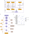
Figure 1
Figure 1
Development of molecular signature and CMap analysis. A, A molecular signature was established through the integration of significant changes between MIBC and NMIBC derived from multiomics data (proteomics and transcriptomics), further enriched with literature-mined data. B, The signature was randomly divided into 10 sets of molecular features and used to query the CMap to predict drugs/compounds affecting the disease’s molecular signature. C, The significant negative enrichment scores for the drugs selected for subsequent in vitro investigations are depicted. GEO, Gene Expression Omnibus; MIBC, muscle-invasive bladder cancer; NMIBC, non muscle-invasive bladder cancer.
Figure 2
Figure 2
Amiodarone decreases proliferation in bladder cancer cell lines. Real-time proliferation assays in UMUC3, HT1197, BFTC905, and RT112 were conducted using the Incucyte S3 system. The cells were treated with increasing concentrations of amiodarone (0–50 μmol/L), and the confluence was measured every 4 hours over 96 hours. A, Cell confluence over time. B, Cell confluence after 96 hours of treatment. Data represent mean ± SEM from at least three independent experiments (one-way ANOVA with Dunnett’s multiple comparisons test; *, P < 0.05; ***, P < 0.001; ****, P < 0.0001). C, Concentration–response curves and IC50 values for amiodarone after 96 hours of treatment.
Figure 3
Figure 3
Amiodarone decreases colony formation capacity in bladder cancer cell lines. Cells seeded in six-well plates were treated with increasing concentrations of amiodarone (0–5 μmol/L). After incubation for 7 to 10 days, the cells were stained with crystal violet, and the plates were scanned. A, Representative images of the colonies. B, Quantification of the colony number. Data are presented as the number of colonies related to the control and represent mean ± SEM from three independent experiments (one-way ANOVA with Dunnett’s multiple comparisons test; **, P < 0.01; ***, P < 0.001; ****, P < 0.0001).
Figure 4
Figure 4
Amiodarone induces apoptosis in bladder cancer cell lines. Cells were treated for 48 or 72 hours with amiodarone (0, 25, and 50 μmol/L for UMUC3; 0, 12.5, and 25 μmol/L for HT1197, BFTC905, and RT112). A, Apoptosis was evaluated using the Caspase-Glo 3/7 Assay and viability using the CellTiter cell proliferation assay. Apoptosis results were normalized to viability. Data are presented as apoptosis related to the control and represent mean ± SEM from at least three independent experiments (one-way ANOVA with Dunnett’s multiple comparisons test; *, P < 0.05; **, P < 0.01; ***, P < 0.001; ****, P < 0.0001). B, Western blot analysis of cPARP expression after 48 hours of treatment with 0, 12.5, and 25 μmol/L amiodarone. GAPDH was used as a loading control. Blots are representative of three independent experiments. Quantification of cPARP expression (normalized by GAPDH) is indicated under each corresponding band. Values are presented as expression related to the control.
Figure 5
Figure 5
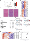
Amiodarone reduces bladder cancer tumor growth in vivo. Tumor growth in BFTC905-bearing NOD/SCID mice treated with 75 mg/kg of amiodarone. A, Tumor volume over time in control (n = 6) and amiodarone-treated (n = 6) tumor-bearing animals during a 3-week cycle treatment. Data represent mean ± SEM (Student t test; **, P < 0.01). B, Body weight of control and amiodarone-treated animals after 27 days of the experiment. Data represent mean ± SEM (Student t test; ****, P < 0.0001). C, Histologic analysis of liver, kidney, and heart sections derived from control and amiodarone-treated animals. Representative hematoxylin and eosin images are shown. D, Heatmap of the top 20 upregulated and top 20 downregulated genes identified by RNA-seq expression analysis of xenograft tumor samples. E, Top 10 significantly deregulated pathways identified by the RNA-seq analysis. F, Gene set enrichment analysis barcode plots for the top five significantly deregulated pathways. G, Heatmap showing the expression of the apoptosis-inducing ligand TNFSF10 and mitochondrially encoded genes of the electron transport chain. H, Dot plot representing the apoptosis gene set activity score derived from RNA-seq gene expression data. FC, fold change.
Figure 6
Figure 6
Amiodarone inhibits mTOR and MAPK pathways. Cells were treated for 24 hours with increasing concentrations of amiodarone (0, 12.5, and 25 μmol/L), and the expression of the proteins AKT, mTOR, S6, and p44/42 MAPK, both phosphorylated (p-) and total, was analyzed by Western blot. GAPDH was used as a loading control. Blots are representative of three independent experiments. Quantification of the protein expression (normalized by GAPDH) is indicated under each corresponding band. Values are presented as expression related to the control. n.d., not determined.

References

    1. American Cancer Society . Cancer Facts & Figures 2024. Atlanta: American Cancer Society; 2024.
    1. Bray F, Laversanne M, Sung H, Ferlay J, Siegel RL, Soerjomataram I, et al. . Global cancer statistics 2022: GLOBOCAN estimates of incidence and mortality worldwide for 36 cancers in 185 countries. CA Cancer J Clin 2024;74:229–63. - PubMed
    1. Sanli O, Dobruch J, Knowles MA, Burger M, Alemozaffar M, Nielsen ME, et al. . Bladder cancer. Nat Rev Dis Primers 2017;3:17022. - PubMed
    1. Tran L, Xiao JF, Agarwal N, Duex JE, Theodorescu D. Advances in bladder cancer biology and therapy. Nat Rev Cancer 2021;21:104–21. - PMC - PubMed
    1. Powles T, Bellmunt J, Comperat E, De Santis M, Huddart R, Loriot Y, et al. . Bladder cancer: ESMO Clinical Practice Guideline for diagnosis, treatment and follow-up. Ann Oncol 2022;33:244–58. - PubMed

MeSH terms