. 2025 May 12;21(5):e1013110.
doi: 10.1371/journal.ppat.1013110. eCollection 2025 May.

Reversible host cell surface remodelling limits immune recognition and maximizes survival of Plasmodium falciparum gametocytes

Affiliations

Reversible host cell surface remodelling limits immune recognition and maximizes survival of Plasmodium falciparum gametocytes

Priscilla Ngotho et al. PLoS Pathog. .

Abstract

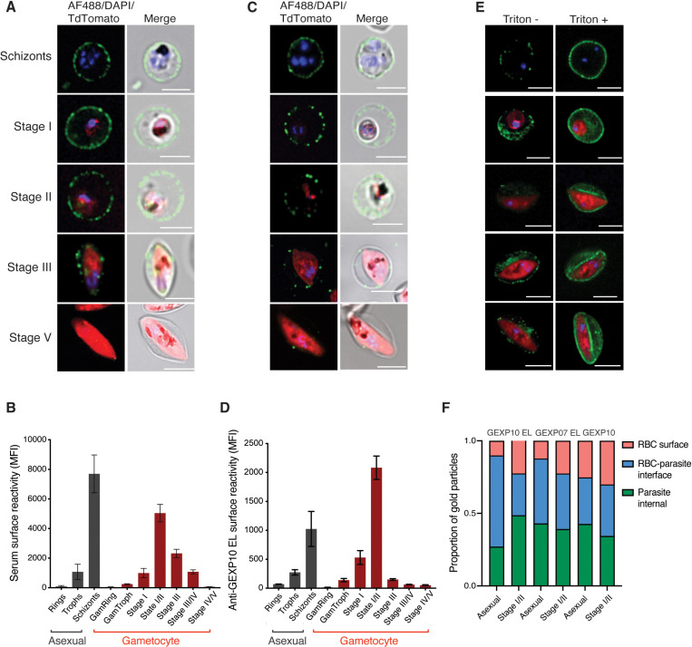
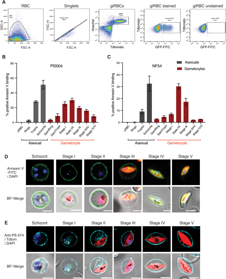
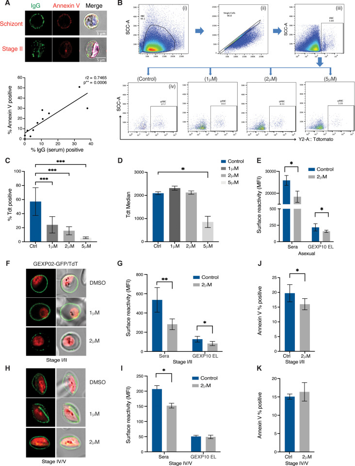
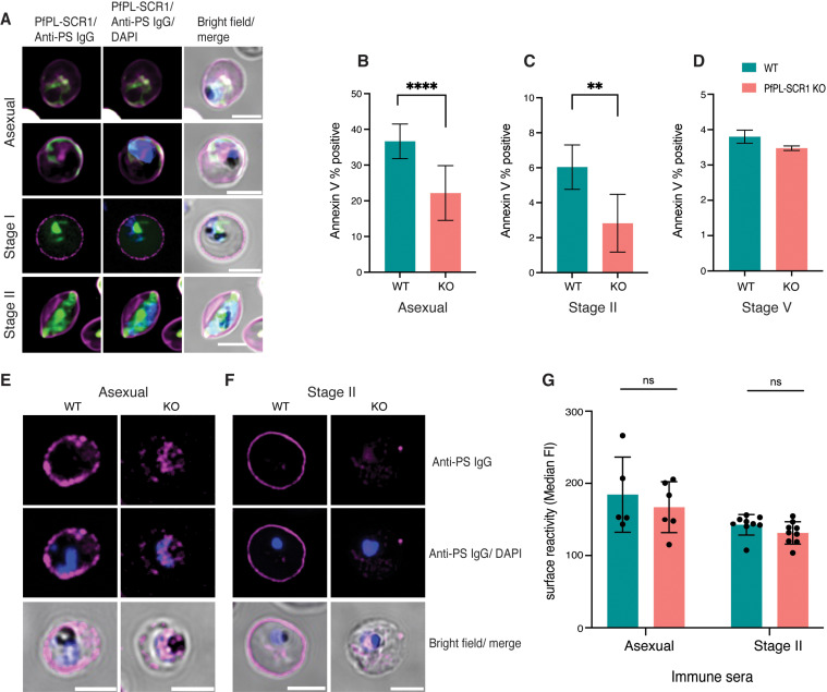
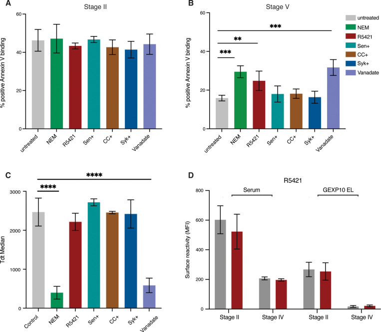
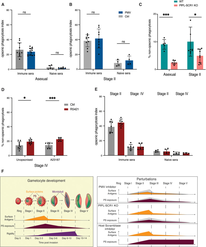
Reducing malaria transmission has been a major pillar of control programmes and is considered crucial for achieving malaria elimination. Gametocytes, the transmissible forms of the P. falciparum parasite, arise during the blood stage of the parasite and develop through 5 morphologically distinct stages. Immature gametocytes (stage I-IV) sequester and develop in the extravascular niche of the bone marrow and possibly spleen. Only mature stage V gametocytes re-enter peripheral circulation to be taken up by mosquitoes for successful onward transmission. We have recently shown that immature, but not mature gametocytes are targets of host immune responses and identified putative target surface antigens. We hypothesize that these antigens play a role in gametocyte sequestration and contribute to acquired transmission-reducing immunity. Here we demonstrate that surface antigen expression, serum reactivity by human IgG, and opsonic phagocytosis by macrophages all show similar dynamics during gametocyte maturation, i.e., peaking in the immature stages and tapering off in mature gametocytes. Moreover, the switch in surface reactivity coincides with reversal in phosphatidylserine (PS) surface exposure, a marker for red blood cell age and clearance. PS is exposed on the surface of a proportion of immature gametocyte-infected RBCs (as well as in late asexual stages) but is removed from the surface in later gametocyte stages (IV-V). Using parasite reverse genetics and drug perturbations, we confirm that parasite protein export into the host cell and phospholipid scramblase activity are required for the observed surface modifications in asexual and sexual P. falciparum stages. Based on these findings we propose that the reversible surface remodelling allows (i) immature gametocyte sequestration in bone marrow followed by (ii) mature gametocyte release into peripheral circulation (and immune evasion due to loss of surface antigens), therefore contributing to mature gametocyte survival in vivo and onward transmission to mosquitoes. Importantly, blocking scramblase activity during gametocyte maturation results in efficient clearance of mature gametocytes, revealing a potential path for transmission blocking interventions. Our studies have important implications for our understanding of parasite biology and form a starting point for novel intervention strategies to simultaneously reduce parasite burden and transmission.


Conflict of interest statement

The authors have declared that no competing interests exist.

Figures

Fig 1
Fig 1. Dynamics of surface antigen exposure in P. falciparum gametocytes.
A,B. Human serum reactivity across asexual and gametocyte development by live microscopy and flow cytometry detection in Pf2004 parasites. (A) Representative images of asexual and gametocytes after live labelling and fluorescence microscopy using immune serum, and (B) flow cytometry quantification of the same samples. C,D. Reactivity of GEXP10 EL antibodies in live cells by (C) fluorescence microscopy and (D) flow cytometry. E. Human serum reactivity in PFA fixed (left) and PFA fixed and Triton X-100 permeabilized cells (right) in asexual and gametocyte stages. F. Quantification of antigen localization based on immuno EM. Asexual stages: 24-36 hpi, GI-IIa: D4, GIIb-III: D6, GIII-IV: D8, GIV-V: D11. Blue: DAPI, green: IgG, red: Tdtomato. B, and D represent the mean from three biological replicates. F, counting a minimum of 30 images per stage and antibody.
Fig 2
Fig 2. Reversible membrane remodelling tracks with antigen exposure during gametocyte development.
A. Example gating strategy for MACS-enriched stage II gametocytes showing annexin V labelled cells in comparison to unstained controls. B-C. Annexin V labelling in live MACS-enriched asexual and gametocyte stages by flow cytometry (B: Pf2004, C: NF54) and fluorescence microscopy (D: Pf2004). E. PS staining using a monoclonal anti-PS antibody in fixed and permeabilized asexual and gametocyte stages (Pf2004). Blue: DAPI, green: Annexin V-FITC, red: Tdtomato, Magenta: anti-PS antibody-647. B, C represent the mean from three biological replicates.
Fig 3
Fig 3. Inhibition of antigen export blocks gametocyte development and surface recognition.
A. Co-staining of surface labelling with human serum and PS with annexin V. Representative images of asexual schizont (top) and stage II gametocytes (bottom). Graph showing correlation of surface labelling and annexin V staining across individual samples (bottom panel). Each point represents the mean of three biological replicates of the combined data from the serum (S1G Fig) and PS exposure (Fig 2A) time course experiments. For each experiment a minimum of 10,000 cells was acquired. B. Flow cytometry data gating strategy. Shown is an example for immature stage II gametocytes, day 5 post induction. Cell population was gated to remove debris (i), followed by single event gating to remove doublets (ii). iRBCs were gated on nuclear dye (iii) and gametocytes on Tdtomato (iv). The Tdtomato % positive cells (gametocytemia) varied based on PMV inhibitor concentration. Left to right: DMSO control, 1μM, 2μM and 5μM PMV inhibitor. C,D. PMV inhibitor arrests early gam development at higher doses based on Tdt MFI quantification. C. Quantification of gametocytemia in MACS enriched parasites by Tdtomato positivity across three PMV inhibitor concentrations. D. Quantification of Tdtomato median fluorescence in Tdtomato positive cells gated in C. E. PMV inhibitor at lower doses blocks antigen export in mature asexual stages (36-44hpi). F. GEXP02 and TdTomato fluorescence upon PMV inhibitor incubation. PMV inhibitor at lower doses blocks antigen export in immature gametocytes. Shown are representative images of immature gametocytes (Stage II). G. Flow cytometry quantification of immune reactivity (human serum and GEXP10 EL) upon PMV inhibitor incubation in immature gametocytes. H-I. PMV inhibitor has less effect on antigen export in mature gametocytes. H. GEXP02 and TdTomato fluorescence upon PMV inhibitor incubation. Shown are representative images of mature gametocytes (Stage IV-V). I. Quantification of immune reactivity (human serum and GEXP10 EL) (I). J, K. Annexin V staining upon PMV inhibitor incubation in immature gametocytes (J) and mature (K) gametocytes. Blue: DAPI, green: IgG (A)/ GEXP02-GFP (F,H), red: Tdtomato. C,D,E,G,I,J,K represent the mean from three biological replicates. Statistics represent paired t-test on inhibitor treated vs untreated (DMSO only) control. p-value <0.0001 ****, p-value <0.001 ***, p-value ≤ 0.01 **, p-value ≤ 0.05 * and p-value > 0.05 ns.
Fig 4
Fig 4. A P. falciparum scramblase affects PS surface exposure.
A. Localisation of PfPL-SCR1-GFP in asexual parasites and immature gametocytes (stage I and stage II) in relation to surface PS. B-D. Effect of PfPL-SCR1 KO on PS exposure in schizonts (B), immature (C) and mature (D) gametocyte stages. E,F. Representative images of live PS staining in PfPL-SCR1 KO parasites using anti-PS antibodies. Shown are asexual schizont stages (E) and Stage II gametocytes (F). G. Serum recognition in the PfPL-SCR1 KO measured by flow cytometry. Blue: DAPI, green: PfPL-SCR1-GFP, Magenta: anti-PS antibody-647. B, C, D, G represent the mean from three biological replicates. Statistics represent paired t-test on PfPL-SCR1 KO vs parental WT line. p-value <0.0001 ****, p-value <0.001 ***, p-value≤ 0.01 **, p-value ≤ 0.05 * and p-value > 0.05 ns.
Fig 5
Fig 5. Blocking PS flipping in gametocytes using scramblase inhibitor.
A. Single dose incubation of compounds tested for effect on immature gametocytes (Stage I-II). Concentration of compounds: NEM (20μM), R5421 (100μM), Senicapoc (6.7μM), CHE (2.5μM), Syk (2.5μM), Vanadate (1μM). B. Single dose of compounds tested for effect on mature gametocytes (Stage IV-V). Concentrations for all compounds as in A. C. Tdtomato reporter median fluorescence in mature gametocytes (Stage IV-V) to determine compound effect on gametocyte maturation. D. Effect of R5421 on human serum and GEXP10 EL antibody surface recognition in immature and mature gametocytes. A-D represent the mean from three biological replicates. Statistics on B-C represent paired t-test on inhibitor treated vs untreated (DMSO only) control. p-value <0.0001 ****, p-value <0.001 ***, p-value ≤ 0.01 **, p-value ≤ 0.05 * and p-value > 0.05 ns.
Fig 6
Fig 6. Phagocytosis of iRBCs upon perturbations.
A-B. Effect of PMV inhibitor on phagocytosis of asexual (A) and immature gametocyte (B) stages using immune serum vs US control serum for opsonisation. C. Effect of PfPL-SCR1 KO PS exposure on iRBC phagocytosis in absence of serum (no opsonisation). Shown are asexual schizonts and ring stages. D. Effect of R5421 on Stage IV-V iRBC non-opsonic phagocytosis in absence of serum and upon addition of the calcium ionophore A23187. E. Effect of R5421 on Stage IV-V iRBC opsonic phagocytosis in presence of serum. F. A model of the remodelling process during gametocyte maturation and effect of the various perturbations. A-E represent the mean from three biological replicates. For the opsonic phagocytosis data, phagocytosis index is determined by subtracting average negative control %DHE and dividing by average positive control %DHE. For the non-opsonised phagocytosis data, % non-opsonic phagocytosis indicates the proportion of DHE positive THP-1 cells (i.e., the proportion of iRBC uptake).

Update of

Similar articles

References

    1. WHO. World malaria report 2021. 2021. PubMed .
    1. Joice R, Nilsson SK, Montgomery J, Dankwa S, Egan E, Morahan B, et al.. Plasmodium falciparum transmission stages accumulate in the human bone marrow. Sci Transl Med. 2014;6(244):244re5. doi: 10.1126/scitranslmed.3008882 - DOI - PMC - PubMed
    1. De Niz M, Meibalan E, Mejia P, Ma S, Brancucci NMB, Agop-Nersesian C, et al.. Plasmodium gametocytes display homing and vascular transmigration in the host bone marrow. Sci Adv. 2018;4(5):eaat3775. doi: 10.1126/sciadv.aat3775 - DOI - PMC - PubMed
    1. Lee RS, Waters AP, Brewer JM. A cryptic cycle in haematopoietic niches promotes initiation of malaria transmission and evasion of chemotherapy. Nat Commun. 2018;9(1):1689. doi: 10.1038/s41467-018-04108-9 - DOI - PMC - PubMed
    1. Obaldia N 3rd, Meibalan E, Sa JM, Ma S, Clark MA, Mejia P, et al.. Bone Marrow Is a Major Parasite Reservoir in Plasmodium vivax Infection. mBio. 2018;9(3):e00625–18. doi: 10.1128/mBio.00625-18 - DOI - PMC - PubMed

MeSH terms

LinkOut - more resources