. 2025 May 12;15(1):16417.
doi: 10.1038/s41598-025-01465-6.

White matter volume and microstructural integrity are associated with fatigue in relapsing multiple sclerosis

Affiliations

White matter volume and microstructural integrity are associated with fatigue in relapsing multiple sclerosis

Alejandra Figueroa-Vargas et al. Sci Rep. .

Abstract

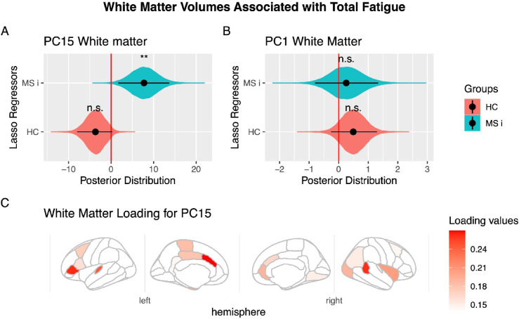
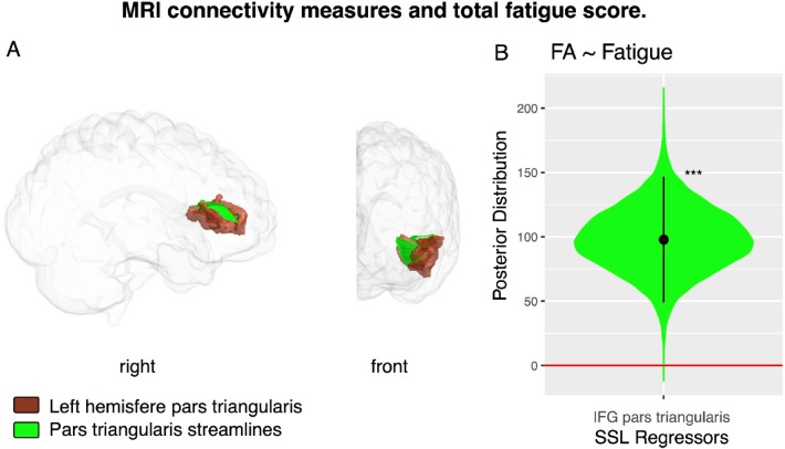
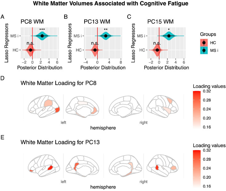
Multiple sclerosis (MS) is a prevalent neurological disorder marked by inflammation and demyelination, with fatigue being one of the most reported and debilitating symptoms. While fatigue occurs across various neurological conditions and even in healthy individuals, the specific mechanisms contributing to fatigue in each context remain unclear. In this study, we conducted a cross-sectional analysis involving 32 people with relapsing MS (PwRMS) and 29 healthy controls who reported fatigue. Participants underwent MRI scans, including T1-weighted and diffusion-weighted imaging. Additionally, the Modified Fatigue Impact Scale was utilized. We employed Bayesian LASSO and Spike-and-Slab LASSO regression models to investigate the hypothesis that fatigue correlates differently with brain structures in PwRMS. Our findings revealed brain regions associated with general and cognitive fatigue. In particular, reduced white matter volume and compromised microstructural integrity in specific areas-such as the cingulate gyrus, inferior frontal gyrus, and the banks of the superior temporal sulcus-showed significant associations with fatigue scores in PwRMS. These results suggest that alterations in specific brain regions may play a critical role in the clinical manifestation of fatigue in MS. Understanding these insights could help differentiate general mechanisms of fatigue from those affecting people with relapsing MS, which may guide future therapeutic strategies.

Keywords: Diffusion tensor imaging (DTI); Fatigue; Frontal pole; Inferior frontal gyrus (IFG); Magnetic resonance imaging (MRI); People with relapsing multiple sclerosis (PwRMS).


Conflict of interest statement

Declarations. Competing interests: The authors declare no competing interests.

Figures

Fig. 1
Fig. 1
White Matter Volumes Associated with Total Fatigue. (A) Principal Component #15 (PC15) demonstrated significant modulation in people with relapsing multiple sclerosis (PwRMS). They showed a significant interaction between PwRMS and healthy controls experiencing fatigue (HC). (B) Principal Component #1 (PC1) showed no significant association with fatigue measures. A-B: The black dot indicates the median, the line shows the 95% high-density interval of the posterior distribution, and the shaded area represents the entire posterior distribution. (C) Loading for PC15. The main white matter volumes are shown according to absolute values of loading of PC15. In the right hemisphere: banks of the superior temporal sulcus (0.26), insula (0.20), lateral occipital (0.19), rostral anterior cingulate (0.19). In the left hemisphere: caudal anterior cingulate (0.27), pars triangularis (0.26), transverse temporal (0.24), cerebellum (0.20) (it is not visible in the figure), cortex entorhinal (0.20), and paracentral (0.18). HC: Healthy control experience fatigue, MC i: Multiple sclerosis interaction, that means the difference in the correlation between HC and patients with multiple sclerosis, rh: right hemisphere, lh: left hemisphere. Statistical significance levels: n.s., no significant difference; *p < 0.05; **p < 0.01; ***p < 0.001.
Fig. 2
Fig. 2
MRI connectivity measures and total fatigue score. (A) Visualization of white matter volumes in the left hemisphere’s Inferior Frontal Gyrus (Pars Triangularis) is shown in brown, with connecting fibers highlighted in green in patients with multiple sclerosis. (B) Posterior distribution of the regressor for Fractional Anisotropy in SWM fibers connecting regions within the Inferior Frontal Gyrus (Pars Triangularis) for the fatigue SSL model. IFG, Inferior Frontal Gyrus; FA, Fractional Anisotropy. Statistical significance levels: n.s., no significant difference; *p < 0.05; **p < 0.01; ***p < 0.001.
Fig. 3
Fig. 3
White Matter Volumes Associated with Cognitive Fatigue, controlled for Physical Fatigue. (A) Principal Component #8 (PC8) demonstrated significant modulation in people with relapsing multiple sclerosis (PwRMS). They showed a significant interaction between PwRMS and healthy controls (HC) experiencing fatigue. (B) Principal Component #13 (PC13) demonstrated significant modulation in people with relapsing multiple sclerosis (PwRMS) and showed a significant interaction between PwRMS and healthy controls experiencing fatigue (HC). (C) Principal Component #15 (PC15) demonstrated significant modulation in people with relapsing multiple sclerosis (PwRMS) and showed a significant interaction between PwRMS and healthy controls experiencing fatigue (HC). A-C: The black dot indicates the median, the line shows the 95% high-density interval of the posterior distribution, and the shaded area represents the entire posterior distribution. (D) Loading for PC #8. The main white matter volumes are shown according to absolute values of loading of PC #8. In the right hemisphere: frontal pole (0.26), caudal middle frontal (0.2), insula (0.17). In the left hemisphere: lateral occipital (0.28), entorhinal (0.24), rostral anterior cingulate (0.21), supramarginal (0.2), parahippocampal (0.18). (E) Loading for PC13. The main white matter volumes are shown according to absolute values of loading of PC13. In the right hemisphere: banks of the superior temporal sulcus (0.30), frontal pole (0.22), parahippocampal (0.22), entorhinal (0.19), isthmus cingulate (0.18). In the left hemisphere: banks of the superior temporal sulcus (0.31), pars orbitalis (0.28), isthmus cingulate (0.27), temporal pole (0.21), pars triangularis (0.21), entorhinal (0.18). HC: Healthy control experience fatigue, MC i: Multiple sclerosis interaction, that means the difference in the correlation between HC and PwRMS, rh: right hemisphere, lh: left hemisphere. Statistical significance levels: n.s., no significant difference; *p < 0.05; **p < 0.01; ***p < 0.001.
Fig. 4
Fig. 4
Connectivity analysis methodology. (A) Raw diffusion-weighted images (DWI) undergo various pre-processing steps to enhance image quality. Following the pre-processing, DTI, FA, and DWI mask images are generated. These outputs are used to create a deterministic whole-brain tractography dataset. (B) The T1w image is co-registered to the DWI space. After co-registration, the cortical parcellation is generated using the HCP pipeline, which uses the Freesurfer mri_synthseg algorithm based on the Desikan Killiany atlas. (C) A structural connectome is generated Using the cortical parcellation and the tractogram to identify and segment the fibers connecting the specific ROIs. The mean FA values of these ROI tracts are then calculated and used for statistical analysis.

References

    1. Walton, C. et al. Rising prevalence of multiple sclerosis worldwide: insights from the atlas of MS, third edition. Mult Scler. J.26, 1816–1821 (2020). - PMC - PubMed
    1. Thompson, A. J., Baranzini, S. E., Geurts, J., Hemmer, B. & Ciccarelli, O. Multiple Scler. Lancet391, 1622–1636 (2018). - PubMed
    1. Kampaite, A. et al. Brain connectivity changes underlying depression and fatigue in relapsing-remitting multiple sclerosis: A systematic review. PLOS ONE. 19, e0299634 (2024). - PMC - PubMed
    1. Meijboom, R. et al. Fatigue in early multiple sclerosis: MRI metrics of neuroinflammation, relapse and neurodegeneration. Brain Commun.6, fcae278 (2024). - PMC - PubMed
    1. Ahvenjärvi, H. et al. Fatigue and health-related quality of life depend on the disability status and clinical course in RRMS. Mult Scler. Relat. Disord. 77, 104861 (2023). - PubMed

MeSH terms

LinkOut - more resources