. 2025 May 12;15(1):16474.
doi: 10.1038/s41598-025-01509-x.

Nrf2 deficiency enhances oxidative stress and promotes susceptibility to tinnitus in mice

Affiliations

Nrf2 deficiency enhances oxidative stress and promotes susceptibility to tinnitus in mice

Huiwen Yang et al. Sci Rep. .

Abstract

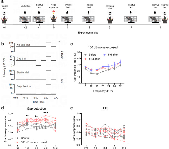
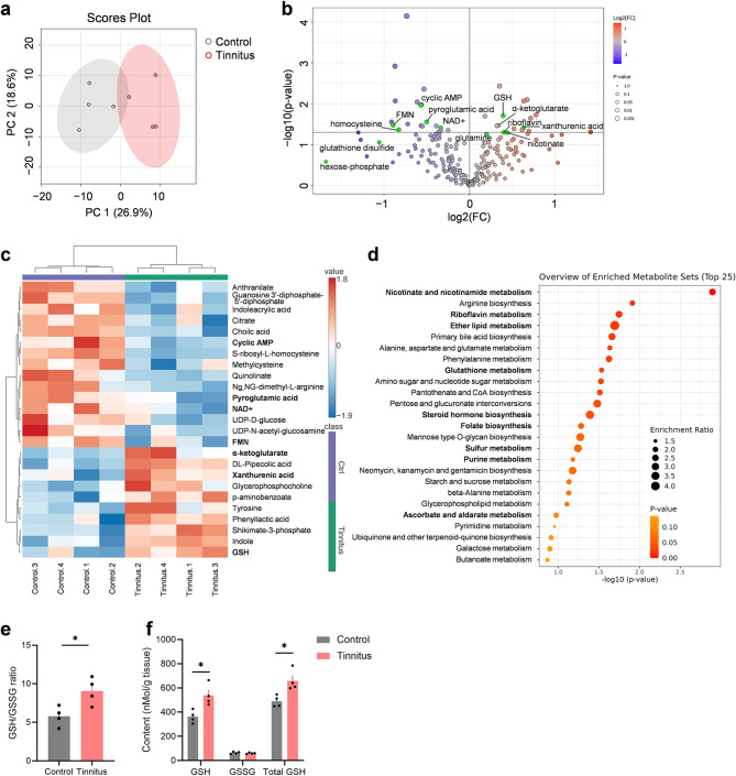
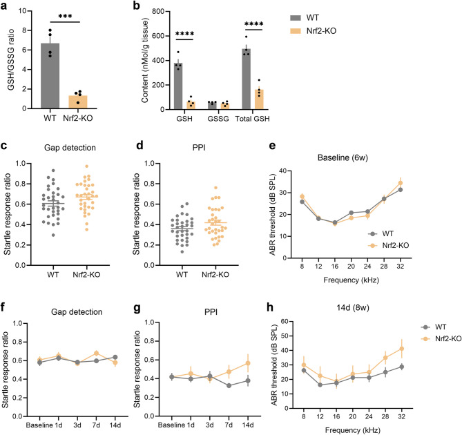
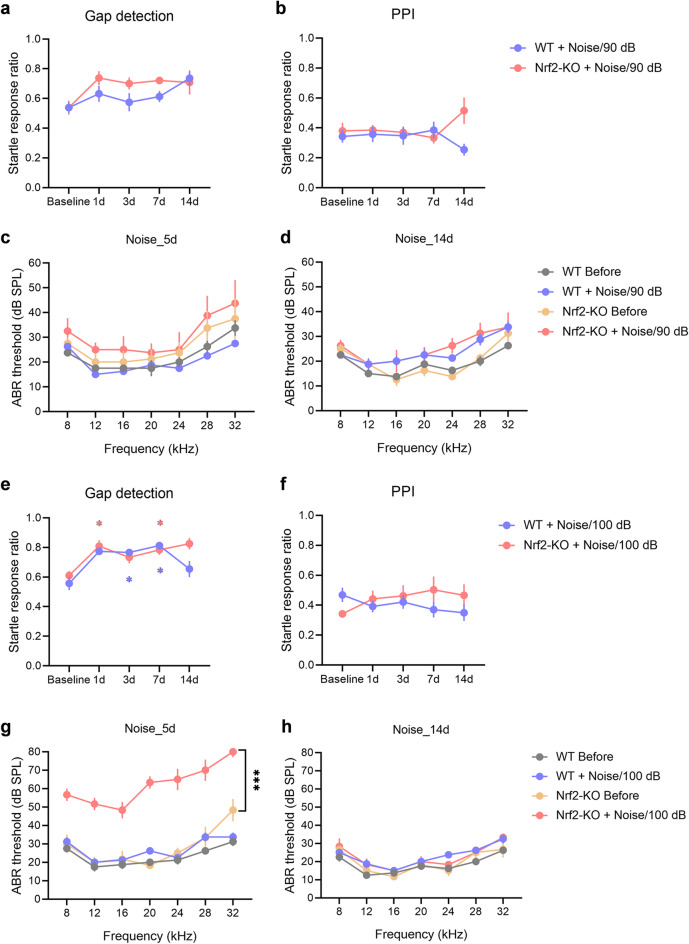
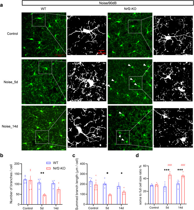
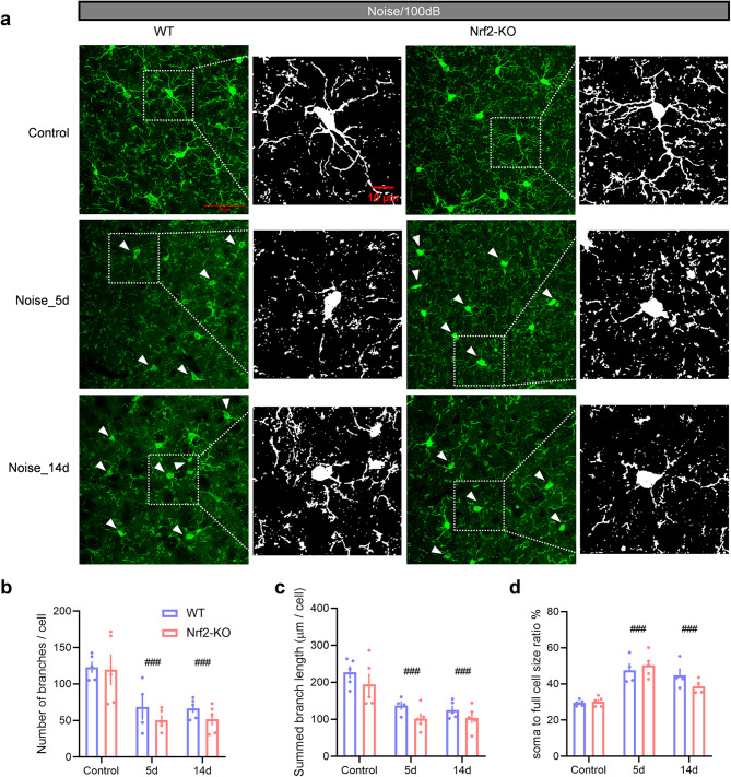
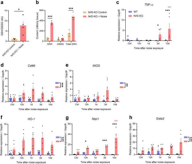
Tinnitus is a prevalent and distressing medical symptom, and no effective pharmacological treatment currently exists. Despite significant advances, tinnitus remains a scientific enigma. To explore the molecular underpinnings of tinnitus, we developed a noise-induced tinnitus model in mice and utilized metabolomics to identify key differences in metabolic pathways. Our results revealed that oxidative stress-related pathways, including glutathione (GSH) metabolism, were significantly enriched in the auditory cortex of mice exhibiting tinnitus-like behavior. To further explore the role of oxidative stress, we examined the involvement of nuclear factor erythroid 2-related factor 2 (Nrf2) in tinnitus by conducting experiments in Nrf2 knockout (Nrf2-KO) mice. While Nrf2-deficient mice did not develop spontaneous tinnitus or hearing loss, they displayed increased susceptibility to prolonged tinnitus-like behavior after noise exposure. This was accompanied by heightened microglial activation, neuroinflammation, and significant alterations in gut microbiota composition, including greater diversity and dysbiosis. Our findings highlight a novel mechanism underlying tinnitus, emphasizing the role of oxidative stress in the auditory cortex and its connection to noise-induced tinnitus. The deficiency of Nrf2 in mice increases their susceptibility to tinnitus, suggesting that Nrf2 may serve as a promising therapeutic target for preventing noise-induced tinnitus.

Keywords: Glutathione; Gut microbiota dysbiosis; Nrf2; Oxidative stress; Tinnitus.


Conflict of interest statement

Declarations. Competing interests: The authors declare no competing interests.

Figures

Fig. 1
Fig. 1
Noise exposure induces tinnitus behaviors without affecting hearing thresholds. (a) Experimental timeline. (b) Schematic representation of the protocol for the gap detection test/GPIAS and PPI. (c) Auditory thresholds of the animals were assessed before noise exposure (gray), 5 days after exposure (blue), and 14 days after exposure (red) (n = 5). Error bars represent SEM. (d-e) Exposure to 100 dB noise results in tinnitus in mice. Tinnitus was assessed using the gap detection test and PPI. Mice showing an increased gap detection ratio and no change in PPI were considered to exhibit behavioral evidence of tinnitus. No significant difference in the gap detection ratio was observed between the noise-exposed group (red) and the control group (gray) before exposure. However, the gap detection ratio of the noise-exposed mice was significantly higher than that of the control group on days 1, 3, and 7 post-exposure, indicating tinnitus-like behavior. By day 14 post-exposure, the gap detection ratio of some mice returned to baseline levels, suggesting the resolution of tinnitus-like behavior (d). The PPI responses of both the noise-exposed and control groups remained stable before and after exposure, with no significant differences observed (e). Each group consisted of 7 mice, with each dot representing a single mouse. The dashed lines between the dots indicate changes in the startle response rate for each individual mouse. ** and *** denote P < 0.01 and P < 0.001, respectively. GPIAS the gap pre-pulse inhibition of acoustic startle test, PPI pre-pulse inhibition, SEM standard error of the mean.
Fig. 2
Fig. 2
Oxidative stress and redox-related pathways are enriched in the tinnitus group. (a) PCA of metabolomics data from the auditory cortex. A significant separation is observed between the tinnitus group (red) and the control group (grey) (n = 4). The ellipse represents the 95% confidence interval of the normal distribution, and each principal component is labeled with its corresponding percentage variance. (b) Volcano plot showing differential metabolites in the tinnitus group compared to the control group, with upregulated metabolites highlighted in red and downregulated metabolites in blue. The threshold for significance is set at a P-value < 0.05. Metabolites involved in oxidative stress and redox processes are highlighted in green. (c) Heatmap of the top 25 differentially regulated metabolites (upregulated in red and downregulated in blue) in the tinnitus group relative to the control group. Values are based on normalized data, as described in the Methods section. (d) Overview of the top 25 enriched metabolite sets related to pathways in the tinnitus group compared to the control group. (e-f) Quantification of glutathione in the auditory cortex. Panel (e) shows the ratio of reduced glutathione (GSH) to oxidized glutathione (GSSG) (GSH/GSSG), while panel (f) displays the levels of GSH, GSSG, and total GSH. Statistical significance was assessed using a non-paired two-tailed t-test. *P < 0.05. Data are presented as mean ± SEM (n = 4). PCA Principal component analysis, SEM standard error of the mean.
Fig. 3
Fig. 3
Nrf2 deficiency results in a significant reduction in GSH levels in the auditory cortex but does not induce spontaneous tinnitus or hearing loss in mice. (a-b) Quantification of GSH in the auditory cortex shows a decreased GSH/GSSG ratio (a) and reduced levels of GSH and total GSH (b) in Nrf2-KO mice (n = 4). (c-e) No significant differences were observed in the gap detection test (c), PPI (d), or ABR thresholds (e) between 6-week-old Nrf2-KO mice and age-matched WT mice. Statistical analysis was performed using an unpaired two-tailed t-test. For behavioral tests, 33 mice were included per group, and for auditory tests, 29 mice per group were used. (f-g) The gap detection test (f) and PPI (g) results in Nrf2-KO mice remained stable throughout a 2-week monitoring period. Statistical analysis was conducted using a two-way repeated measures ANOVA (n = 7). (h) No significant differences were found in ABR thresholds between 8-week-old Nrf2-KO and WT mice (n = 4). Error bars represent the SEM. Statistical significance is indicated by ***P < 0.001 and ****P < 0.0001. Nrf2 nuclear factor erythroid 2-related factor 2, GSH glutathione, Nrf2-KO Nrf2 knockout, WT wild-type, PPI pre-pulse inhibition, ABR auditory brainstem response, ANOVA analysis of variance, SEM standard error of the mean.
Fig. 4
Fig. 4
Nrf2-KO mice exhibit increased susceptibility to noise-induced tinnitus. (a-b) Gap detection tests (a) and PPI (b) were assessed over a 14-day period following exposure to 90 dB SPL noise (n = 7). Statistical analysis was conducted using a genotype × noise exposure two-way repeated measures ANOVA. (c-d) ABR thresholds were evaluated 5 days (c) and 14 days (d) post-exposure to 90 dB SPL noise, with statistical comparisons made using multiple t-tests (n = 4). (e-f) Gap detection tests (e) and PPI (f) were also monitored over 14 days after exposure to 100 dB SPL noise (n = 7). Asterisk (*) indicates statistically significant differences compared to baseline values (P < 0.05), with colors representing the respective animal group. (g-h) ABR thresholds were assessed 5 days (g) and 14 days (h) after 100 dB SPL noise exposure (n = 3 for the Nrf2-KO group and n = 4 for the WT group). All error bars represent the SEM. ***P < 0.001. Nrf2-KO nuclear factor erythroid 2-related factor 2 knockout, PPI pre-pulse inhibition, ANOVA analysis of variance, ABR auditory brainstem response, WT wild-type, SEM standard error of the mean.
Fig. 5
Fig. 5
Microglial deramification is observed in Nrf2-KO mice after 90 dB SPL noise exposure, but not in WT mice. (a) Representative images of IBA1-stained microglia in the auditory cortex of Nrf2-KO and WT mice under control conditions and following noise exposure. In the control group, microglia displayed a ramified morphology, indicative of a resting state. After 5 and 14 days of noise exposure, some microglia in the Nrf2-KO group exhibited activated morphology (white arrows), whereas no morphological changes were observed in the WT group. (b-d) Microglial morphological alterations were quantified as indicators of activation. Five days post-exposure, the number of microglial branches significantly decreased in the Nrf2-KO group (b). At both 5 and 14 days after noise exposure, the total branch length of microglia in the Nrf2-KO group was significantly reduced (c), and the ratio of the cell body to total cell size increased (d), suggesting enhanced microglial activation. No significant differences were observed in the microglial activation index in the WT group (n = 4 or 5). Scale bars = 50 μm and 10 μm. Data are expressed as mean ± SEM. *, **, and *** denote P < 0.05, P < 0.01, and P < 0.001, respectively, for comparisons between Nrf2-KO and WT groups. ### indicates P < 0.001 compared to control groups, with color coding representing the group being compared. Nrf2-KO nuclear factor erythroid 2-related factor 2 knockout, WT wild-type, IBA1 ionized calcium-binding adapter molecule 1, SEM standard error of the mean.
Fig. 6
Fig. 6
Microglial deramification in both Nrf2-KO and WT mice following 100 dB SPL noise exposure. (a) Representative images of IBA1-stained microglia in the auditory cortex of Nrf2-KO and WT mice under control conditions, and at 5- and 14-days post-exposure. Both groups showed activated microglia at 5- and 14-days post-exposure (white arrowheads). (b-d) From 5 to 14 days after exposure, both Nrf2-KO and WT mice exhibited significant reductions in the number of microglial branches (b), total branch length (c), and an increase in the soma-to-cell size ratio (d). Activation indices were also elevated in both groups. No significant differences were observed between the Nrf2-KO and WT groups (n = 4 or 5). Scale bars = 50 μm and 10 μm. Error bars represent the SEM. ### indicates P < 0.001 compared to control. Nrf2-KO nuclear factor erythroid-2-related factor 2 (Nrf2) knockout, WT wild-type, IBA1 ionized calcium-binding adapter molecule 1, SEM standard error of the mean.
Fig. 7
Fig. 7
Noise exposure induces enhanced neuroinflammation in the auditory cortex of Nrf2-KO mice. (a-b) Quantification of GSH in the auditory cortex reveals an increase in the GSH/GSSG ratio (a) and elevated levels of GSH and total GSH (b) in Nrf2-KO mice following noise exposure. Unpaired two-tailed t-tests were performed (n = 3 for the control group and n = 4 for the Nrf2-KO group). (c) TNF-α mRNA levels do not show a significant increase in WT mice after noise exposure, but are elevated in Nrf2-KO mice on days 3 and 10 post-exposure. (d-e) Expression of the microglial markers CD86 and iNOS is exclusively elevated in Nrf2-KO mice. (f-h) The expression of Nrf2 downstream genes HO-1, Nqo1, and Gsta2 is significantly increased in Nrf2-KO mice (n = 4–6 mice per time point). Error bars represent the SEM. *P < 0.05, **P < 0.01, ***P < 0.001 indicate statistical significance; ### denotes P < 0.001 compared to the control group, with the color of # representing the group being compared. Nrf2-KO nuclear factor erythroid-2-related factor 2 knockout, GSH glutathione, TNF-α tumor necrosis factor-alpha, iNOS inducible nitric oxide synthase, HO-1 heme oxygenase 1, Nqo1 NAD(P)H dehydrogenase quinone 1, Gsta2 glutathione S-transferase alpha 2, SEM standard error of the mean.

Similar articles

References

    1. Jarach, C. M. et al. Global prevalence and incidence of tinnitus: A systematic review and Meta-analysis. JAMA Neurol.79, 888–900 (2022). - PMC - PubMed
    1. Mølhave, M., Udholm, S., Hawton, K., Ovesen, T. & Erlangsen, A. Association between hospital-diagnosed tinnitus and suicide: A nationwide Danish longitudinal study. J. Psychosom. Res.185, 111879 (2024). - PubMed
    1. Henton, A. & Tzounopoulos, T. What’s the buzz? The neuroscience and the treatment of tinnitus. Physiol. Rev.101, 1609–1632 (2021). - PMC - PubMed
    1. Baguley, D., McFerran, D., Hall, D. & Tinnitus Lancet382, 1600–1607 (2013). - PubMed
    1. Lockwood, A. H., Salvi, R. J., Burkard, R. F. & Tinnitus N Engl. J. Med.347, 904–910 (2002). - PubMed