

Morphological variations of the interatrial septum and potential implications in equine cardiology
- PMID: 40355652
- PMCID: PMC12069587
- DOI: 10.1038/s41598-025-01387-3
Morphological variations of the interatrial septum and potential implications in equine cardiology
Abstract
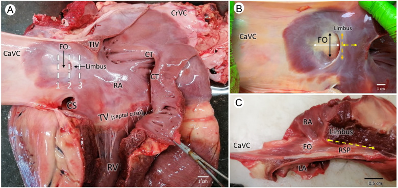
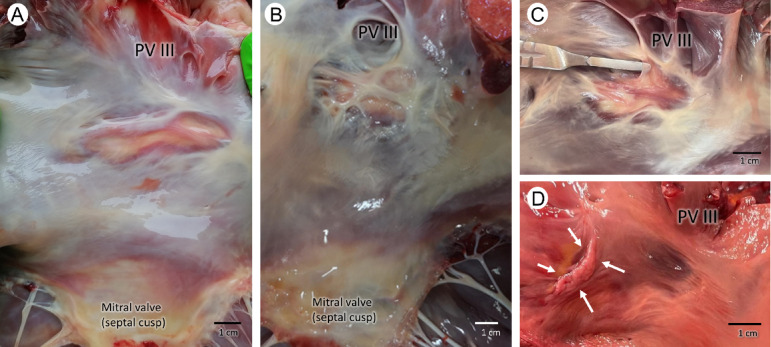
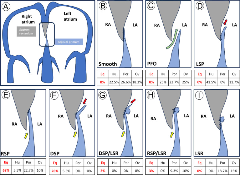
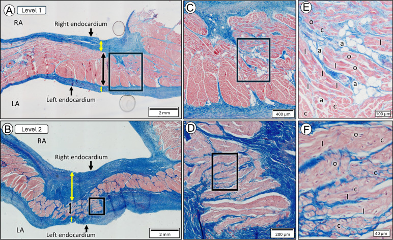
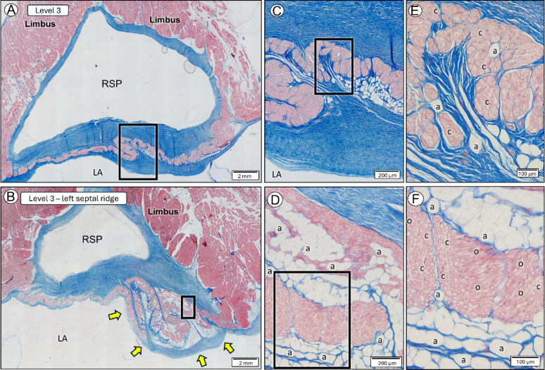
The interatrial septum morphology, shaped by the septum primum and secundum fusion, results in the formation of the fossa ovalis (FO) and its limbus. Incomplete fusion can lead to a patent foramen ovale (PFO), while complete fusion may produce septal ridges and pouches (SPs), with SPs in humans linked to ischemic stroke and atrial arrhythmias. In horses, atrial tachycardia and fibrillation often originate near the FO. This study examines adult equine interatrial septum morphology to enhance understanding the region and guide electrophysiological interventions for equine cardiac arrhythmias. Post-mortem examinations of 62 adult equine hearts, assessed the interatrial septum morphology from both right and left sides, measuring the dimensions of the FO and the craniocaudal length, and dorsoventral height of the SPs. Histological analysis at selected septal locations evaluated the wall's thickness and composition. Significant morphological variations were observed, particularly the consistent presence of right-sided SP. The septum wall comprises three layers, with the central layer containing cardiomyocytes in varied orientations, interspersed with fibroadipose tissue, features potentially contributing to atrial arrhythmias. Understanding the equine interatrial septum morphology is important for optimizing transseptal puncture outcomes, by facilitating accurate intracardiac echocardiography interpretation, guiding precise puncture site selection and improving procedural safety and efficacy.
Keywords: Arrhythmia; Fossa ovalis; Horse; Ridge; Septal pouch; Transeptal puncture.
© 2025. The Author(s).
Conflict of interest statement
Declarations. Competing interests: The authors declare no competing interests.
Figures
References
-
- Holda, M. K., Holda, J., Koziej, M., Piatek, K. & Klimek-Piotrowska, W. Porcine heart interatrial septum anatomy. Ann. Anat.217, 24–28. 10.1016/j.aanat.2018.01.002 (2018). - PubMed
-
- Krishnan, S. C. & Salazar, M. Septal pouch in the left atrium: a new anatomical entity with potential for embolic complications. JACC Cardiovasc. Interv. 3, 98–104. 10.1016/j.jcin.2009.07.017 (2010). - PubMed
-
- Holda, M. K. et al. Atrial septal pouch - Morphological features and clinical considerations. Int. J. Cardiol.220, 337–342. 10.1016/j.ijcard.2016.06.141 (2016). - PubMed
MeSH terms
LinkOut - more resources
Full Text Sources
