. 2025 May 12;15(1):16457.
doi: 10.1038/s41598-025-99128-z.

FOSL1 promotes keratinocyte migration and wound repair by modulating the IL17 signaling pathway

Affiliations

FOSL1 promotes keratinocyte migration and wound repair by modulating the IL17 signaling pathway

Haoran Mao et al. Sci Rep. .

Abstract

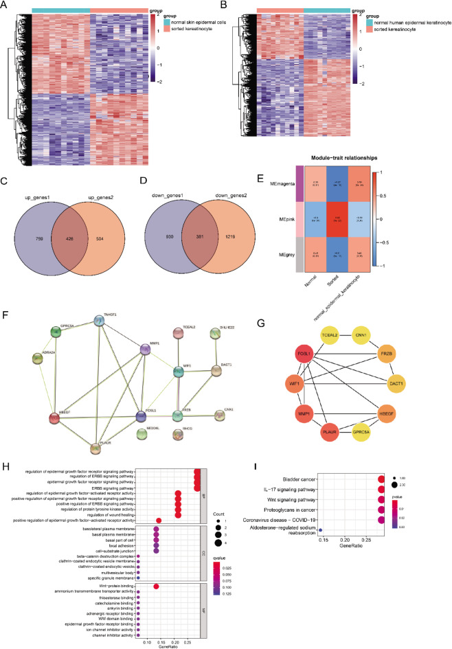
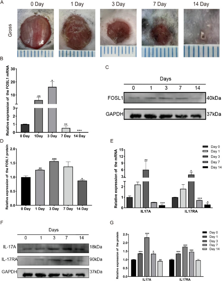
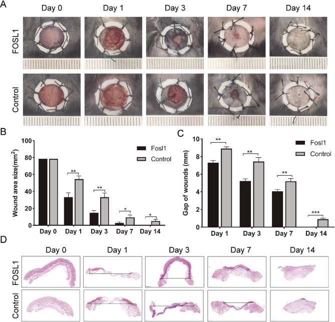
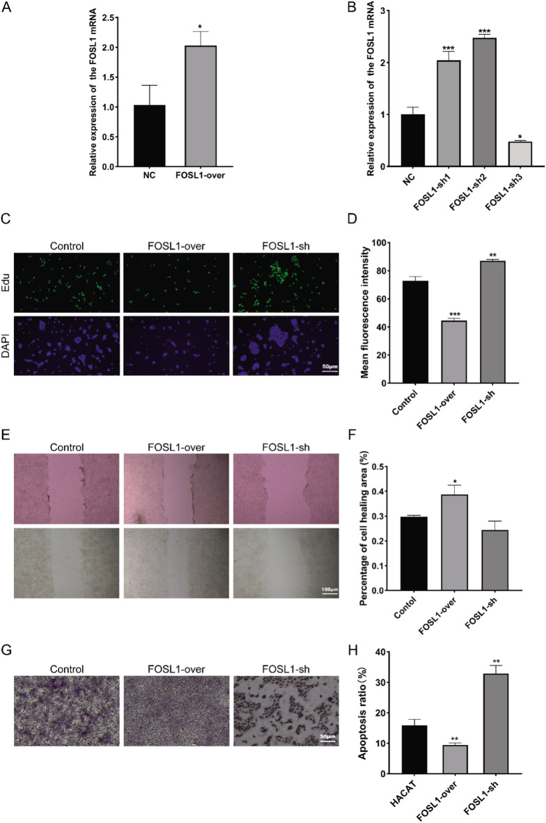
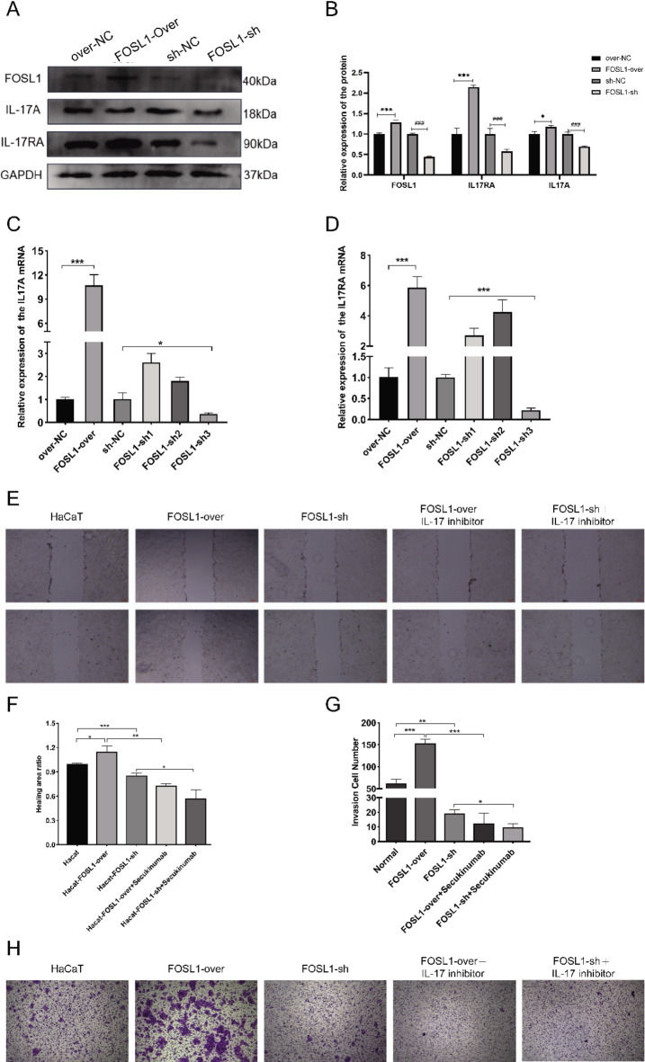
Keratinocytes, the most important cell type constituting the epidermis, migrate to restore the epithelial barrier during wound healing and are a crucial step in wound healing. This study utilized bioinformatics analysis of comprehensive expression datasets of aberrantly expressed genes in wound healing to identify the abnormal expression of the critical transcription factor Fos-like antigen-1 (FOSL1), which is involved in various diseases. Currently, there is limited research on the role of FOSL1 in wound healing, and its molecular mechanisms remain unclear. This study explores the role and regulatory mechanisms of FOSL1 in the wound-healing process. A comprehensive expression dataset of abnormal genes in wound repair was constructed by bioinformatics analysis. Mouse trauma models and mouse wound splint models were constructed to verify the role of FOSL1 in vivo. Real-time quantitative polymerase chain reaction (qRT-PCR), immunoblot, immunofluorescence staining, and HE staining were used to confirm the analysis, and FOSL1 was used as the target in the wound healing process. At the cellular level, using 5'-ethynyl-2'-deoxyuridine (EdU) assay, Transwell assay, Migration assay, western blotting and immunofluorescence, FOSL1 promoted the molecular mechanism of wound repair by regulating the proliferation and migration of keratinocytes through IL-17 signaling pathway. Bioinformatics analysis revealed differential expression of FOSL1 during wound healing. In the mouse back wound model, qRT-PCR, western blotting (WB), and immunofluorescence staining showed significant upregulation of FOSL1 and IL-17 expression during wound tissue healing, indicating a close association between FOSL1 and mouse wound healing. In the mouse wound splinting model, subcutaneous injection of recombinant FOSL1 protein contributed to wound surface healing. Overexpression of FOSL1 in HaCaT cells promoted their proliferation and migration abilities. When IL-17 inhibitor was added to HaCaT cells, both FOSL1 overexpression and knockdown inhibited the proliferation and migration abilities of HaCaT cells. Thus, this study confirms that FOSL1 promotes keratinocyte proliferation and migration through the IL-17 signaling pathway, facilitating wound healing in epidermal wound repair. The results of this study indicate that FOSL1 plays a key role in epidermal wound healing, and elucidate a new molecular mechanism by which FOSL1 promotes keratinocyte proliferation and migration through IL-17 signaling pathway in epidermal wound repair, thereby promoting wound healing.

Keywords: FOSL1; IL-17; Keratinocytes; Wound healing.


Conflict of interest statement

Declarations. Competing interests: The authors declare no competing interests. Ethics approval: Yes, the first animal experiment was approved by the Experimental Animal Ethics Committee of Jinan University with the approval number IACUC-20220719-07. The second animal experiment was approved by the Guangzhou Myers Biological Animal Center Experimental Committee with the approval number IACUC-MIS20230074. In conducting the project, we followed the policies of the Nature Portfolio journal and the ARRIVE Guidelines 2.0.

Figures

Fig. 1
Fig. 1
(A, B) Screening of differential genes during wound healing. (C, D) Cluster heat map analysis of the differentially expressed genes of normal skin epidermal cells and sorted kereatinocytes and normal human epidermal keratinocyte and sorted kereatinocyte from the GSE30355 dataset. (E) Intersection of up-regulated and down-regulated genes during wound healing. (F) WCDNA analysis using MEpink module of kereatinocytes and genes in the hub gene module. MEpink module for protein interaction network analysis (G) FOSL1 and protein interaction network analysis derived from other DEGs (H), The GO enrichment analysis of the key genes in Figure (G, I), KEGG enrichment analysis of the key genes in panel (G).
Fig. 2
Fig. 2
FOSL1 was upregulated in wound tissues. (A) Macroscopy view of wound healing on days 0, 1, 2, 3, 7, 14 mice. Scale bar: 1 mm. (B) A mouse back full-thickness skin resection trauma model in days 0, 1, 2, 3,7, The FOSL1 mRNA expression was determined by qPCR in 14. (C) A mouse back full-thickness skin resection trauma model in days 0, 1, 2, 3, 7, 14 FOSL1 protein expression was determined by Western Blot. (D) Quantification of C. All bars represent mean ± SD (n = 6). (E) qPCR detection of IL17A and IL17RA mRNA expression in traumatic epidermal tissue. (F, G) Western Blot test for the expression of IL17A and IL17RA proteins in traumatic epidermal tissue; Quantification of F. All bars represent mean ± SD (n = 3). *, P < 0.05; **, P < 0.01; ***, P < 0.001.
Fig. 3
Fig. 3
FOSL1 promotes wound healing. (A) Macroscopy view of wound healing on days 0, 1, 3, 7 and 14 in FOSL1 and Control mice. (B, C) Quantification of (AD). All bars represent mean ± SD (n = 3). (D) Wound healing as monitored by histological staining of skin sections at the wound edges at days 0, 1, 3, 7 and 14 after injury in WT, FOSL1 mice.*, P < 0.05; *, P < 0.01; ***, P < 0.001.
Fig. 4
Fig. 4
Expression of FOSL 1 promoted HACAT cell proliferation and decreased the apoptosis of HACAT. (A, B) Expression levels of FOSL 1 overexpression and knockdown mRNA were determined by qPCR. (C, D) Cell apoptosis was assessed via EdU staining assay. Green represents EdU staining and blue represents nuclear staining. Scale bar: 50 μm. (E, F) migration of FOSL1 HaCat cells for overexpression or knockdown by Transwell assays. (G, H) immunohistochemica. *, P < 0.05; *, P < 0.01; ***, P < 0.001.
Fig. 5
Fig. 5
FOSL 1 promoted the expression of IL-17A / IL-17A. (A, B) The FOSL1/IL-17A/IL-17RA/GAPDH level was examined by Western blot. (C, D) The IL-17A and IL-17RA mRNA level was examined by qPCR. Data are shown as mean ± S.E. (E, F) Wound healing assays were shown in HaCat cells after transfection with FOSL1-Over/shRNA and IL-17 inhibitor or NC. Cell migration was observed using a microscope (original magnification × 100). (G, H) Transwell assays were used to detect cell invasion capacities of HaCaT cells after transfection with FOSL1-Over/shRNA and IL-17 inhibitor or NC (dyeing with crystal violet; original magnification, × 200).*, P < 0.05; *, P < 0.01; ***, P < 0.001.

Similar articles

References

    1. Eming, S. A., Martin, P. & Tomic-Canic, M. Wound repair and regeneration: mechanisms, signaling, and translation. Sci. Transl. Med.6(265), 265sr266 (2014). - PMC - PubMed
    1. Werner, S. & Grose, R. Regulation of wound healing by growth factors and cytokines. Physiol. Rev.83(3), 835–870 (2003). - PubMed
    1. Pastar, I. et al. Epithelialization in wound healing: A comprehensive review. Adv. Wound Care (New Rochelle)3(7), 445–464 (2014). - PMC - PubMed
    1. Zhang, L., Xiang, J., Zhang, F., Liu, L. & Hu, C. Mscs can be a double-edged sword in tumorigenesis. Front. Oncol.12, 1047907 (2022). - PMC - PubMed
    1. Sobolev, V. V. et al. Role of the transcription factor Fosl1 in organ development and tumorigenesis. Int. J. Mol. Sci.23(3), 1521 (2022). - PMC - PubMed

LinkOut - more resources