. 2025 May 13;8(1):737.
doi: 10.1038/s42003-025-08183-9.

Gasdermin-D pores induce an inactivating caspase-4 cleavage that limits IL-18 production in the intestinal epithelium

Affiliations

Gasdermin-D pores induce an inactivating caspase-4 cleavage that limits IL-18 production in the intestinal epithelium

J K Bruce et al. Commun Biol. .

Erratum in

Abstract

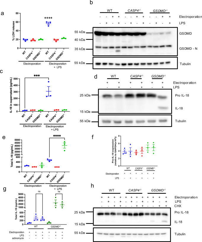
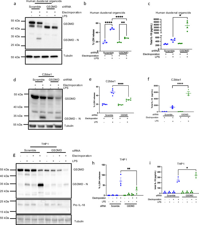
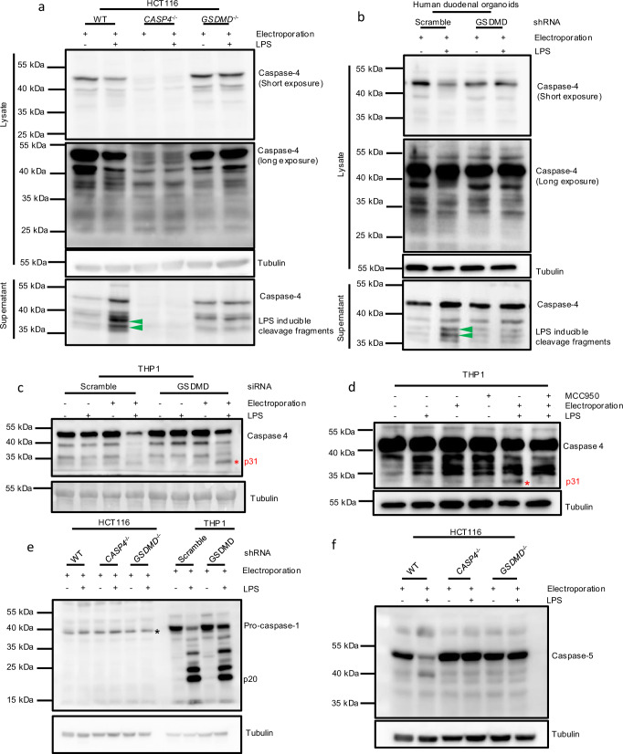
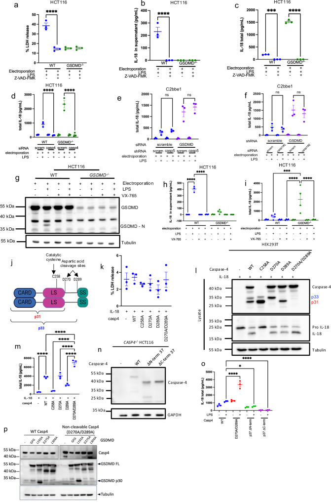
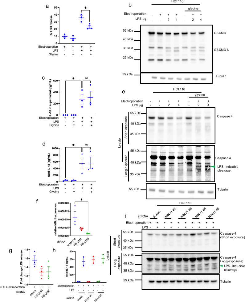
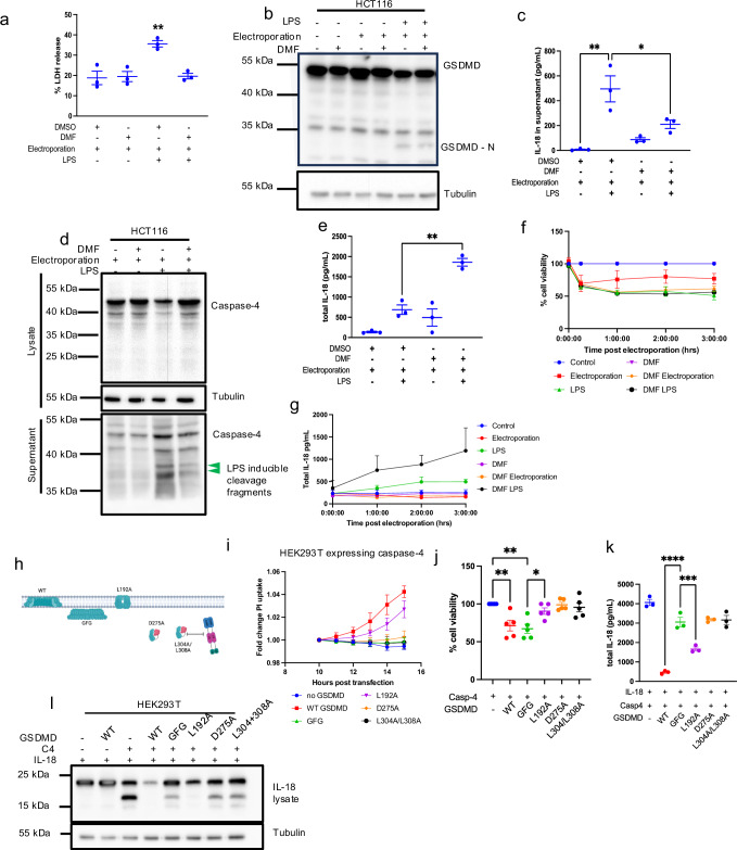
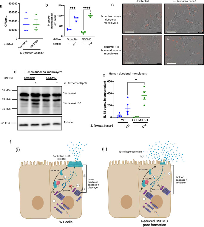
Intestinal epithelial-derived IL-18 is critical for homeostatic intestinal barrier function and is secreted through Gasdermin D (GSDMD) pores. Inflammasome activation is a prerequisite for both IL-18 maturation and GSDMD pore formation. However, GSDMD pores also cause pyroptotic cell death, which could be detrimental to the intestinal epithelial barrier. How epithelial cells balance the need to secrete IL-18 and to maintain barrier integrity remains poorly understood. In human intestinal epithelial cell lines and in primary human epithelial intestinal organoids, but not in immune cells, GSDMD plasma membrane pore formation by LPS electroporation and by gram-negative bacterial infection induced a non-conventional p37 caspase-4 fragment that was associated with reduced levels of mature IL-18. By contrast, limiting GSDMD plasma membrane pores pharmacologically and via point-mutagenesis prevented caspase-4 cleavage and increased IL-18 production, suggesting that p37 caspase-4 cleavage may regulate IL-18 maturation in the intestinal epithelium. In support, co-expression of caspase-4 cleavage mutants and IL-18 in HEK293T cells revealed that non-cleavable caspase-4 produced more mature IL-18 than cleaved caspase-4. Overall, these studies suggest that epithelial inflammasomes encode feedback pathways that control the balance between cytokine secretion and cell death. This may be an important mechanism to ensure homeostatic IL-18 production in the intestinal epithelium.


Conflict of interest statement

Competing interests: The authors declare no competing interests.

Figures

Fig. 1
Fig. 1. GSDMD limits IL-18 maturation in human intestinal epithelial cells.
HCT116 human epithelial cells were electroporated with 2 μg LPS as indicated. Cells were harvested 3 h post electroporation. Cell lysis (a) was measured using LDH cytotoxicity assay. GSDMD (b) and IL-18 cleavage (d) were measured by western blot of whole cell lysates. IL-18 secretion was measured by ELISA in cell free supernatant (c). Total IL-18 (e, g) was measured by lysing cells into supernatant then conducting ELISA. Pro-IL-18 transcript levels were measured by qPCR (f). To block transcription, cells were pre-treated with actinomycin for 1 h prior to electroporation (g). To block translation, cells were incubated with cycloheximide (CHX) for 6 h prior to electroporation (h). Representative of at least 3 experiments, data displayed as mean ±  S.E.M. ***p < 0.005, ***p < 0.001.
Fig. 2
Fig. 2. Overproduction of IL-18 is enhanced in intestinal epithelial cells.
Human duodenal organoids (ac), C2bbe1 intestinal epithelial cells (df) and THP1 human monocytes (gi) were electroporated with 2 μg LPS as indicated. Cell lysis was measured by LDH assay (b, e, h), GSDMD expression and cleavage was measured by western blot (a, d, h), total IL-18 was measured by lysing cells into supernatant then conducting ELISA (c, f, i). Representative of at least 3 experiments, data displayed as mean ± S.E.M. ****p < 0.001, **p < 0.01, *p < 0.05.
Fig. 3
Fig. 3. Blunted LPS-induced caspase-4 processing correlates with IL-18 hyperproduction in GSDMD–/– epithelial cells.
HCT116 epithelial cells (a, e, f), Human duodenal organoids (b) or human THP1 monocytes (c, d) were electroporated with 2 μg LPS. For (d) THP1 cells were co-treated with NLRP3 inhibitor MCC950. Inflammatory caspase activation was measured by western blot on whole cells lysates and supernatant where indicated. For (e) star indicates non-specific band. See Supplementary Fig. 1 for further explanation. Representative of at least 3 experiments.
Fig. 4
Fig. 4. Non-cleaved caspase-4 matures IL-18 in human intestinal epithelial cells.
HCT116 epithelial cells were treated with 2 μM of pan-caspase inhibitor Z-VAD-FMK (ac), siRNA targeting caspase-4 (d) or with caspase-1/4 specific inhibitor VX-765 (gi). C2bbe1 epithelial cells were transduced with shRNA or transfected with siRNA as indicated to generate double -caspase-1 or -5 and GSDMD knockdowns (e, f). For inflammasome activation, cells were electroporated with 2 μg LPS. HEK293T cells (km) or CASP4–/– HCT116 cells were transfected with caspase-4 expression vectors indicated in (j). HEK293T cells were harvested 16 h post transfection (km, p). HCT116 cells were electroporated with LPS 16 h post transfection (n, o). Cell lysis was measured by LDH cytotoxicity assay (a, k). Western blots were conducted on whole protein lysates (g, l, n, p). IL-18 release was measured by ELISA on supernatant alone (b, h) or by lysing cells into supernatant prior to ELISA (c, d, e, f, i, m, o). Representative of at least 3 experiments except for panel (p) (N = 1), data displayed as mean ± S.E.M. ****p < 0.001, ***p < 0.005.
Fig. 5
Fig. 5. Cell lysis does not regulate caspase-4 cleavage and IL-18 production.
ae HCT116 cells were electroporated then placed in media with 10 mM glycine to prevent cell lysis. In panels (fi), HCT116 cells were silenced for NINJ1 expression using shRNA constructs, as indicated, before LPS electroporation. Cell lysis was measured by LDH cytotoxicity assay (a, g). GSDMD (b) and caspase-4 (e, i) cleavage was measured by western blots of whole cell lysates. IL-18 was measured by ELISA on supernatants (c), or on cells lysed into supernatant (d, h). The effect of NINJ1 shRNA constructs 1 and 5 on NINJ1 expression in HCT116 cells was determined by qRT-PCR (f). Green arrow indicates LPS dependent caspase-4 cleavage products. Representative of at least 3 experiments (except for (h) where n = 2), data displayed as mean ± S.E.M. *p < 0.05.
Fig. 6
Fig. 6. Blocking GSDMD pore formation inhibits caspase-4 cleavage and increases IL-18 production but does not prevent cell death.
a–g HCT116 cells were pre-treated with 50 μM dimethylfumerate (DMF) for 1 h then electroporated with LPS and placed back into media containing 50 μM DMF. h–l GSDMD and mutants indicated in schematic (h) were generated. GFG contains point mutations at W48 and W50, that prevent insertion of pores into the plasma membrane. L192A is cleaved and forms pores with less efficiency that WT GSDMD, D275A cannot be cleaved, L304A/L308A cannot bind the caspase-4 exosite. Mutants were transiently transfected into HEK293T cells along with WT caspase-4 and IL-18. Cell lysis was measured by LDH (a), GSDMD, caspase-4 or IL-18 cleavage was measured by western blots on whole cell lysates or supernatant as indicated (b, e). IL-18 was measured by ELISA on supernatant alone (c), or on cells lysed into supernatant (e, g, k). Cellular viability was measured by CellTitre-Glo assay® at indicated timepoints for (f) or 16 h following transfection for (j). PI uptake was measured by calculating the mean fluorescence intensity (MFI) of each condition compared to non-transfected control (i). Representative of at least 3 experiments, data displayed as mean ± S.E.M. ****p < 0.001***p < 0.005, **p < 0.01, *p < 0.05. Panel (h) was created with Biorender.
Fig. 7
Fig. 7. GSDMD regulates IL-18 production and caspase-4 cleavage during Shigella infection in primary human monolayers.
Human duodenal organoids were trypsinised into single cells and seeded onto culture plates pre-coated with 1:40 Matrigel:PBS. Cells were infected with S. flexneri ∆ ospc3 at an MOI of 100 for 4 h. CFUs in infected cells were enumerated (a), PI uptake was monitored using incucyte live cell imager and PI uptake was normalised per area confluency (b), images of monolayers taken 4 h post infection taken at 20x (i) scramble, uninfected; (ii) scramble, Shigella flexneri ∆ ospc3, (iii) GSDMD KD uninfected, (iv) GSDMD KD Shigella flexneri ∆ ospc3. Scale bars (indicting 200 micrometres) are depicted for each micrograph (c). Caspase-4 was measured in western blot of whole cell lysates (d), IL-18 secretion into the supernatant was measured by ELISA on cell free supernatants (e). A schematic of caspase-4 regulation (f). In (f) (i) LPS-induced caspase-4 activation (1) leads to activation of GSDMD and IL-18 (2). GSDMD pores lead to caspase-4 cleavage (3) and this stops IL-18 processing (4). In (f)(ii), during conditions of reduced GSDMD –mediated plasma pore formation, caspase-4 activation (1) leads to IL-18 processing. A lack of efficient pore formation reduces inhibitory feedback on caspase-4 (3). This leads to continued activity of caspase-4 on IL-18 and IL-18 hypersecretion (4). All experiments are representative of at least 3 experiments. Data displayed as mean ± S.E.M. *p < 0.05, ****p < 0.001. Panel (f) created with Biorender.

References

    1. Winsor, N., Krustev, C., Bruce, J., Philpott, D. J. & Girardin, S. E. Canonical and noncanonical inflammasomes in intestinal epithelial cells. Cell Microbiol21, e13079 (2019). - PubMed
    1. Sborgi, L. et al. GSDMD membrane pore formation constitutes the mechanism of pyroptotic cell death. EMBO J.35, 1766–1778 (2016). - PMC - PubMed
    1. Shi, J. et al. Cleavage of GSDMD by inflammatory caspases determines pyroptotic cell death. Nature526, 660–665 (2015). - PubMed
    1. Evavold, C. L. et al. The pore-forming protein gasdermin D regulates interleukin-1 secretion from living macrophages. Immunity48, 35–44.e36 (2018). - PMC - PubMed
    1. Chen, X. et al. Pyroptosis is driven by non-selective gasdermin-D pore and its morphology is different from MLKL channel-mediated necroptosis. Cell Res.26, 1007–1020 (2016). - PMC - PubMed

MeSH terms

LinkOut - more resources